MUCOPOLYSACCHARIDOSES
Key features
A group of inherited disorders, primarily autosomal recessive, characterized by deficiencies of specific lysosomal enzymes involved in the catabolism of glycosaminoglycans (GAGs), also referred to as mucopolysaccharides
Excessive amounts of the GAGs are found in various tissues and lead to coarse facies, intellectual disability, hepatosplenomegaly, skeletal abnormalities, and corneal clouding
Hunter syndrome (X-linked recessive) has distinctive skin-colored to white papules over the scapulae (pebbling)
Screening tests include examination of the urine, peripheral leuko- cytes, and dermal fibroblasts for the presence of excessive GAGs followed by genetic mutational analysis
Clinical Features and Pathology
The mucopolysaccharidoses are due to mutations in several genes that encode enzymes involved in the catabolism of glycosaminoglycans (GAGs). Examples of these GAGs include dermatan sulfate, heparan sulfate, and chondroitin sulfate. The storage of excessive amounts of GAGs in various tissues can lead to coarse facies, intellectual disability, hepatosplenomegaly, skeletal abnormalities (dysostosis multiplex), joint stiffness, cardiovascular disease, and corneal clouding (Table 48.3).
Cutaneous manifestations are nonspecific (e.g. hypertrichosis, thickening of the skin), except for the presence of skin-colored to white papules or nodules over the scapulae (“pebbling”) in patients with Hunter syndrome, which is inherited in an X-linked recessive fashion. The lesions measure 1–10 mm in diameter and may become confluent; they can extend from the posterior axillary line to the midline. Less common sites are the arms, the lateral upper chest, and the buttocks. Extensive dermal melanocytosis (i.e. Mongolian spots) can occur on the trunk and extremities in patients with Hunter or Hurler syndrome.
Screening tests include examination of the urine for excessive amounts of GAGs and examination of circulating leukocytes or dermal fibroblasts for vacuoles or granules that can be demonstrated via special stains (see Table 46.1). The peripheral blood smear should be fixed in absolute methanol and fixation of the skin biopsy specimen in absolute alcohol is preferred. Specific enzyme assays or genetic mutational analysis can then be performed; prenatal diagnosis is possible.
In biopsy specimens from all types of mucopolysaccharidosis (MPS), metachromatic granules are seen within the cytoplasm of fibroblasts and in some cases in sweat glands, the external root sheath of hair follicles, and epidermal keratinocytes. The latter may have a pale distended cytoplasm which displaces the nucleus to one side. Biopsy specimens of diffusely thickened skin demonstrate fragmentation and hyalinization of collagen and increased mucin. Extracellular deposits of metachromatic material and mucin deposition are seen in the papulonodular lesions of Hunter syndrome. By electron microscopy, fibrillogranular material is seen within lysosomes in all types of MPS.
Treatment
Although supportive interventions (e.g. home oxygen, physical therapy for joint stiffness, cardiac valve replacement) play an important role in the treatment of MPSs, additional therapeutic options – in particular, enzyme replacement therapy (ERT) and hematopoietic stem cell transplantation (HSCT) offer long-term hope for affected individuals. Currently available ERT includes α-L-iduronidase (laronidase; Aldurazyme™) for MPS I, iduronate-2-sulfatase (idursulfase; Elaprase™)
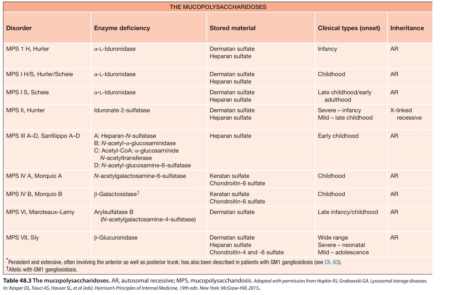
Table 48.3 The mucopolysaccharidoses. AR, autosomal recessive; MPS, mucopolysaccharidosis. Adapted with permission from Hopkin RJ, Grabowski GA. Lysosomal storage diseases. In: Kasper DL, Fauci AS, Hauser SL, et al (eds). Harrison’s Principles of Internal Medicine, 19th edn. New York: McGraw-Hill, 2015.
for MPS II, N-acetylgalactosamine 4-sulfatase (galsulfase; Naglazyme™) for MPS VI, and β-glucuronidase (vestronidase-α; Mepsevii™) for MPS VII. ERT is effective in controlling pulmonary insufficiency, decreased joint mobility and hepatosplenomegaly, but does not improve bone or valvular heart disease. Nor does it affect the intellectual disability, as the enzymes cannot pass the blood–brain barrier.
Allogeneic HSCT has been used successfully in patients with MPS I, II, IVA, VI, and VII. Results vary depending upon the age of the patient and the disease stage at the time of the procedure, with better results when performed at an early age. Joint mobility, cardiopulmonary function, hearing, vision, and coarse facies can be improved with allogeneic HSCT, in addition to improvement of CNS impairment in MPS I, II, and VII. Substrate reduction therapy and gene therapy represent future treatment options. An example of the latter is administration of hematopoietic stem and progenitor cells transduced ex vivo with an α-l-iduronidase-encoding lentiviral vector after myeloablative conditioning29a.