PATHOGENESIS
Mutations in any of the genes encoding the enzymes in the heme biosynthetic pathway can lead to a pathologic accumulation and excess excretion of porphyrins and/or porphyrin precursors, as a result of enzyme dysfunction. Except for patients with acquired porphyria cutanea tarda, all forms of porphyria are inherited as monogenetic traits (see Table 49.3).
Cutaneous Findings
To date, no single factor can explain the various photosensitivity patterns evoked by porphyrins plus visible and UV radiation. Nonetheless, a number of cellular and soluble factors are thought to be involved, including reactive oxygen species, certain cell types (e.g. erythrocytes, mast cells, polymorphonuclear cells, fibroblasts), and soluble mediators (e.g. components of the complement system or factor XII-dependent pathways, eicosanoids), as well as matrix metalloproteinases. Most likely, interactions between these factors contribute to the development of cutaneous lesions.
Within the Soret band (400 to 410 nm), porphyrins absorb light energy most efficiently and enter into an excited molecular state. There is also less prominent absorption of adjacent UV wavelengths and other minor visible bands. These light-excited porphyrins can then in turn transfer this absorbed energy to oxygen molecules thereby generating highly reactive oxygen species. Cellular and tissue damage induced by photoactivated porphyrins is believed to result primarily from the formation of reactive singlet oxygen and free radicals, with subsequent lipid peroxidation and protein cross-linking.
The type of cellular damage depends on the solubility and tissue distribution of the porphyrins. Accumulation of water-soluble uro- and coproporphyrins leads to blistering, as is seen in most of the cutaneous porphyrias (e.g. porphyria cutanea tarda, variegate porphyria). In contrast, accumulation of lipophilic protoporphyrins leads to an immediate cutaneous burning sensation after exposure to
the appropriate wavelengths of light, accompanied by erythema and edema, as seen in erythropoietic protoporphyria.
Acute Porphyric Attack
Two porphyria variants, ALA dehydratase deficiency porphyria and acute intermittent porphyria, are not associated with cutaneous findings (see Table 49.1). The dysfunctional enzymes in these two porphyrias are involved in early steps of heme biosynthesis, and their substrates, ALA and porphobilinogen (PBG), respectively, are non-phototoxic porphyrin precursors (see Fig. 49.1). However, both acute intermittent porphyria and ALA dehydratase deficiency porphyria can manifest with lifethreatening acute neurologic attacks (see Table 49.2). It appears that ALA and PBG, which are excreted in massive amounts from the liver during an acute attack, are extremely neurotoxic. Lacking appropriate barrier protection, the autonomic and peripheral nervous systems are particularly susceptible to the toxic effects.
Of note, porphyrin abnormalities are also observed in the setting of lead poisoning, sideroblastic and hemolytic anemia, iron deficiency, renal failure, cholestasis, liver disease, and gastrointestinal hemorrhage. However, associated photosensitivity has only been documented in rare cases of sideroblastic anemia.
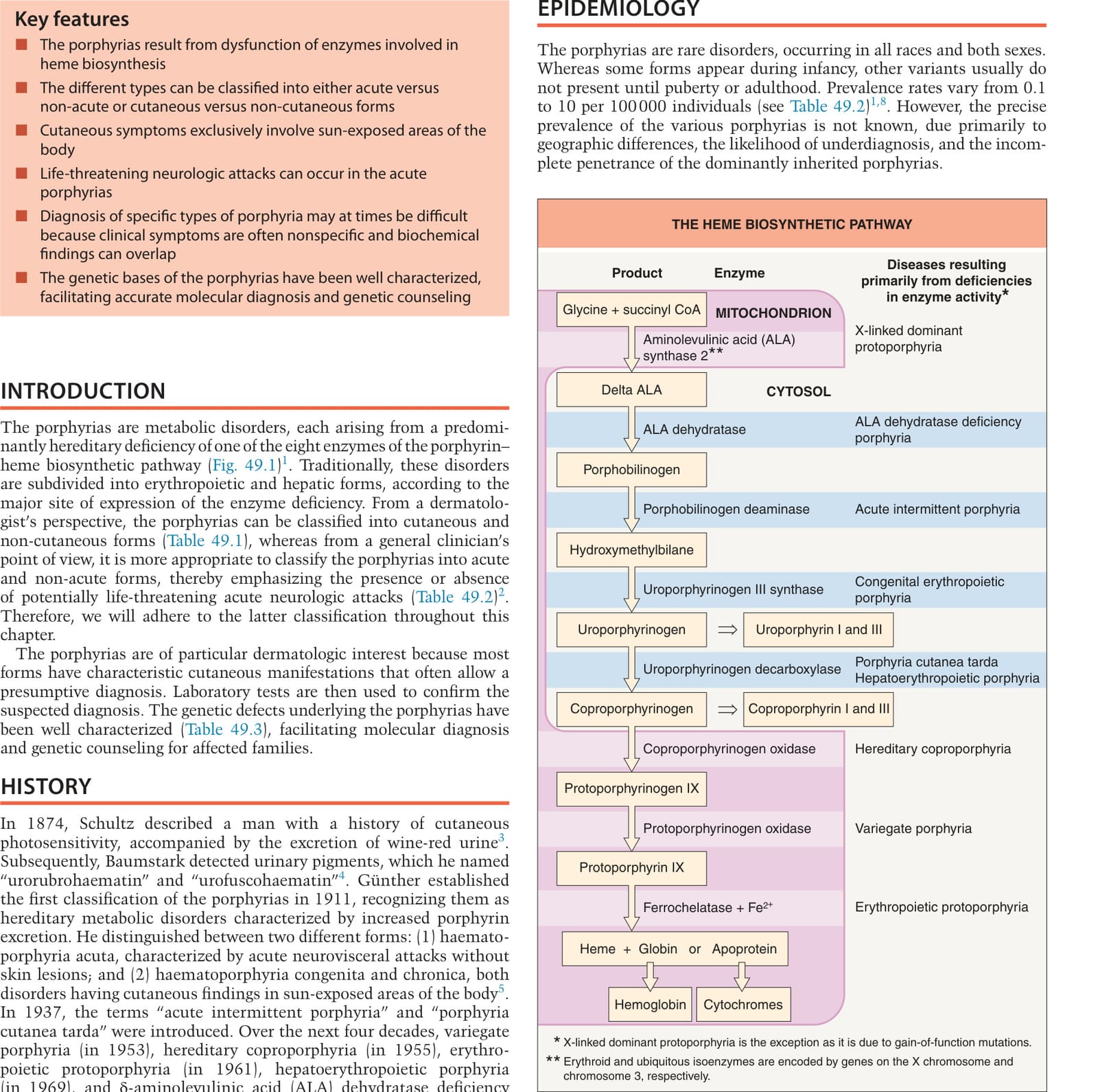
Fig. 49.1 The heme biosynthetic pathway. ALA, aminolevulinic acid, also referred to as δ-aminolevulinic acid or 5-aminolevulinic acid.
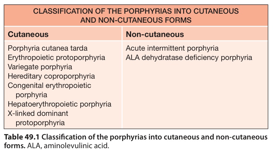
Table 49.1 Classification of the porphyrias into cutaneous and non-cutaneous forms. ALA, aminolevulinic acid.
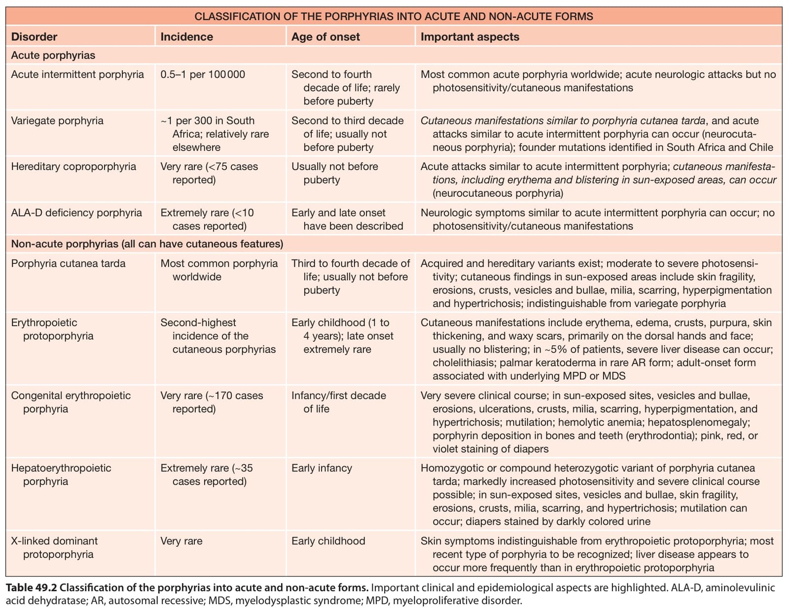
Table 49.2 Classification of the porphyrias into acute and non-acute forms. Important clinical and epidemiological aspects are highlighted. ALA-D, aminolevulinic acid dehydratase; AR, autosomal recessive; MDS, myelodysplastic syndrome; MPD, myeloproliferative disorder.
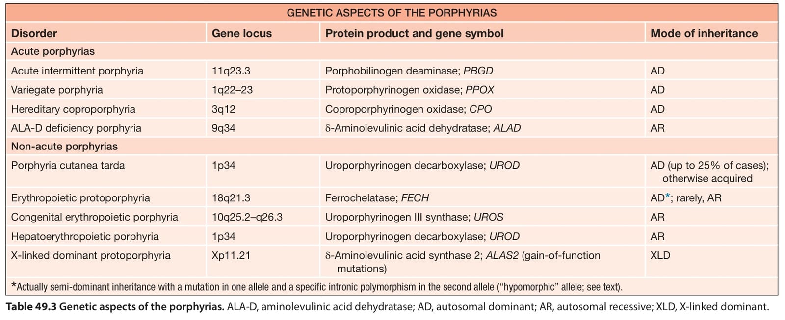
Table 49.3 Genetic aspects of the porphyrias. ALA-D, aminolevulinic acid dehydratase; AD, autosomal dominant; AR, autosomal recessive; XLD, X-linked dominant.