THE NON-ACUTE PORPHYRIAS
The non-acute porphyrias consist of porphyria cutanea tarda (PCT), erythropoietic protoporphyria (EPP), congenital erythropoietic porphyria (CEP), hepatoerythropoietic porphyria (HEP), and X-linked dominant protoporphyria (XLDPP). All these porphyrias present primarily with cutaneous findings (see Tables 49.1 & 49.2). While histologic examination of involved skin is not required to confirm a presumptive diagnosis of any of the cutaneous porphyrias, a biopsy specimen may have been obtained to exclude other entities in the differential diagnosis. Occasionally, the pathologist may identify histologic features that support the diagnosis of cutaneous porphyria even though the clinician suspected another disorder, e.g. epidermolysis bullosa acquisita.
Porphyria Cutanea Tarda (PCT)
Worldwide, PCT is the most common type of porphyria. It results from decreased activity of uroporphyrinogen decarboxylase, the fifth enzyme in heme biosynthesis (see Fig. 49.1).
At least two types of PCT can be distinguished: (1) a sporadic (acquired) variant, designated type I PCT, in which the dysfunctional enzyme is exclusively expressed in the liver; and (2) an autosomal dominant familial (hereditary) variant, designated type II PCT, in which the catalytic enzymatic defect is detected in all tissues (see Table 49.3). In most countries, the ratio of type I to type II PCT is currently estimated to be approximately 3 : 1 to 4 : 1. Interestingly, Elder reported several families with typical clinical and biochemical features of overt PCT but with normal uroporphyrinogen decarboxylase activity in their red blood cells. This variant of the disease has been designated type III PCT. Indeed, there is increasing evidence that some facets of the etiology of PCT have yet to be completely elucidated.
The cutaneous manifestations of PCT include increased photosensitivity and skin fragility as well as blistering, erosions, crusts, milia, and scars in sun-exposed sites (Fig. 49.3; see Table 49.2). Additionally, postinflammatory hyperpigmentation, hypertrichosis (Fig. 49.4), scarring alopecia, and morpheaform and sclerodermoid changes (Fig. 49.5) can be observed.
Histologic examination of vesiculobullae commonly reveals subepidermal cell-poor blisters with a characteristic festooning of dermal papillae; the latter is most likely due to the deposition of PAS-positive
glycoproteins in and around the wall of vessels localized in the upper dermis (Fig. 49.6). Direct immunofluorescence microscopy often demonstrates immunoglobulins (mainly IgG; less commonly IgM), complement, and fibrinogen at the dermal–epidermal junction and around blood vessels of the papillary dermis. As opposed to autoimmune bullous diseases, these immunoglobulin deposits are presumed to be non-antigen-specific. In sclerotic lesions, there is dermal fibrosis as well as PAS-positive deposits around blood vessels.
Biochemically, increased urinary excretion of uroporphyrin (type I isomers > type III isomers), hepta-carboxylated porphyrins (type III isomers > type I isomers), and coproporphyrin can be detected, as well as increased excretion of isocoproporphyrin in the feces; the latter finding is exclusive to PCT and hepatoerythropoietic porphyria (see Table 49.4). Historically, a presumptive clinical diagnosis of PCT was followed by an examination of the patient’s urine, both under Wood’s lamp illumination (in the dark) and after exposure to natural light. Due to the excessive excretion of porphyrins, the urine of PCT patients turns red to brown after several hours of exposure to natural light and it has a pink to red fluorescence when exposed to a UVA light source.
However, it has to be borne in mind that these bedside observations are neither sensitive nor specific diagnostic tests.
A wide variety of triggering factors (including hepatotoxins) have been reported to precipitate the clinical manifestations of PCT, among them alcohol (most common), estrogens, polychlorinated hydrocarbons, dialysis in patients with renal failure, iron, inheritance of specific mutations in HFE which underlie classic hemochromatosis, and viral infections such as hepatitis C and HIV. Interestingly, homozygosity for HFE mutation C282Y has been found to be associated with an earlier onset of disease in both sporadic and familial PCT, with the effect being more marked in familial PCT.
Erythropoietic Protoporphyria (EPP)
EPP is due to a semi-dominantly inherited deficiency of ferrochelatase, the last enzyme in the heme biosynthetic pathway (see Fig. 49.1).
Clinically, EPP is characterized by cutaneous photosensitivity that typically manifests early in life, i.e. during early childhood. Acute photosensitivity episodes are characterized by intense burning,
stinging, and pruritus of sun-exposed skin, particularly on the nose, cheeks and dorsal hands, followed by erythema, edema, crusts, petechiae, and then wax-like scarring (Fig. 49.7 & 49.8; see Table 49.2); blistering usually does not occur. Skin symptoms can develop
Subtle scarring on the nose and linear scars on the upper cutaneous lip in a 6-year-old child. B Extensive scarring of the cheeks and linear scars on the upper cutaneous lip and chin in an adult. A, Courtesy Gillian Murphy, MD; B, Courtesy Luis Requena, MD.
within minutes of sun exposure, often starting early in the springtime, continuing throughout the summer, and diminishing during fall and winter. Window glass does not protect the patient. Patients suffering from EPP often report that the only way to relieve the burning and stinging sensations is to cool the affected skin under cold water. In patients with EPP who have not had recent sun exposure, the only detectable cutaneous findings may be scars on the nose and lips (see Fig. 49.8). In rare patients with recessively inherited EPP, palmar keratoderma can be seen.
Adult-onset EPP, also referred to as late-onset EPP, is very unusual, and the atypical age of onset often delays the diagnosis. To date, all reported individuals with late-onset EPP have had a history of either a myeloproliferative disorder or myelodysplastic syndrome. Interestingly, in these patients the hematologic disease is thought to possibly give rise to somatic hematopoietic mutations or clones that result in abnormal porphyrin metabolism and systemic protoporphyrin accumulation. Thus, this disorder should be considered in adults with new-onset, otherwise unexplained photosensitivity, especially if they have an underlying hematologic disorder.
Histologically, vacuolization of epidermal cells is seen in acute lesions. Intercellular edema may also be present, as well as vacuolization and lysis of endothelial cells within superficial dermal blood vessels. In older lesions (e.g. areas of waxy scarring), eosinophilic PAS-positive deposits are observed around blood vessels (Fig. 49.9). There can be a resemblance to the amorphous protein depositions seen in lipoid proteinosis. Ultrastructurally, thickening and degeneration of capillary basement membranes is seen.
Biochemically, EPP is characterized by an increase in free protoporphyrin levels in erythrocytes, plasma, feces, and other tissues such as the liver (see Table 49.4). Historically, microscopic examination of a blood smear via UV illumination was performed to detect erythrocyte fluorescence. However, this test is not particularly sensitive or specific. The greatest concern in patients with EPP is a rapid accumulation of protoporphyrin within the liver and biliary system, with the development of cholestasis. This can lead to hepatic damage and progressive (even fatal) liver failure. Of note, liver function tests may remain normal until late in the course of the disease.
The genetic basis for the development of cutaneous symptoms in EPP has been elucidated. Only those individuals who inherit a heterozygous ferrochelatase gene mutation involving one parental allele (allele A) and a specific intronic polymorphism (IVS3–48 T/C) of the other parental allele (allele B) develop skin symptoms. This latter specific polymorphism in trans constitutes a “hypomorphic” allele and leads to abnormal modulation of splicing. As a result, the aberrantly spliced mRNA is degraded by a nonsense-mediated decay mechanism, producing a decreased steady-state level of mRNA. Combined, a mutation in allele A plus the polymorphism in allele B lead to a marked reduction in ferrochelatase activity that is ~15%–25% of normal.
Certainly, the identification of the molecular mechanisms under-lying the photosensitivity observed in EPP has to be considered a milestone in porphyria research. Still, the development of protoporphyrin-induced hepatic disease and the molecular mechanisms under-lying the phenotype associated with severe liver injury are not yet well understood.
X-linked Dominant Protoporphyria (XLDPP)
XLDPP is characterized by gain-of-function mutations in ALAS2 on the X chromosome. This gene encodes the erythroid tissuespecific isoform of the first enzyme in the heme biosynthetic pathway, δ-aminolevulinic acid synthase 2. The resultant increase in synthetase activity eventually leads to an overproduction of protoporphyrin IX and a clinical phenotype that is indistinguishable from EPP. However, the percentage of patients at risk for potentially fatal liver disease appears to be higher than in classic EPP. Biochemical profiling demonstrates higher total protoporphyrin levels in erythrocytes (as compared to EPP due to ferrochelatase deficiency), with ~40% being zinc protoporphyrin.
Congenital Erythropoietic Porphyria (CEP)
Synonym: Günther disease
With ~170 cases reported to date, CEP is a very rare, autosomal recessively inherited disorder that results from markedly decreased catalytic activity of uroporphyrinogen III synthase, the fourth enzyme in heme biosynthesis (see Fig. 49.1). CEP manifests shortly after birth with severe cutaneous photosensitivity, blistering, erosions, crusts, and ulcerations, followed by extensive scarring and deformation, primarily of the hands (Fig. 49.10A; see Table 49.2). On the face, a loss of eyebrows and eyelashes, as well as severe mutilation of cartilaginous structures (e.g. the nose), are frequently observed. Furthermore, erythrodontia (Fig. 49.10B), acro-osteolysis, and skeletal abnormalities (e.g. demineralization) are common clinical features. In addition, a variable degree of hematologic involvement, ranging from mild forms of hemolytic anemia to intrauterine hydrops fetalis and hepatosplenomegaly, can be observed. Pink, red, or violet staining of diapers can serve as an early clue to the diagnosis. Biochemically, there is increased urinary excretion of uroporphyrin I and coproporphyrin I plus elevated levels of coproporphyrin I in the stool (see Table 49.4).
Hepatoerythropoietic Porphyria (HEP)
HEP, the homozygous (or compound heterozygous) variant of hereditary PCT, is an extremely rare disease that is due to a pronounced decrease in uroporphyrinogen decarboxylase activity (see Table 49.3). Clinically, HEP usually presents during early childhood, with dark urine in the diapers often the initial clinical finding. Subsequently, severe cutaneous photosensitivity develops, associated with blistering, pruritus, hypertrichosis, hyperpigmentation, and scleroderma-like scarring (Fig. 49.11; see Table 49.2). If the clinical course is severe, the symptoms closely resemble those observed in CEP. However, unlike the latter, HEP is not usually associated with hematologic abnormalities (e.g. severe anemia).
Urinary levels of uroporphyrin and hepta-carboxylated porphyrins are elevated, as are fecal levels of coproporphyrin and isocoproporphyrin. Increased levels of zinc-chelated protoporphyrin within erythrocytes can also be seen (see Table 49.4).
Pseudoporphyria
Pseudoporphyria (also referred to as pseudoporphyria cutanea tarda, bullous dermatosis of dialysis, or therapy-induced bullous photosensitivity) encompasses those conditions that clinically resemble PCT. However, in contrast to PCT, no biochemical abnormalities in porphyrin metabolism are detected (see Fig. 49.2). Skin fragility, erosions, blisters, and scarring have a predilection for the dorsal aspect of the hands (Fig. 49.12), the face, and the extensor surfaces of the legs. Pseudoporphyria is commonly encountered in patients suffering from stage 4 or 5 chronic kidney disease or in those undergoing renal dialysis (hemodialysis more often than peritoneal dialysis). It is also seen in association with the ingestion of specific drugs, including nonsteroidal anti-inflammatory drugs (e.g. naproxen, nabumetone, ketoprofen), furosemide, antibiotics (e.g. nalidixic acid, tetracycline), and retinoids (Table 49.5), and it can occur in association with the use of tanning beds. In the latter situation, vesicles may be seen on the palms.
Histologic findings are indistinguishable from those found in lesions of PCT. Patients who require renal dialysis may develop PCT or pseudoporphyria. Even if they are anuric, it is important to remember that the diagnosis of PCT can be established via plasma or stool studies (see Table 49.4). Treatment is easier for drug-induced pseudoporphyria,
as the major recommendation is discontinuation of exposure to the suspected precipitant. Sun protection is advised for all patients.
Differential Diagnosis
PCT must be distinguished from other types of cutaneous porphyria that manifest with blistering. These include mild variants of CEP and HEP and importantly (since they can be associated with life-threatening acute attacks), variegate porphyria (VP) and hereditary coproporphyria (HCP). Furthermore, pseudoporphyria (see above), epidermolysis bullosa acquisita, polymorphous light eruption, and phototoxic and bullous drug eruptions have to be excluded. These latter diseases can easily be differentiated from PCT by porphyrin analysis. If no porphyrin abnormalities are detected, histologic examination of lesional skin (routine and immunofluorescence) can aid in establishing the diagnosis.
In EPP, the differential diagnosis consists of solar urticaria, phototoxic or photoallergic contact dermatitis and drug reactions, polymorphous light eruption, hydroa vacciniforme, and lipoid proteinosis.
CEP has to be differentiated from HEP and the rare homozygous (or compound heterozygous) variants of VP and HCP as well as STING-associated vasculopathy with onset in infancy (SAVI; see Table 45.7). Mild variants can sometimes mimic PCT. Likewise, the most important entities in the differential diagnosis of HEP are CEP and the rare aforementioned homozygous or compound heterozygous variants of VP and HCP.
Treatment
A causal therapeutic strategy would consist of either specific enzyme replacement therapy or gene therapy. However, such therapeutic modalities are not currently available.
The avoidance of UV light exposure, sun-protective clothing, window films, and regular application of broad-spectrum sunscreens are crucial, both prophylactically and therapeutically. However, because the primary wavelengths that induce the porphyrias are in the visible 400–410 nm range, sunscreens are limited in their photoprotective efficacy, with the exception of titanium dioxide and zinc oxide (see Ch. 132). Tinted sunscreens containing iron oxide are also protective against visible light exposure.
Patients with PCT should be encouraged to eliminate triggering factors such as alcohol ingestion and estrogen therapy. In patients with iron overload, successful treatment can be achieved by repeated phlebotomy (venesection) of ~500 ml blood every 2 weeks, as tolerated by the patient; some authors recommend weekly venesections of 300 ml blood. When required, maintenance phlebotomy is performed at less frequent intervals.
Therapeutic phlebotomy reduces iron stores, thereby improving heme synthesis by reducing iron-induced inhibition of uroporphyrinogen decarboxylase activity. While the therapeutic goal is to reduce serum ferritin levels to the lower limit of the reference range, care should be taken to avoid inducing anemia. Phlebotomy usually leads to resolution of skin fragility and blistering within 2–4 months. However, normalization of urinary porphyrin concentrations will usually take longer (about 12 months).
Another therapeutic option is low-dose hydroxychloroquine. This antimalarial is thought to work by accelerating hepatic elimination and urinary excretion of porphyrins, and it may also inhibit porphyrin synthesis. Standard therapy consists of 100 mg of hydroxychloroquine twice weekly, and complete remission can be expected within 6–9 months (Table 49.6); scored tablets should be prescribed. Administration of higher doses (as are used in cutaneous lupus erythematosus) can lead to hepatotoxicity. Hydroxychloroquine can be combined with phlebotomy to induce a remission more rapidly. The genetic background of PCT patients, with respect to the presence of HFE mutations, appears to play a critical role in the outcome of hydroxychloroquine treatment. Whereas heterozygosity for mutation C282Y and compound heterozygosity for HFE mutations did not compromise the therapeutic response, PCT patients homozygous for C282Y failed to respond to antimalarial therapy and retained high serum iron and ferritin levels as well as transferrin saturation.
In a pilot trial, deferasirox led to improvement of cutaneous findings in 8 of 10 patients with PCT who completed 6 months of treatment. Treatment of underlying HCV infection with direct-acting antivirals also leads to improvement.
In some patients with EPP, β-carotene minimizes burning, stinging, and photosensitivity reactions. Although β-carotene has no effect on the protoporphyrin levels in erythrocytes, it reduces photosensitivity by quenching the formation of free radicals that occurs during the cutaneous photoreaction. The administered doses range from 30 to 90 mg/day in children and from 60 to 180 mg/day in adults, with desired maximum plasma levels of 600–800 mcg/dl. Afamelanotide, an analogue of melanocyte stimulating hormone ([Nle, D-Phe]-α-MSH), was FDA-approved for the prevention of EPP phototoxicity in 2019, and it received regulatory approval in Europe in 2016. In a multi-center, randomized, placebo-controlled trial involving 168 patients with EPP, those treated with afamelanotide (16 mg every 60 days as a resorbable implant) experienced increased tolerance to sun exposure and improved quality of life. In a case series, narrowband UVB phototherapy, including home phototherapy, was shown to be of benefit after 15–20 treatments. Cholestyramine or charcoal can be considered in order to reduce enterohepatic recirculation of porphyrins and bile acids. Overall, current therapeutic options for EPP are more limited than for PCT.
Patients with CEP require surveillance for anemia and skin infections. Frequent blood transfusions can suppress erythropoiesis, thereby decreasing porphyrin production and photosensitivity. Concomitant administration of deferoxamine or deferasirox can reduce the resulting iron overload. Bone marrow or hematopoietic stem cell transplantation leads to marked reduction of porphyrin levels and photosensitivity and has been reported to be curative.
No specific treatment options are currently available for patients with HEP and XLDPP. Careful patient education regarding sun protection is very important.
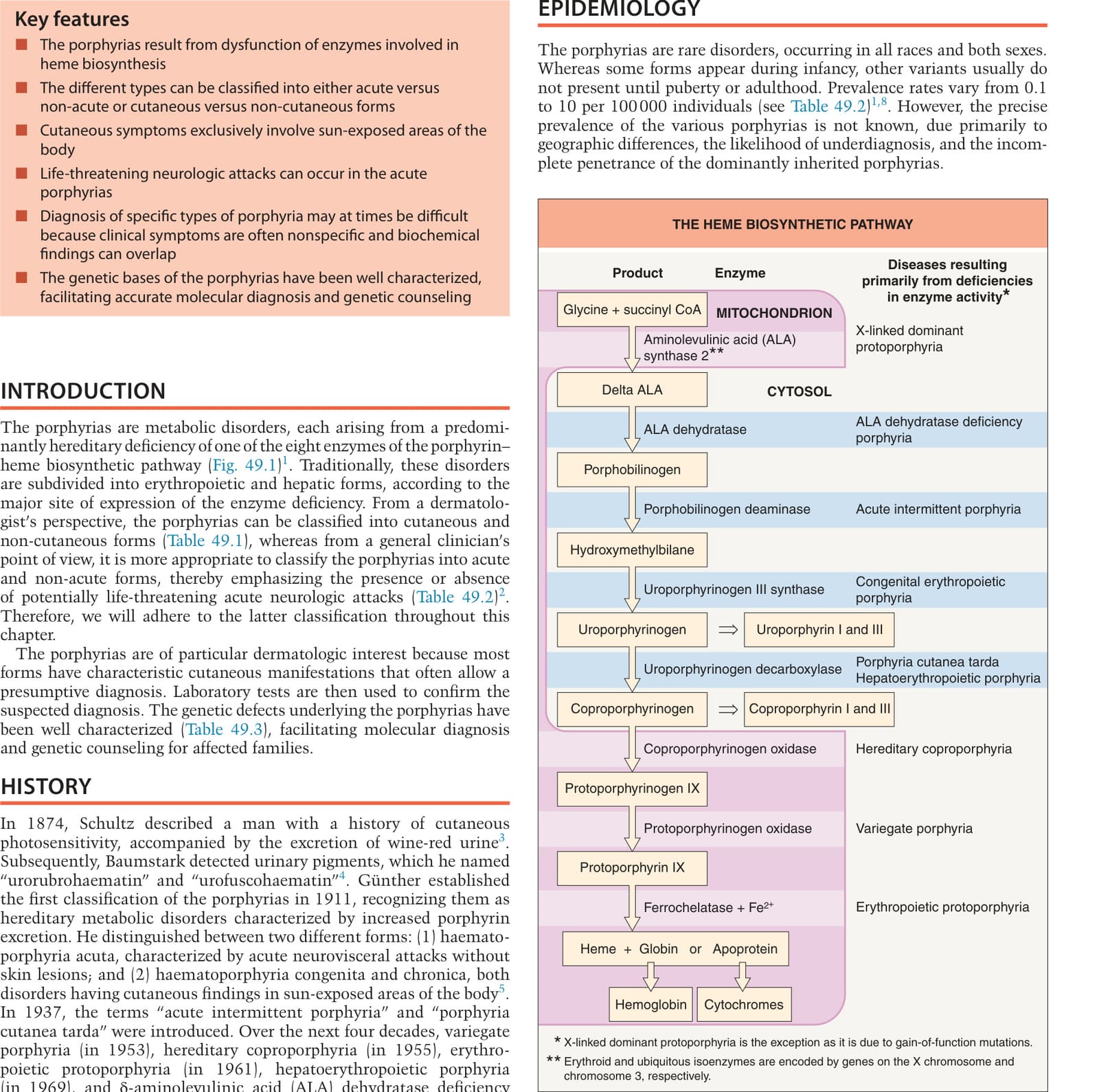
Fig. 49.1 The heme biosynthetic pathway. ALA, aminolevulinic acid, also referred to as δ-aminolevulinic acid or 5-aminolevulinic acid.
**Fig. 49.2 Approach to the diagnosis of porphyrias based upon clinical signs and symptoms. Increased zinc protoporphyrin can be seen in iron deficiency anemia, heavy metal intoxication, and anemia of chronic disease. AIP, acute intermittent porphyria; ALA, aminolevulinic acid; EBA, epidermolysis bullosa acquisita; EPP, erythropoietic protoporphyria; HCP, hereditary coproporphyria; PBG, porphobilinogen; PCT, porphyria cutanea tarda; VP, variegate porphyria; XLDPP, X-linked dominant protoporphyria. *24-hour collection. **Increased free protoporphyrin. *Increased free and zinc protoporphyrins; ~40% zinc protoporphyrin.
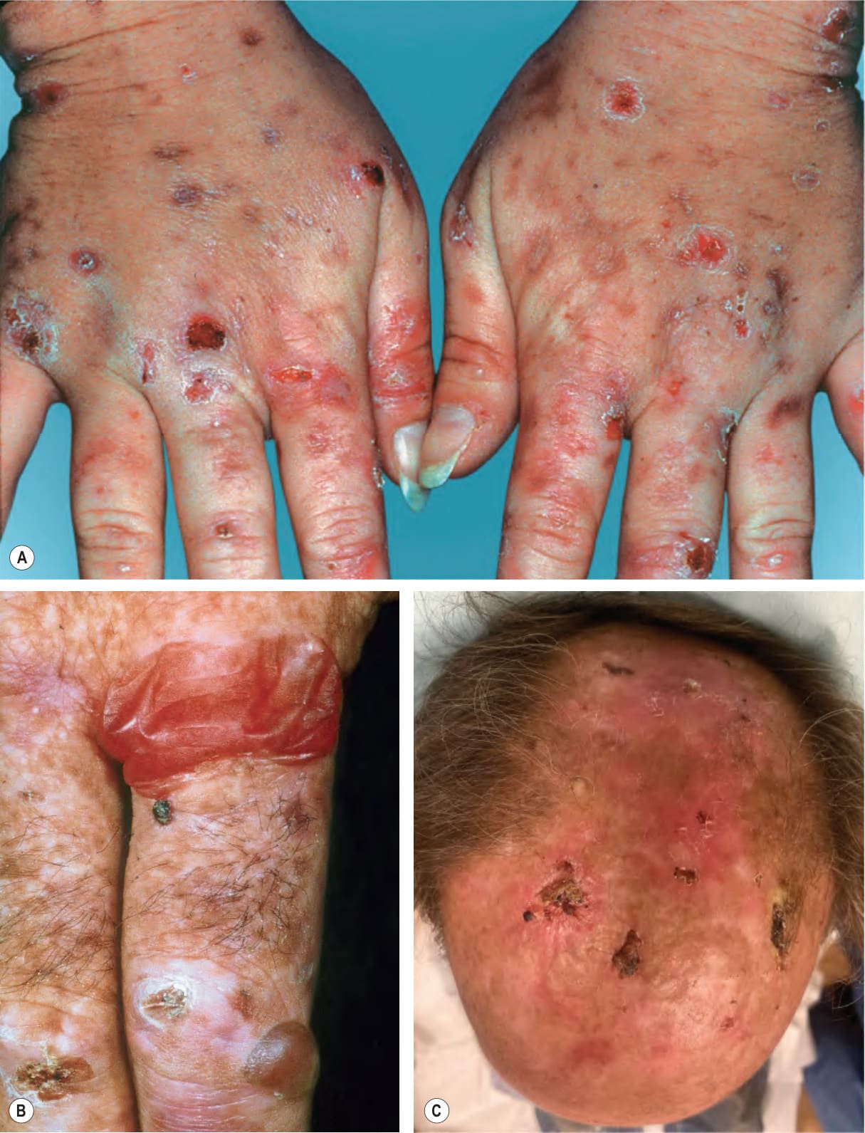
Fig. 49.3 Porphyria cutanea tarda.A Marked fragility with multiple hemorrhagic crusts, erosions, and milia as well as scars. B Flaccid hemorrhagic bulla and tense vesicle with clear fluid on the forefinger, accompanied by crusts and scars. C Extensive scarring of the bald scalp with multiple large hemorrhagic crusts in a patient with hepatitis C viral infection. C, Courtesy Jeffrey P. Callen, MD.
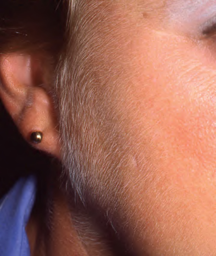
Fig. 49.4 Porphyria cutanea tarda. Hypertrichosis of the face. Courtesy Jeffrey P. Callen, MD.
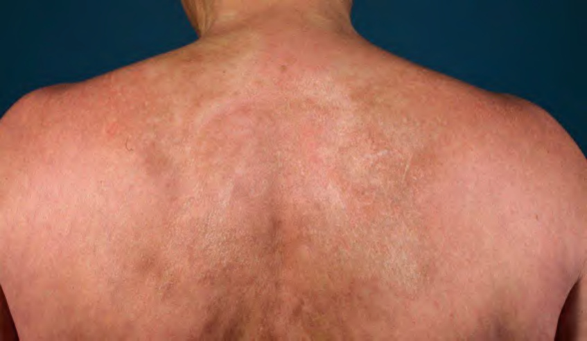
Fig. 49.5 Porphyria cutanea tarda – sclerodermatous presentation. The patient had shiny, yellow–white to brown firm plaques on the neck and upper back.
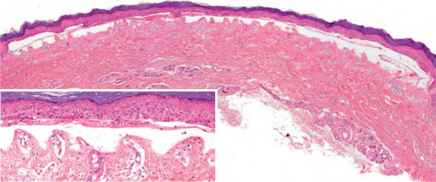
Fig. 49.6 Porphyria cutanea tarda – histologic features. Subepidermal blister of acral skin with a minimal (“cell-poor”) dermal inflammatory infiltrate. Festooning of the dermal papillae is seen (inset). Courtesy Lorenzo Cerroni, MD.
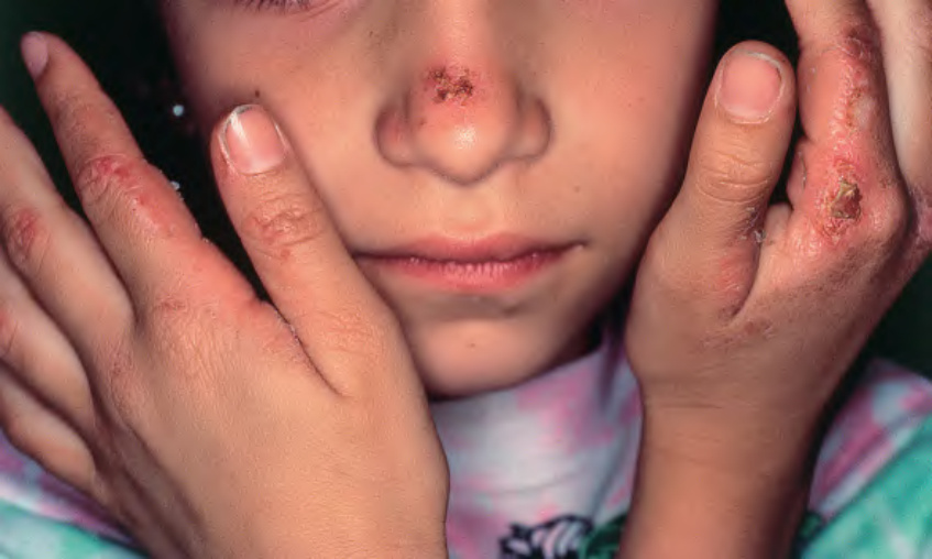
Fig. 49.7 Erythropoietic protoporphyria. Erythema, edema, and hemorrhagic crusts on the nose as well as the fingers in a young girl.
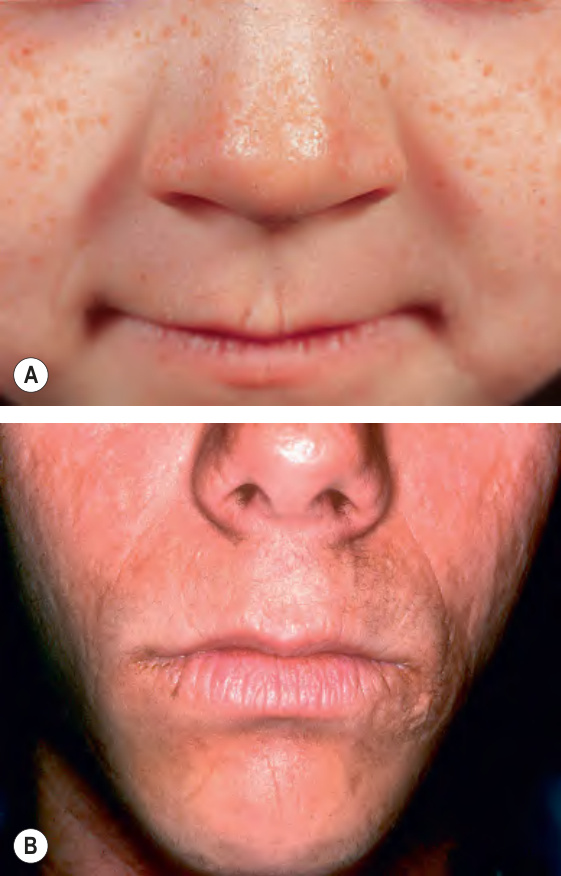
Fig. 49.8 Erythropoietic protoporphyria.A
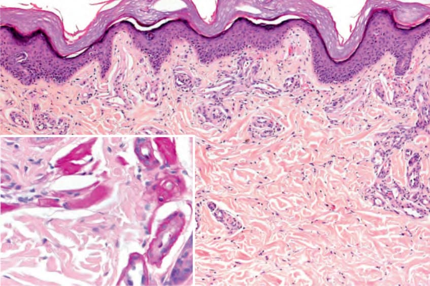
Fig. 49.9 Erythropoietic protoporphyria – histologic features. Eosinophilic deposits around blood vessels are more pronounced than in porphyria cutanea tarda. These deposits are highlighted by a PAS stain (inset). Courtesy Lorenzo Cerroni, MD.
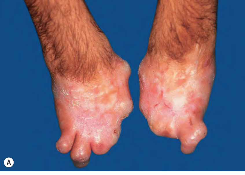
Fig. 49.10 Congenital erythropoietic porphyria.A Severe mutilation of the hands due to scarring. B Erythrodontia due to massive protoporphyrin deposition in the teeth. A, Courtesy José Mascaro, MD.
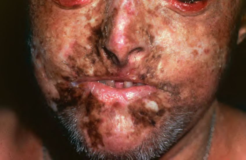
Fig. 49.11 Hepatoerythropoietic porphyria. Hypertrichosis and severe scarring are seen, resulting in a clinical appearance similar to congenital erythropoietic porphyria. Courtesy José Mascaro, MD.
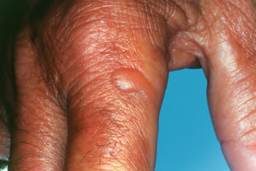
Fig. 49.12 Dialysis-associated pseudoporphyria. Vesicles formed on the dorsal aspects of the hands in this patient with chronic renal failure. Courtesy Kalman Watsky, MD.
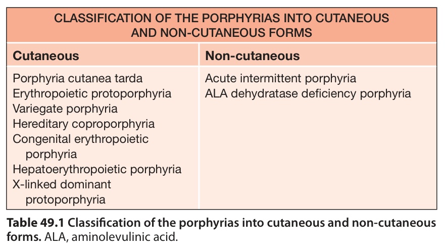
Table 49.1 Classification of the porphyrias into cutaneous and non-cutaneous forms. ALA, aminolevulinic acid.
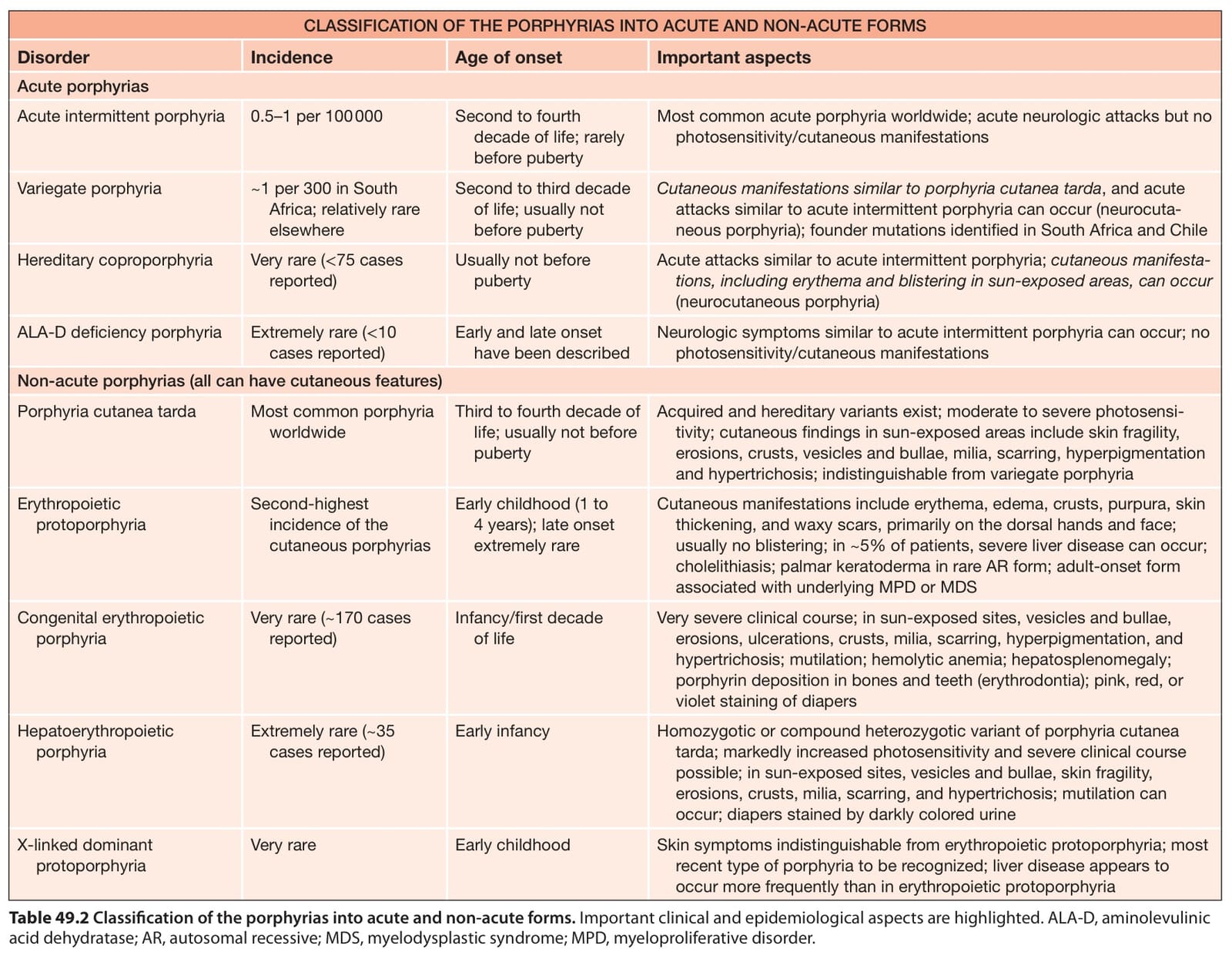
Table 49.2 Classification of the porphyrias into acute and non-acute forms. Important clinical and epidemiological aspects are highlighted. ALA-D, aminolevulinic acid dehydratase; AR, autosomal recessive; MDS, myelodysplastic syndrome; MPD, myeloproliferative disorder.
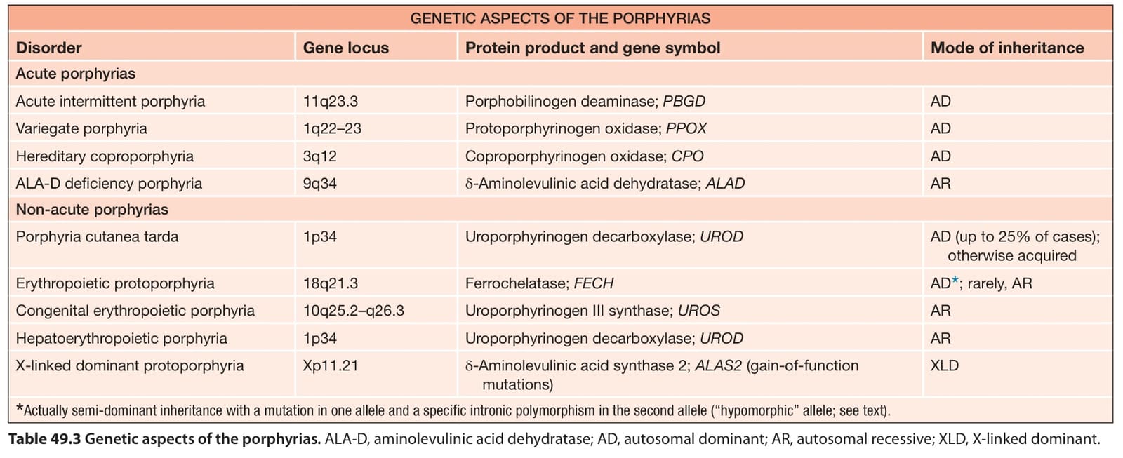
Table 49.3 Genetic aspects of the porphyrias. ALA-D, aminolevulinic acid dehydratase; AD, autosomal dominant; AR, autosomal recessive; XLD, X-linked dominant.
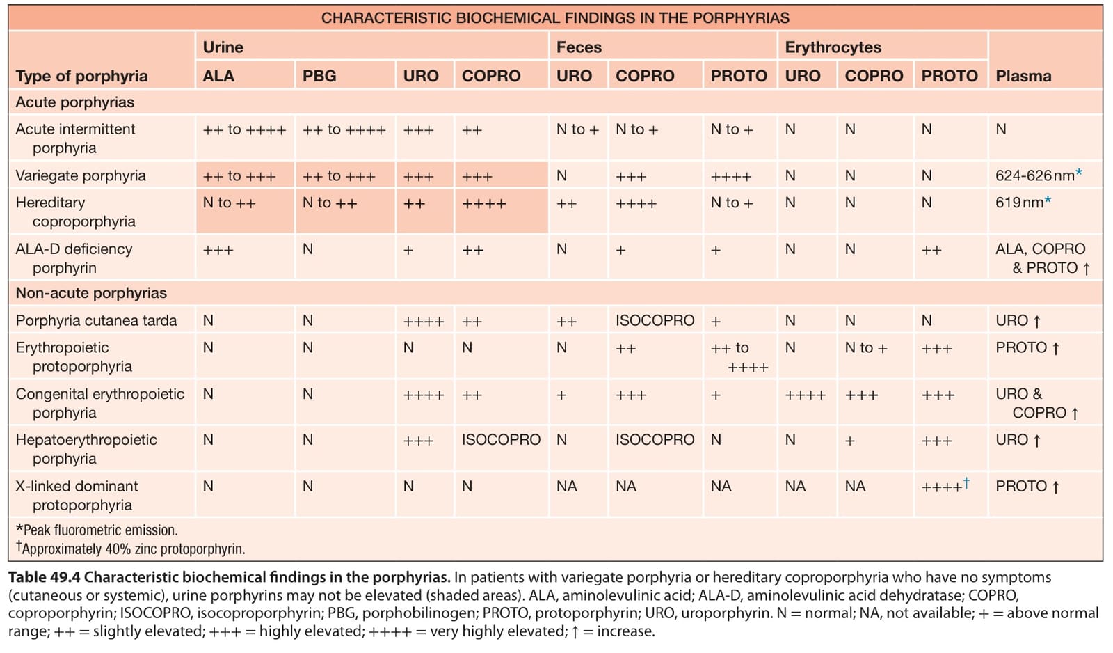
Table 49.4 Characteristic biochemical findings in the porphyrias. In patients with variegate porphyria or hereditary coproporphyria who have no symptoms (cutaneous or systemic), urine porphyrins may not be elevated (shaded areas). ALA, aminolevulinic acid; ALA-D, aminolevulinic acid dehydratase; COPRO, coproporphyrin; ISOCOPRO, isocoproporphyrin; PBG, porphobilinogen; PROTO, protoporphyrin; URO, uroporphyrin. N = normal; NA, not available; += above normal range; ++= slightly elevated; +++= highly elevated; ++++= very highly elevated; ↑= increase.
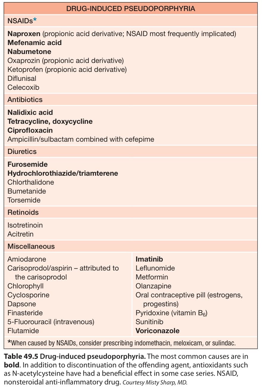
Table 49.5 Drug-induced pseudoporphyria. The most common causes are in bold. In addition to discontinuation of the offending agent, antioxidants such as N-acetylcysteine have had a beneficial effect in some case series. NSAID, nonsteroidal anti-inflammatory drug. Courtesy Misty Sharp, MD.
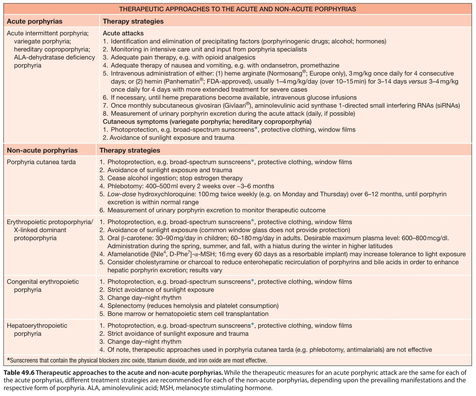
Table 49.6 Therapeutic approaches to the acute and non-acute porphyrias. While the therapeutic measures for an acute porphyric attack are the same for each of the acute porphyrias, different treatment strategies are recommended for each of the non-acute porphyrias, depending upon the prevailing manifestations and the respective form of porphyria. ALA, aminolevulinic acid; MSH, melanocyte stimulating hormone.