THE ACUTE PORPHYRIAS
The acute porphyrias consist of acute intermittent porphyria (AIP), variegate porphyria (VP), hereditary coproporphyria (HCP), and δ-aminolevulinic acid dehydratase (ALA-D) deficiency porphyria (see Tables 49.1 & 49.2).
Patients with acute porphyrias can present with a broad range of clinical findings, including persistent colicky abdominal pain, nausea and vomiting, obstipation, tachycardia, hypertension, seizures, paresthesias, para- and tetraplegia, and acute psychosis (Table 49.7). Unfortunately, this spectrum of fairly nonspecific symptoms can mimic a number of other diseases and the porphyrias are often not included in the differential diagnosis, especially if neurologic symptoms prevail. Acute attacks can be precipitated by a variety of factors, including porphyrinogenic drugs (e.g. antimalarials, tetracyclines, sulfonamides), alcohol, hormonal changes, recurrent or chronic infections, and reduced caloric intake due to fasting or diets. The latter may be a result of enhanced expression of the transcriptional coactivator PGC-1α.
In addition to the manifestations outlined in Table 49.7, patients with VP and HCP may have cutaneous findings that are clinically indistinguishable from those of PCT. As a result, VP and HCP are also referred to as neurocutaneous porphyrias.
A consortium of porphyria specialists from several European porphyria centers has established the European Porphyria Network (EPNET). The goals of the EPNET are to set standards and provide guidelines for the diagnosis and treatment of porphyrias. On the EPNET (porphyria.eu)
and the American Porphyria Foundation (porphyriafoundation.com) websites, important information is available for both patients and physicians.
Acute Intermittent Porphyria (AIP)
Worldwide, AIP is the most common type of acute porphyria. This autosomal dominantly inherited disorder is characterized by a deficiency of porphobilinogen deaminase, the third enzyme in heme biosynthesis (see Fig. 49.1).
AIP will not be discussed in detail here because patients do not have skin manifestations. It usually presents after puberty with acute porphyric attacks (see Table 49.7) that may be precipitated by the various trigger factors outlined above. Elevated urinary levels of the porphyrin precursors ALA and PBG can be detected during an acute attack: ALA levels range from 5 to 20 times normal and PBG levels are increased to as high as 20- to 50-fold normal.
Variegate Porphyria (VP)
Synonyms: South African porphyria Mixed porphyria
VP is characterized by an autosomal dominantly inherited deficiency of protoporphyrinogen oxidase, the seventh enzyme in the pathway of heme biosynthesis (see Fig. 49.1).
Cutaneous and neuropsychiatric symptoms can occur separately or simultaneously in affected individuals. Clinically, the skin findings in VP cannot be differentiated from those observed in PCT. Likewise, the acute attacks observed in VP are identical to those encountered in AIP.
During acute attacks, elevated urinary levels of ALA and PBG are encountered. However, during periods of remission, urinary ALA and PBG levels may be within normal range (see Table 49.4 [shaded areas]). Therefore, additional biochemical analyses of fecal porphyrins are mandatory in order to establish the diagnosis of VP. Elevated levels of stool protoporphyrin and coproporphyrin can be detected, with the concentration of protoporphyrin usually being higher than that of coproporphyrin. It is noteworthy that this abnormal biochemical stool profile can also be detected during periods of remission between attacks. On the other hand, a plasma fluorescence emission peak of 624–626 nm is seen only in symptomatic patients, including those with cutaneous involvement.
Hereditary Coproporphyria (HCP)
HCP is a very rare autosomal dominantly inherited disease, characterized by a deficiency of coproporphyrinogen oxidase, the sixth enzyme in the porphyrin–heme biosynthetic pathway (see Fig. 49.1). The clinical symptoms are similar to those encountered in VP. However, in contrast to the latter, the concentrations of coproporphyrin in the stool are usually higher than those of protoporphyrin (see above).
δ-Aminolevulinic Acid Dehydratase (ALA-D) Deficiency Porphyria
Synonyms: Plumboporphyria Doss porphyria
This autosomal recessively inherited porphyria variant is extremely rare. As fewer than 10 cases have been reported worldwide, ALA-D deficiency porphyria does not have an important clinical presence. The disease can present during early childhood (as well as in adulthood) with acute neurologic symptoms that resemble those encountered in AIP.
Differential Diagnosis
When patients with VP or HCP present with cutaneous manifestations, the differential diagnosis is identical to that of PCT (see above). If acute neurologic attacks prevail, a broad range of gastrointestinal, neurologic, and psychiatric diseases have to be excluded, including an “acute abdomen”, peripheral neuropathies, and viral infections. A more detailed discussion can be found in textbooks of internal medicine.
Treatment
Cutaneous symptoms
Patients with VP or HCP must minimize outdoor light exposure by wearing sun-protective clothing and applying broad-spectrum sunscreens. In contrast to PCT, phlebotomy seems to be of no benefit as are chloroquine and hydroxychloroquine. Furthermore, the latter antimalarials belong to the group of drugs considered to be potential inducers of acute porphyric attacks. Thus, their use is not recommended.
Acute porphyric attack
An acute porphyric attack is a potentially life-threatening event associated with a significant mortality rate (up to 5%). It requires rapid therapeutic intervention to prevent complications such as paralysis, respiratory failure, and coma. Treatment should consist of the following consecutive measures (see Table 49.6): 1.Precipitating factors, e.g. porphyrinogenic drugs, must be identified and eliminated. If necessary, patients should be initially monitored in an intensive care unit. 2.Systemic symptoms such as abdominal pain, nausea, and vomiting should be treated symptomatically, e.g. by administration of opioid analgesics and ondansetron or promethazine, respectively. 3.The most important therapeutic step for a patient with an acute attack is prompt intravenous administration of heme preparations such as heme arginate (Normosang® [Orphan Europe/Recordati Rare Diseases]) or hemin (FDA-approved; Panhematin® [Recordati Rare Diseases]). Heme arginate reduces the overproduction of porphyrin precursors via a negative feedback mechanism in which the increased activity of ALA synthase, the rate-limiting enzyme in heme biosynthesis, is repressed. Of note, heme preparations have the disadvantage of being unstable, and thrombophlebitis is a fairly common side effect. Historically, acute attacks were treated primarily with glucose infusions. However, given the availability of effective heme preparations (see above), glucose should only be administered adjunctively or as a temporary measure if heme arginate or hematin are not immediately available.
In 2019 and 2020, the FDA and European Medicines Agency approved givosiran (Givlaari® [Alnylam Pharmaceuticals]) for the treatment of adults with acute (hepatic) porphyria. Givosiran is a solution of small interfering RNAs (siRNAs) that are injected subcutaneously once monthly. These siRNAs promote the degradation of aminolevulinic acid synthase 1 (ALAS1) mRNA within liver cells,
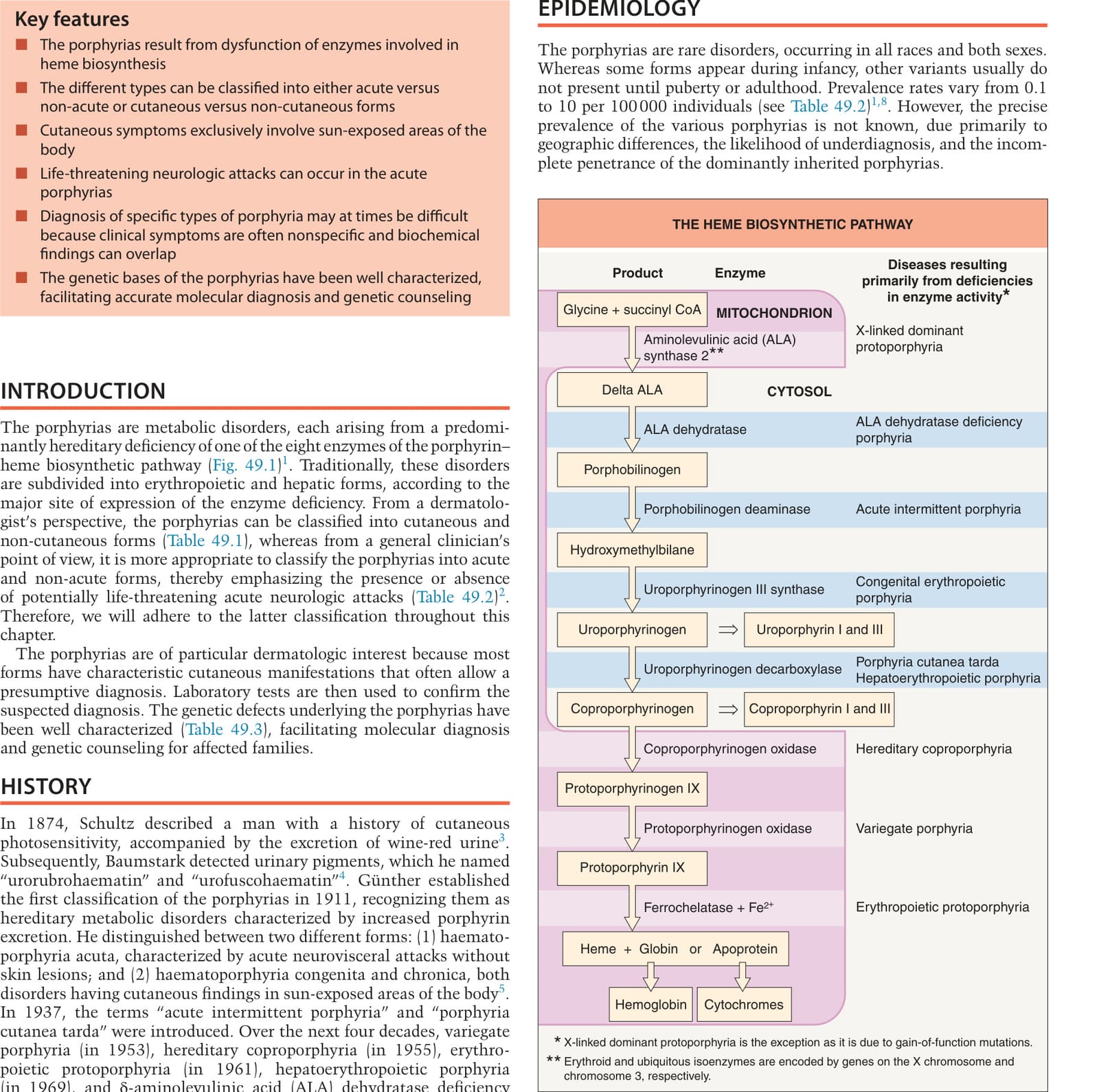
Fig. 49.1 The heme biosynthetic pathway. ALA, aminolevulinic acid, also referred to as δ-aminolevulinic acid or 5-aminolevulinic acid.
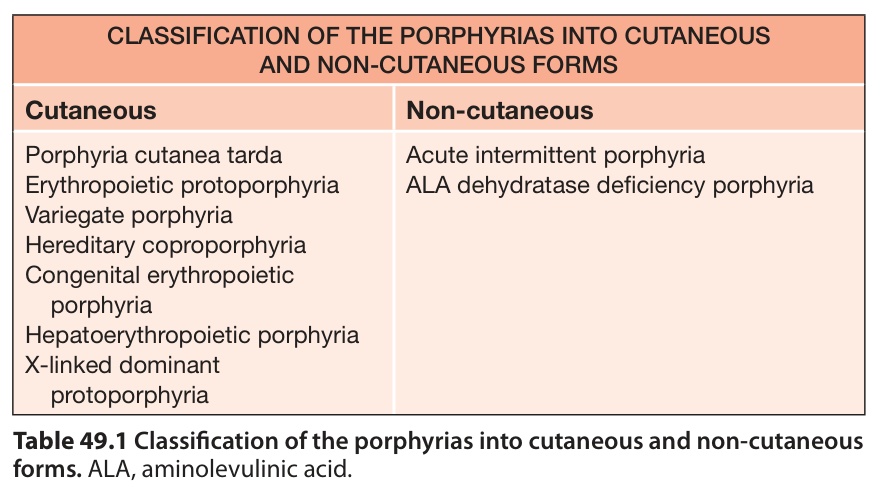
Table 49.1 Classification of the porphyrias into cutaneous and non-cutaneous forms. ALA, aminolevulinic acid.
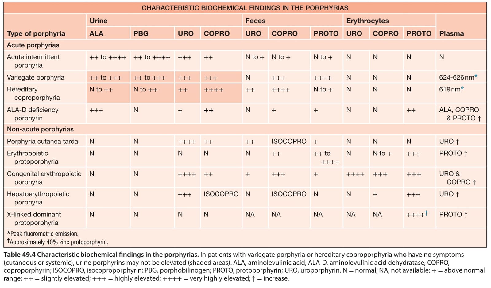
Table 49.4 Characteristic biochemical findings in the porphyrias. In patients with variegate porphyria or hereditary coproporphyria who have no symptoms (cutaneous or systemic), urine porphyrins may not be elevated (shaded areas). ALA, aminolevulinic acid; ALA-D, aminolevulinic acid dehydratase; COPRO, coproporphyrin; ISOCOPRO, isocoproporphyrin; PBG, porphobilinogen; PROTO, protoporphyrin; URO, uroporphyrin. N = normal; NA, not available; += above normal range; ++= slightly elevated; +++= highly elevated; ++++= very highly elevated; ↑= increase.
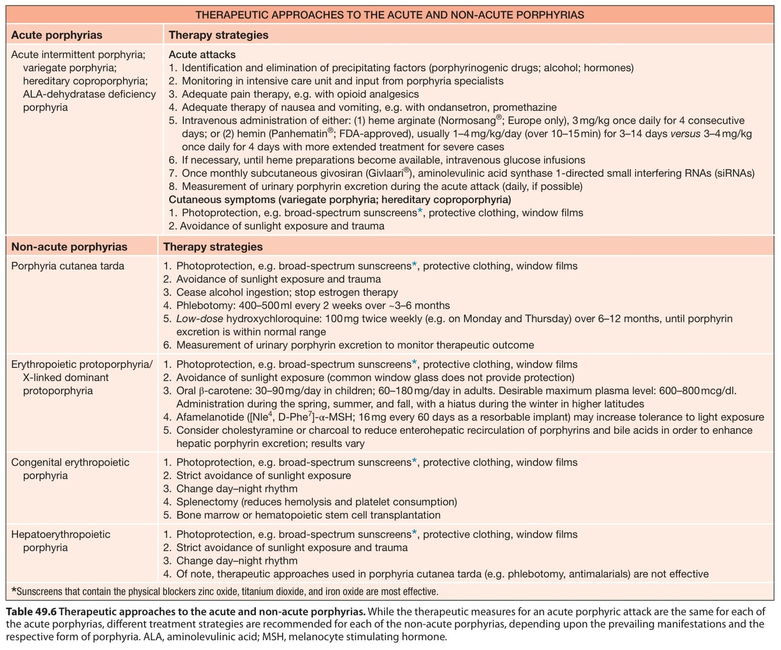
Table 49.6 Therapeutic approaches to the acute and non-acute porphyrias. While the therapeutic measures for an acute porphyric attack are the same for each of the acute porphyrias, different treatment strategies are recommended for each of the non-acute porphyrias, depending upon the prevailing manifestations and the respective form of porphyria. ALA, aminolevulinic acid; MSH, melanocyte stimulating hormone.
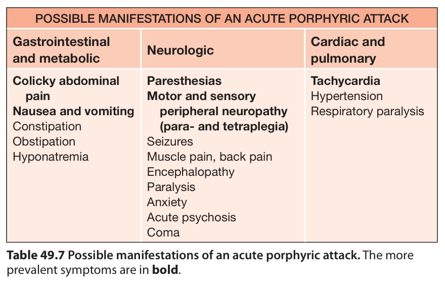
Table 49.7 Possible manifestations of an acute porphyric attack. The more prevalent symptoms are in bold.