CLINICAL FEATURES
Weight-for-height scores and height-for-age scores are utilized to evaluate malnutrition; scores are compared to the standard deviations (SD) from the median value for the reference population. An individual is considered to have moderate malnutrition if the scores are between the 2nd to 3rd (lower) SD and severe malnutrition if below the 3rd SD, with or without symmetrical edema. Cutaneous manifestations in part reflect the fact that epidermal maturation (from basal cell layer to stratum corneum) occurs over a period of 10–14 days. Deficiencies,
especially of macronutrients, can interfere with this process, resulting in skin that is dry and thin in appearance with associated epidermal atrophy. Prolonged deficiency can reduce both protein production, including that of dermal collagen and muscle, and the amount of subcutaneous fat. As a result, patients may develop dermal atrophy, muscle wasting, and/or lax skin.
Additional clinical findings of marasmus and kwashiorkor, such as dyschromia (hypo- and hyperpigmentation), desquamation, and erosions, are reviewed in Table 51.1. Patients may also have cutaneous manifestations from a deficiency in one or more micronutrients, and these are outlined in Table 51.2. Mucocutaneous clues to the possibility of a nutritional deficiency are summarized in Fig. 51.2.
nutritional disorder. Risk factors are similar to those outlined for marasmus in Table 51.1. Courtesy Karynne O. Duncan, MD.
Protein–Energy Malnutrition
Worldwide, protein–energy malnutrition (or undernutrition) is the most prevalent nutritional deficiency. The two major forms of severe acute malnutrition (SAM) are: (1) marasmus, also referred to as wasting or malnutrition without edema (Figs. 51.3 & 51.4); and (2) kwashiorkor, also referred to as edematous malnutrition. Patients with marasmic kwashiorkor have features of both wasting and edema. According to the WHO, on a global basis, SAM affects almost 20 million young children (<5 years of age).
Marasmus (SAM without edema) can occur in individuals of all ages and results from a prolonged inadequate intake of total calories (energy).
These patients frequently develop recurrent infections due to their inability to mount a normal immune response.
Kwashiorkor (SAM with edema) is a more acute form of childhood and adult protein–energy malnutrition (see Table 51.1). A characteristic finding is peripheral edema or even anasarca in association with hypoalbuminemia (Fig. 51.5). One consequence of this edema is a body weight that is higher than is seen with marasmus, and it may be 60%–80% of expected weight (rather than <40%). Some patients will have overlapping features of both disorders and this is referred to as marasmic kwashiorkor. In at-risk populations, kwashiorkor can develop after a child is weaned from breast milk and begins a diet rich in carbohydrates but deficient in protein. In addition to peripheral edema, patients with kwashiorkor also develop desquamation and erosions that have been likened to “enamel paint” and “flaky paint” (Fig. 51.6).
Based upon clinical observations, it was thought that kwashiorkor was due to a relative deficiency in protein intake. However, more recently, this proposed pathogenesis has been questioned based upon several findings. For example, protein intake was found to be similar in a group of children, irrespective of whether they had marasmus or kwashiorkor. In addition, kwashiorkor can develop within just a few weeks of an acute, life- threatening illness. A current theory is that kwashiorkor is related to physiologic stresses secondary to an acute or chronic illness
(including infections), with the stress leading to an increase in protein and energy requirements at a time when intake is frequently limited. This would help to explain why kwashiorkor often occurs in the setting of an acute illness superimposed upon a chronic state of milder malnutrition. It is hypothesized that the stress of the acute illness blocks the proteinsparing response that is normally in effect during states of starvation. There is also increasing evidence for a relationship between the gut microbiome and the development of malnutrition, including kwashiorkor. A deficiency of anti-inflammatory taxa and an increase in inflammogenic bacteria were noted in the gut microbiome of affected children.
Both kwashiorkor and marasmus can have secondary causes such as “rice milk” diets or chronic diarrhea (see Table 51.1). The cutaneous and systemic abnormalities associated with primary protein–energy malnutrition are often reversible following nutritional therapy. However, if the malnutrition is prolonged, this may preclude normal physical and/or mental development.
Severe acute malnutrition is less common in adults, but can occur in the setting of critical illnesses requiring hospitalization, where catabolism exceeds anabolism. Protein loss and hypoalbuminemia may also develop in patients with exfoliative erythroderma (see Ch. 10). Delayed wound healing is one of the important consequences of protein deficiency.
Essential Fatty Acid Deficiency
Essential fatty acids (EFAs) are unsaturated fatty acids that must be obtained from an exogenous source because they cannot be synthesized by the human body. Linoleic acid and alpha-linolenic acid, which
There is intense edema of the buttocks and lower extremities in addition to areas of erythema with erosions and desquamation. The edema is a reflection of hypoalbuminemia. Courtesy Ramón Ruiz-Maldonado, MD.
represent omega-6 and omega-3 fatty acids, respectively, are major types of EFAs. These fatty acids are present in plant oils (e.g. canola, flaxseed, soybean) and in fish oils. EFAs have multiple functions, both structural and synthetic, including prostaglandin formation, phospholipid membrane integrity, energy storage, and proper lamellar granule formation. Humans can convert linoleic acid into arachidonic acid. From 15% to 30% of the skin’s fatty acids are EFAs and cutaneous as well as systemic manifestations of EFA deficiency are outlined in Table 51.1.
While an isolated deficiency of EFAs is uncommon, it may occur following parenteral nutrition without lipid supplementation and with overly aggressive low-fat diets. In most instances, however, EFA deficiency is associated with other nutritional deficiencies. The presence of decreased plasma levels of linoleic and linolenic acids plus increased levels of eicosatrienoic acid (normally undetectable) establishes the diagnosis of EFA deficiency (see Table 51.1).
Data from the American Heart Association suggest that obtaining 5%–10% of energy from omega-6 fatty acids (e.g. linoleic acid) reduces the risk of cardiovascular disease. However, there is still controversy regarding the recommended upper limit of consumption of EFAs, as epidemiologic studies suggest that an excess of linoleic acid may contribute to cardiovascular disease, cancer, and inflammation.
Vitamins and Trace Elements
Micronutrients represent a diverse array of dietary components that are necessary to sustain health. The physiologic roles of micronutrients are as varied as their composition. Several micronutrients function as cofactors for enzymes, while others act as biochemical substrates or hormones; in addition, there are some whose function has not been well defined. Under normal circumstances, the recommended average daily dietary intake for each micronutrient is measured in milligrams or even smaller quantities. This distinguishes micronutrients from macronutrients (carbohydrates, fats, proteins) as well as the macrominerals (calcium, magnesium, phosphorus).
Vitamins
Vitamins are required constituents of the human diet as they are essential for development and maintenance of bodily functions. However, they do not have a direct role as an energy source. Vitamins are categorized as either fat-soluble (A, D, E, K) or water-soluble (all others) (see Table 51.2). While none of the fat-soluble vitamins appear to serve as cofactors for enzymes, most of the water-soluble vitamins have this function. In addition, symptomatic vitamin excess is more likely with fat-soluble vitamins (see below). Lastly, the use of vitamins
as therapeutic interventions in dermatology includes topical vitamin D analogues for psoriasis (see Ch. 129), oral nicotinamide for autoimmune blistering diseases and non-melanoma skin cancer prevention, oral or topical retinoids to treat acne and disorders of cornification (see Ch. 126), and topical vitamin C to try to reverse photodamage.
Tables 51.2 and 51.3 review the major fat- and water-soluble vitamins, including their functions and recommended daily dosages, as well as the risk factors for vitamin deficiencies and associated signs and symptoms.
Excessive ingestion of vitamins most commonly occurs in individuals seeking potential (and often unproven) “anti-aging” or antineoplastic effects. High doses of fat-soluble vitamins may lead to harmful side effects, including hepatic toxicity, nephrolithiasis, and peripheral neuropathy (Table 51.4). They may also have unanticipated effects at high doses in certain populations. For example, in the β-Carotene and Retinol Efficacy Trial (CARET), a large cancer prevention trial, patients at high risk for lung cancer were given supplements containing β-carotene and vitamin A. Not only did these supplements fail to prevent lung cancer, they were actually associated with increased rates
of lung cancer. The reported association of oral isotretinoin and topical tretinoin with increased mortality in smokers remains controversial.
Toxicity due to high doses of water-soluble vitamins (i.e. B vitamins, vitamin C) is rarely seen, as these compounds are rapidly excreted via the urine. While not an excess issue, ingestion of biotin to possibly improve hair and nails can interfere with biotin-streptavidin-based cardiac troponin, β-hCG, parathyroid, and thyroid (TSH, T3, T4) immunoassays.
Vitamin D plays a role in the absorption of calcium and phosphorus, helping to maintain normal serum levels of these minerals (Fig. 51.11). Vitamin D also functions as an antiproliferation and prodifferentiation hormone, but its exact role is not known. Although a possible association of vitamin D deficiency with cutaneous disorders such as alopecia areata, atopic dermatitis, and psoriasis has been suggested, to date there are no known cutaneous signs of vitamin D deficiency. In addition, the Institute of Medicine (IOM) found no strong evidence to support a role for vitamin D supplementation for any medical condition other than bone health.
With the exception of individuals with sarcoidosis, the preferred method for assessing vitamin D status is measurement of total serum levels of 25-hydroxyvitamin D. While secondary hyperparathyroidism can influence the 1,25-dihydroxyvitamin D assay, this is not the case with the 25-hydroxyvitamin D assay. In addition, 25-hydroxyvitamin D has a longer half-life (2 weeks) and therefore is thought to better reflect body stores. Currently, although a consensus is lacking, vitamin D deficiency is defined by most experts as a serum level of 25-hydroxy vitamin D that is <12 ng/ml (30 nmol/l), and vitamin D insufficiency as circulating levels of 12–20 ng/ml (30–50 nmol/l) (see Table 51.2).
Based upon these levels, it is estimated that one billion people worldwide have either deficient or insufficient levels of vitamin D. Depending upon the study, ~50%–60% of nursing home residents, the elderly, and hospitalized patients in the US are deficient in vitamin D. Decreased levels of 25-hydroxyvitamin D (<20 ng/ml) were also observed in half of US Hispanic or Black adolescents and White preadolescent girls. In addition, ~50% of Black and 55% of White infants have vitamin D deficiency, while over 90% of infants in Iran, Turkey, and India have vitamin D deficiency.
While the IOM recommends a daily intake of vitamin D of 400 IU/ day (0–12 months of age), 600 IU/day (1–70 years of age), and 800 IU/ day (>70 years of age), some experts have advocated 1000 IU/day for children or adolescents and 1500–2000 IU/day for adults (unless there are contraindications). All of these doses are below the age-dependent, tolerable, upper daily limits of 1000–4000 IU for infants and children (up to 18 years old) and 4000 IU for adults, with the range reflecting the current lack of a consensus regarding the upper daily limit. Vitamin D (cholecalciferol; animal diet source) is viewed as nutritionally superior to vitamin D (ergocalciferol; plant source) and
therefore is regarded as the preferred form for supplementation and for fortifying foods. Although it is difficult to obtain enough vitamin D from dietary sources alone, patients should be made aware of vitamin D-rich foods such as fatty fish, beef liver, cheese, egg yolks, mushrooms that have been exposed to ultraviolet light, and fortified foods.
An endogenous source of vitamin D production also exists, initiated with the conversion of 7-dehydrocholesterol to provitamin D when skin is exposed to ultraviolet B radiation (see Fig. 51.11). It is thought that typically over 90% of vitamin D is produced via UVB exposure; however, the skin’s ability to synthesize vitamin D diminishes with age and it is reduced in individuals with darker skin phototypes. The balance between sun protection (to prevent photodamage and cutaneous malignancies) and the risk of vitamin D deficiency has become a matter of debate. As effects of vitamin D, beyond calcium homeostasis and bone mineralization, have become increasingly recognized, the debate has intensified. For example, immunomodulatory effects via the innate immune system have been reported, as has an association between vitamin D deficiency and an increased risk of several internal malignancies. While there are studies that refute the latter as well as no direct evidence linking sunscreen use with vitamin D deficiency, it is prudent to recommend that patients who are limiting sun exposure and/or have risk factors for the development of vitamin D deficiency ingest the daily recommended amount of vitamin D (see Tables 51.2 & 51.3).
Trace elements
Trace elements and minerals constitute ~3% of body weight at birth and 4% in adults. Based upon animal studies, 15 trace elements have been identified as essential for health: iron, zinc, copper, chromium, selenium, iodine, fluoride, manganese, molybdenum, cobalt, nickel, tin, silicon, vanadium, and arsenic (in very small doses). There is compelling evidence that the first 10 (in italics) are essential nutrients in humans. They serve multiple cellular functions including as cofactors for enzymes and as prosthetic groups in metalloproteins. An example of the latter is the complexing of iron and protoporphyrin IX via ferrochelatase to form heme. Deficiency syndromes related to essential trace elements, other than iron, zinc, copper, iodine or cobalt, were not recognized until more recently. Explanations include the exceedingly small requirements for these trace elements, their ubiquitous nature in foodstuffs, and the lack of routine laboratory assays. Only trace elements of dermatologic importance will be discussed in this chapter.
Zinc is one of the most important trace elements in humans, playing a critical role in the function of more than 200 zinc-dependent metalloenzymes that regulate lipid, protein, and nucleic acid synthesis and degradation. Zinc can be found in human breast milk, animal-based foods, shellfish, legumes, and green leafy vegetables. There is evidence to suggest that zinc plays a role in enhancing wound healing as well as immune function and this may explain the poor wound healing and
increased susceptibility to cutaneous infections observed in patients with chronic zinc deficiency. Due to its antioxidant properties, zinc might protect the skin against UV-induced damage.
Patients with zinc deficiency can develop erythema, scale-crusts and erosions, especially periorally, acrally, and in the anogenital region (Fig. 51.12); occasionally, vesicles or bullae are seen as well as psoriasiform plaques. Zinc deficiency may also lead to alopecia, paronychia, onychodystrophy, blepharitis, conjunctivitis, stomatitis, and angular cheilitis. Diarrhea, depression (apathy), and dermatitis (erosive) are sometimes considered the triad of zinc deficiency, but the complete triad is seen in only 20% of patients. Superimposed cutaneous infections with Candida spp. and staphylococci are very common. Patients are typically irritable and sleep poorly; children with chronic zinc deficiency may experience growth retardation and/or develop hypogonadism. If treatment is not initiated, death may ensue.
Zinc deficiency can be either acquired or inherited as an autosomal recessive disorder, known as acrodermatitis enteropathica (AE). The latter is due to mutations in the gene that encodes the transmembrane
zinc transporter SLC39A4 which is expressed in the intestine and kidney. Clinical manifestations usually appear within 1 to 2 weeks after weaning from breast milk, or at 4 to 10 weeks of age if bottle-fed. Of note, transient neonatal zinc deficiency can develop in breastfed babies when breast milk contains low levels of zinc due to dysfunction of SLC30A2, the zinc transporter in the breast (in the mother). In addition, infants and children with cystic fibrosis can develop zinc deficiency (Fig. 51.13). This deficiency should be considered in children who have treatment-resistant seborrheic dermatitis or anogenital dermatitis.
There are several risk factors for developing acquired zinc deficiency, including alcohol use disorder, anorexia nervosa, diets high in mineralbinding phytate (Middle Eastern diets), and vegan diets. Of note, vegan diets can also lead to low levels of long-chain n-3 (omega-3) fatty acids, calcium, vitamin D, and vitamin B. Intestinal malabsorption often results in multiple deficiencies, including zinc. In addition, zinc deficiency can be seen in association with pregnancy, HIV disease, chronic renal failure, sickle cell disease, and drugs (e.g. penicillamine). Especially when combined with other risk factors, drugs such as diuretics and antacids can result in zinc deficiency while quinolones and tetracyclines can reduce absorption of zinc supplements.
The histologic finding of epidermal necrosis (see below) plus low serum alkaline phosphatase (a zinc-dependent enzyme) and zinc levels suggest zinc deficiency; the normal reference range for zinc is 70–150 mcg/dl (10.7–22.9 mcmol/l). Reduced zinc levels can occasionally lag behind cutaneous manifestations. Some patients with necrolytic acral erythema also have reduced zinc levels (and improve with zinc supplementation) while others have associated hepatitis C viral infections. Likewise, zinc levels can be normal or reduced in patients with necrolytic migratory erythema (NME) as well as EGFR inhibitorinduced NME-like eruptions.
Cutaneous and systemic manifestations respond dramatically to supplementation with elemental zinc (1–2 mg/kg/day in the acquired forms and 3 mg/kg/day for AE). There are several forms of commercially available zinc supplements including zinc sulfate, zinc gluconate, and zinc acetate, and they contain variable amounts of elemental zinc, e.g. 220 mg zinc sulfate tablets contain 50 mg of elemental zinc. Patients with persistent malabsorption syndromes and AE require lifelong zinc supplementation, along with regular serum zinc determinations. Of note, ingestion of excessive amounts of zinc can decrease copper absorption and lead to copper deficiency.
Copper is an essential trace element that is required for the function of a number of enzymes, e.g. tyrosinase, lysyl oxidase. In the bloodstream, 90% of copper is associated with ceruloplasmin, and the remainder is bound to other plasma proteins, primarily albumin. Acquired copper deficiency is rare, but it has been reported in infants receiving milk low in copper, in protein–energy malnutrition, and as a consequence of excessive zinc intake. Symptoms include anemia, neutropenia, and failure to thrive. Cutaneous findings are limited to rare reports of pigmentary dilution of the skin and hair.
Menkes disease, also known as kinky hair disease, is an X-linked recessive condition characterized by defective copper absorption with low copper levels in the blood, liver, and hair. Affected infants may appear normal and develop normally until 2 to 3 months of age, when they gradually manifest failure to thrive, lethargy, hypothermia, and hypotonia. In addition to seizures and developmental delay, patients may also have anemia and bony abnormalities (similar to those in scurvy). Arteriography demonstrates tortuosity and elongation of arteries, a reflection of immature elastin. Decreased activity of several enzymes, including cytochrome C oxidase (in the brain), lysyl oxidase (in connective tissues and blood vessels), and ascorbic acid oxidase (in bones), may account for the associated clinical findings.
Pudgy cheeks, a cupid’s bow of the upper lip, and horizontal eyebrows comprise the typical facies in Menkes disease. However, a more obvious and characteristic finding is alopecia with abnormal hair shafts. There are 180° twists of the hair (pili torti), segmental shaft narrowing (monilethrix), and brush-like swellings of the hair shaft (trichorrhexis nodosa). Hairs are light in color, sparse (Fig. 51.14), fragile, and kinky. Patients may also have diffuse cutaneous pigmentary dilution due to decreased activity of tyrosinase, a copper-dependent enzyme. In addition, obligate female carriers may have patches of swirled
hypopigmentation or pili torti along the lines of Blaschko, as a result of lyonization.
The clinical features, low serum levels of copper and ceruloplasmin, and microscopic hair shaft findings establish the diagnosis. Infants with Menkes disease have a poor prognosis, with a life expectancy of 3 to
5 years and progressive deterioration leading to death. Treatment with copper histidine is unsuccessful in the majority of patients, but those with mutations leading to reduced, but not absent, copper transport may be more likely to respond to early intervention. Mutations in ATP7A, which encodes a copper-transporting ATPase, lead to Menkes
disease (as well as occipital horn syndrome; see Ch. 97); therefore, prenatal diagnosis is possible.
Copper toxicity can be acquired or inherited. The acquired form usually results from the ingestion of excessive amounts of copper (e.g. milk boiled in eroded copper pots) and this leads to gastrointestinal symptoms and, occasionally (in predisposed individuals), childhood cirrhosis. The inherited form is Wilson disease, an autosomal recessive disorder characterized by an accumulation of copper within internal organs, in particular the liver, cornea, and brain. Affected individuals have mutations in ATP7B which also encodes a copper-transporting P-type ATPase. Dysfunction of this protein leads to an impairment of both intrahepatic trafficking and biliary excretion of copper.
Because daily copper intake exceeds the body’s requirements, an effective means of excreting excess copper is crucial. This is accomplished by ATP7B, which mediates both copper secretion into plasma (coupled with ceruloplasmin synthesis) and its excretion into bile. When ATP7B is dysfunctional, the presence of excess copper within tissues induces free-radical reactions and lipid peroxidation. Resultant hepatic damage leads to steatosis, inflammation, cirrhosis, and eventually liver failure.
The diagnosis of Wilson disease is established by the detection of low serum ceruloplasmin, increased urinary copper excretion, increased hepatic copper content, and/or genetic testing. The clinical hallmarks of Wilson disease are hepatomegaly, cirrhosis, Kayser–Fleischer corneal rings, and neurologic symptoms (dysarthria, dyspraxia, ataxia, and parkinsonian-like extrapyramidal signs).
The chelating agents penicillamine and trientine, which facilitate excretion of copper, are approved for the treatment of Wilson disease. Oral zinc acetate can be prescribed for pre-symptomatic patients or as maintenance therapy, as it blocks intestinal absorption via induction of copper-binding metallothionein within enterocytes, thereby preventing serosal transfer.
Selenium, in the form of selenocysteine, is an essential component of several enzymes, including those involved in thyroid hormone metabolism and protection against oxidative damage (e.g. glutathione peroxidase). Deficiency of selenium typically results in a cardiomyopathy as well as muscle pain and weakness due to muscle degeneration; there may also be signs and symptoms of hypothyroidism. Cutaneous manifestations include hypopigmentation of the skin and hair (considered early signs), leukonychia, and xerosis, all of which respond to selenium replacement. There was also a report of erythematous scaly papules and plaques involving the cheeks, hips, thighs, and popliteal fossae, along with erosions in the diaper area. Elevated serum levels of creatine kinase and transaminases are often present in selenium deficiency as well.
Selenium deficiency has been reported in patients receiving total parenteral nutrition and in those who live in areas where the soil content of selenium is poor. Low serum levels of selenium and glutathione peroxidase activity are indicative of selenium deficiency. The recommended replacement dosage in selenium-deficient patients is a maximum of 100–200 mcg/day, and the Recommended Dietary Allowance (RDA) is 55 mcg/day for adults and 15 mcg/day for infants.
Although controversial, over-supplementation can occur due to reported beneficial cardiovascular and anticancer effects of selenium. If supplements are taken, they should not exceed 200 mcg/day. Excessive selenium ingestion (e.g. supplements, Brazil nuts, selenium-enriched green tea) can produce selenosis in humans, leading to a peripheral neuropathy, dermatitis, facial flushing, alopecia, discolored or brittle nails, nausea and vomiting as well as a garlic-like breath odor. The use of selenium sulfide shampoo in large areas of eroded or ulcerated skin may lead to excessive absorption, with loss of appetite and tremor.
Anorexia Nervosa and Bulimia (Nervosa)
Anorexia nervosa and bulimia nervosa are eating disorders characterized by starvation, excessive exercise, self-induced vomiting, and/or abuse of medications (e.g. laxatives, diuretics). Both can result in primary and secondary nutritional deficiencies. Anorexia nervosa is a persistent unwillingness or inability to maintain adequate body weight, whereas patients with bulimia typically binge-eat then purge or fast. These disorders occur most commonly in adolescent girls and young adult women, but can affect both sexes and all age groups. Skin disorders, either real or perceived, are common in these patients and women in particular report a greater dissatisfaction with the appearance of their skin when compared to a similar cohort.
The dietary changes and starvation associated with anorexia nervosa and bulimia can lead to telogen effluvium, lanugo-like hair, xerosis, generalized pruritus, carotenoderma (see Fig. 51.10), and hyperpigmentation. In addition, patients can develop acrocyanosis, perniosis, and livedo reticularis. Signs of specific nutritional deficiencies (e.g. pellagra, scurvy) may be present, as well as poor wound healing, seborrheic dermatitis, and edema. Additional clues to the diagnosis include petechiae, interdigital intertrigo, paronychia, and particularly calluses or scars on the knuckles or dorsal surface of the hand (Russell’s sign) due to repeated self-induced vomiting over prolonged periods of time (Fig. 51.15). Patients with bulimia may also have enlarged salivary glands and erosion of their tooth enamel. Psychiatric and nutritional support must be instituted as severe malnutrition is common.
Obesity
Obesity, whether acquired or inherited, is defined as a body mass index (BMI; measured in kg/m), that is >30; the latter has been found to correlate with high levels of the adipokine leptin. Genetic disorders such as Prader–Willi, Bardet–Biedl, Alström, and Wilson–Turner syndromes have been associated with childhood-onset obesity. In addition, endocrine disorders such as Cushing disease, Cushing syndrome, and insulin resistance include obesity amongst their clinical manifestations.
Acquired obesity is at epidemic proportions in both adults and children in industrialized nations. Although paradoxical, it is a functional malnutrition due to the consumption of high-caloric foods that are low in nutrients. Obesity is a systemic disease, as evidenced by an increased incidence of hypertension, hyperglycemia and hyperlipidemia, components of the metabolic syndrome (see Table 53.5), as well as atherosclerotic cardiovascular disease and premature death. Cutaneous manifestations are nonspecific, but some of the more common findings, such as acanthosis nigricans, appear to be related to insulin resistance (see Fig. 51.15).
Bariatric Surgery and Gastrointestinal Malabsorption
The increase in acquired obesity has led to an increase in the number and types of bariatric surgery performed. Due to subsequent alterations in gastrointestinal absorption, post-bariatric surgery patients are at risk for developing nutritional deficiencies. Two of the more commonly performed surgeries are laparoscopic adjustable gastric banding and the Roux-en-Y gastric bypass. Because the normal absorptive surface is left intact with the former, nutrient deficiencies in these patients are rare. On the other hand, the Roux-en-Y gastric bypass procedure reduces stomach size and, as a result, there are lower levels of intrinsic factor, gastrin, hydrochloric acid, and pepsinogen, which lead to a reduction in protein digestion. The Roux-en-Y procedure also decreases the physical grinding of food that typically helps to release vitamins and minerals. Abnormally low levels of micronutrients, such as iron, zinc, copper, and vitamins A, E, B (folate), and B, may be seen in these patients, and long-term monitoring is required, as it may take months or years for signs and symptoms to appear.
In disorders such as Crohn disease that are associated with gastrointestinal malabsorption, at least 50% of patients may have low plasma concentrations of vitamin C, copper, niacin, and zinc.
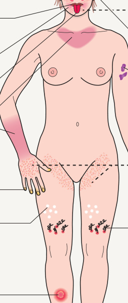
Fig. 51.2 Mucocutaneous clues that suggest a possible
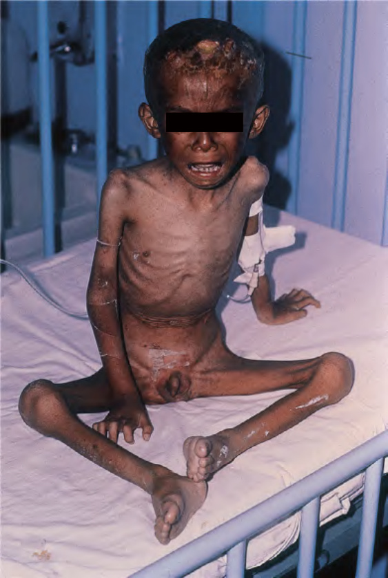
Fig. 51.3 Marasmus. This child is emaciated and has obvious hyperpigmentation. Both erosions and desquamation are present on the scalp. Courtesy Ramón Ruiz-Maldonado, MD.
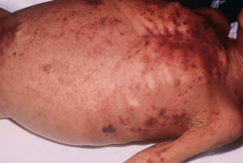
Fig. 51.4 Marasmus. Multiple sites of purpura are seen. Courtesy Ramón Ruiz-Maldonado, MD.
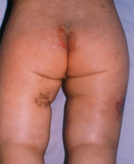
Fig. 51.5 Kwashiorkor.
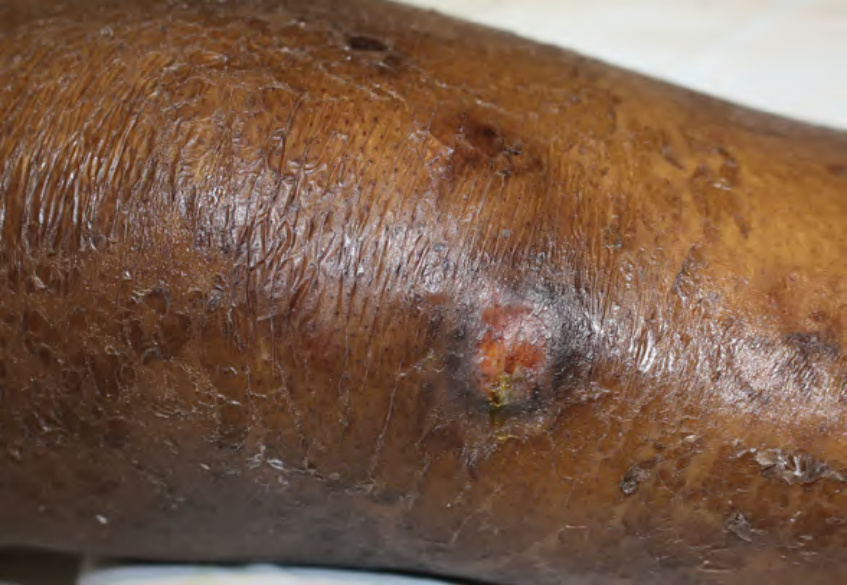
Fig. 51.6 Kwashiorkor. Edema, ulceration, and shellac-like scale that resembles peeling paint.
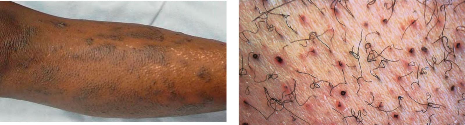
Fig. 51.7 Phrynoderma of vitamin A deficiency. Multiple clusters of follicular papules with central keratotic plugs. Histologically, keratinous plugs within follicles, hyperkeratosis, and atrophy of sebaceous glands are seen. It must be differentiated from pityriasis rubra pilaris and keratosis pilaris.
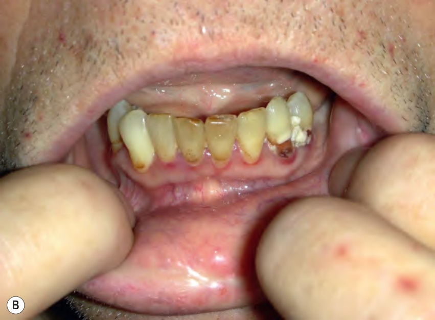
Fig. 51.8 Scurvy.A Corkscrew hairs and perifollicular hemorrhage on the lower extremities. B Gingivitis and gingival erosions. B, Courtesy Jeffrey P. Callen, MD.
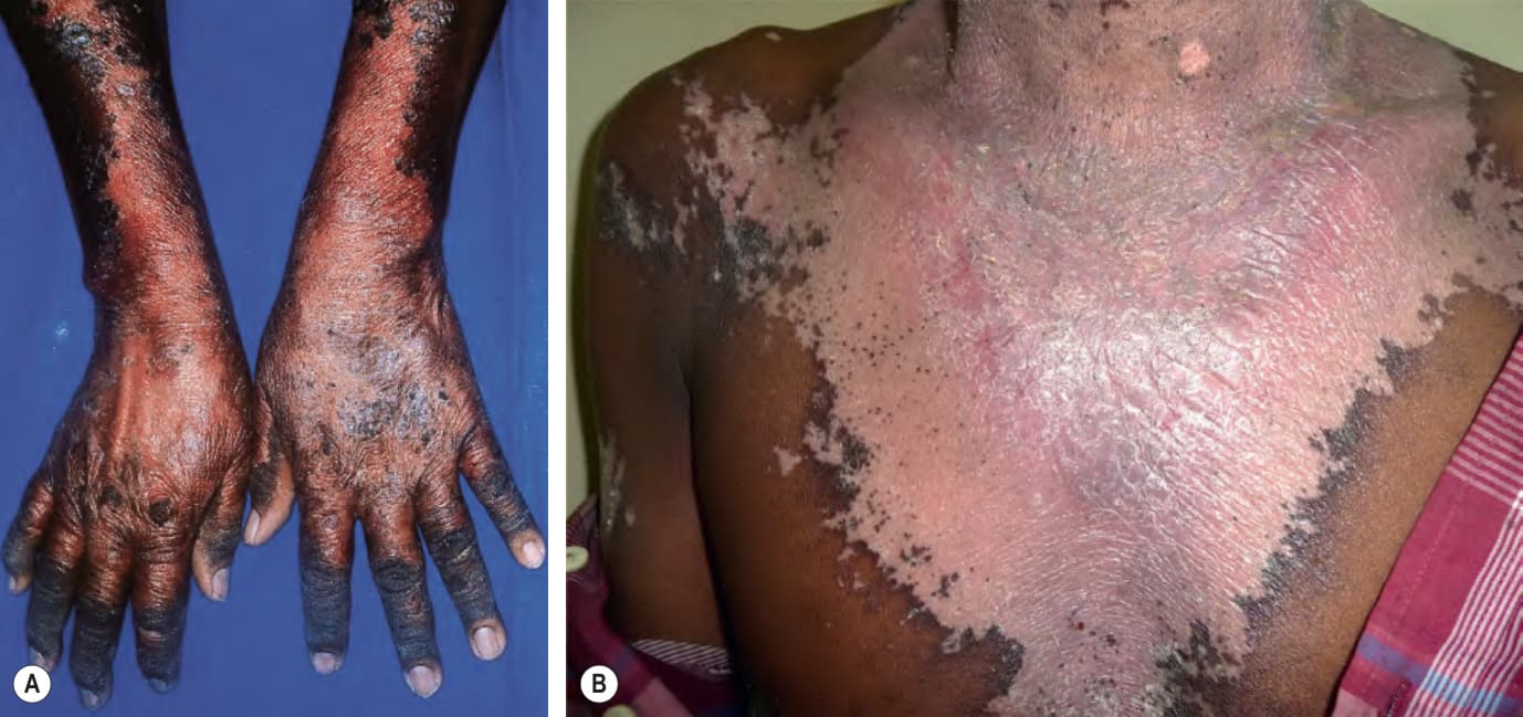
Fig. 51.9 Pellagra.A Hyper pigmentation and desquamation of the dorsal aspects of the hands and forearms. The shellaclike scale is seen best on the forearms. B Striking hypopigmentation with peripheral desquamation of the sun-exposed area of the chest in a man from sub-Saharan Africa. B, Courtesy Rosemarie Moser, MD.
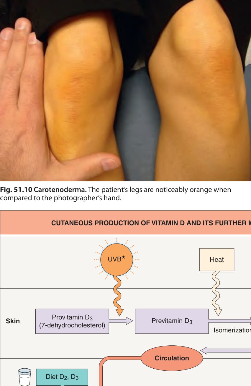
Fig. 51.10 Carotenoderma. The patient’s legs are noticeably orange when compared to the photographer’s hand.
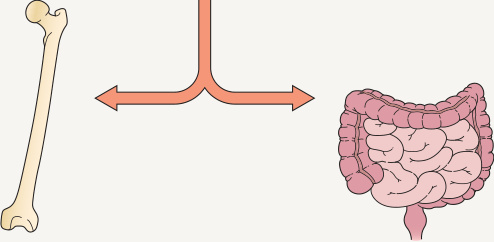
**Fig. 51.11 Cutaneous production of vitamin D and its further metabolism. During exposure to ultraviolet B radiation, 7-dehydrocholesterol within the skin is converted to previtamin D, which is then immediately converted to vitamin D in a heatdependent process. Of note, the heat from excessive sunlight exposure can degrade previtamin D and vitamin D into inactive photoproducts. Both forms of vitamin D (D and D) are biologically inactive and they require activation in the liver and then the kidney. After binding to carrier proteins, vitamin D is transported to the liver, where it is enzymatically hydroxylated to 25-hydroxyvitamin D [25(OH)D], the major circulating form of vitamin D. 25-hydroxyvitamin D is then converted into its active form, 1,25-dihydroxyvitamin D [1,25(OH)D], within the kidney by the enzyme 1α-hydroxylase. Of interest, this final hydroxylation step can also occur in keratinocytes when the enzyme CYP27B1 is upregulated in response to wounding or by Toll-like receptor (TLR) activation from microbial-derived ligands. Serum levels of phosphorus, calcium, and fibroblast growth factor 23 can either increase or decrease renal production of 1,25(OH)D. 1,25(OH)D decreases its own synthesis via feedback inhibition and decreases the synthesis and secretion of parathyroid hormone by the parathyroid glands. 1,25(OH)D also enhances intestinal calcium absorption in the small intestine by interacting with the vitamin D receptor–retinoic acid X receptor complex (VDR-RXR) to enhance the expression of the epithelial calcium channel and calbindin-D 9 K, a calcium-binding protein. In addition, 1,25(OH)D is recognized by its receptor in osteoblasts, leading to a series of events that maintain calcium and phosphorus levels in the blood which in turn promotes mineralization of the skeleton. *Most effective wavelength = 300 ± 5 nm. Measurement of this form most commonly done to assess vitamin D status.
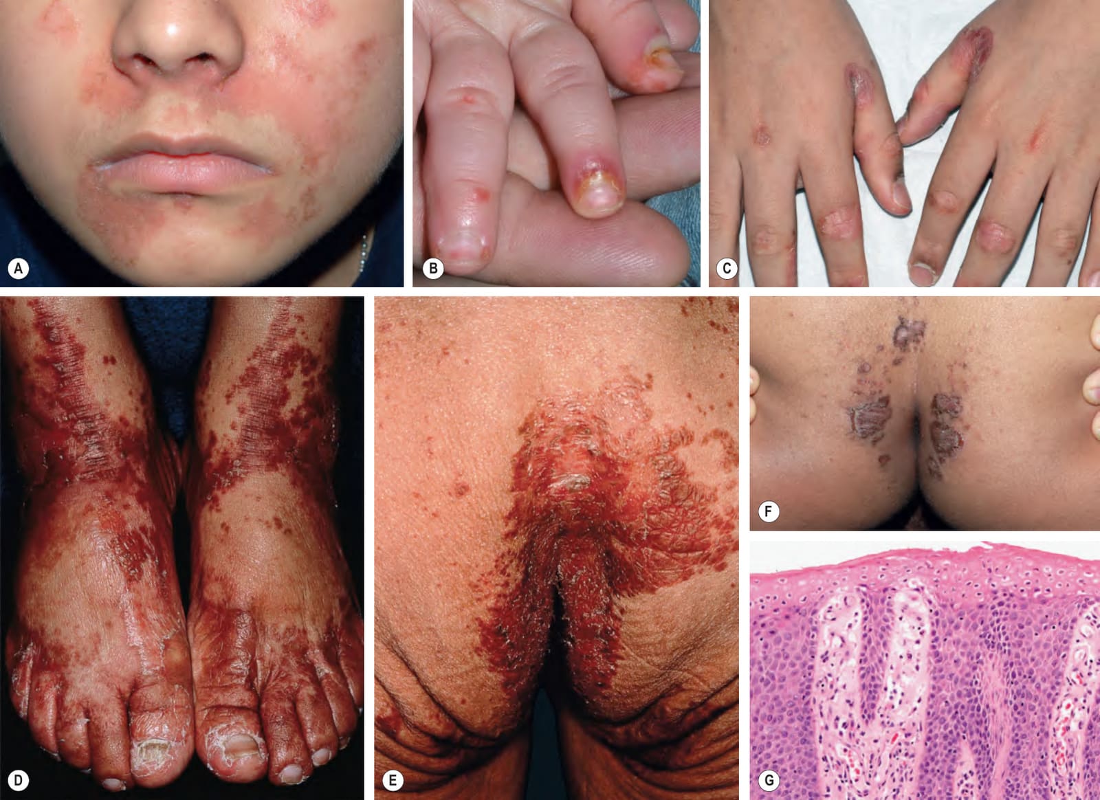
Fig. 51.12 Zinc deficiency – inherited and acquired forms. Erythema with erosions and scale-crust that is often shellac-like favors the perioral (A), acral (B–D), and anogenital (E, F) regions. Pustular paronychia can also be seen (B). The degree of involvement can vary from subtle to striking and may mimic other types of “nutritional dermatitis” (see Fig. 51.13), necrolytic migratory erythema, and more common disorders such as seborrheic dermatitis and psoriasis. The superficial erosions are a reflection of necrosis of the upper portion of the epidermis (G). A–C,F, Courtesy Julie V. Schaffer, MD; G, Courtesy Lorenzo Cerrroni, MD.

Fig. 51.13 Differential diagnosis of “nutritional dermatitis” (in addition to exogenous protein–energy malnutrition).Courtesy Julie V. Schaffer, MD; Photomicrograph Courtesy Luis Requena, MD.

Fig. 51.14 Menkes disease. This child has the characteristic pale skin and sparse kinky hair. Courtesy Ramón Ruiz-Maldonado, MD.
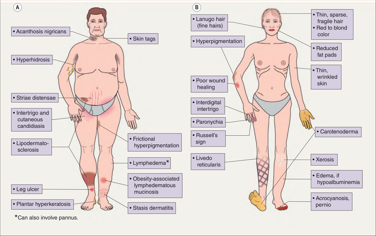
Fig. 51.15 Cutaneous findings in obese individuals (A) and patients with anorexia nervosa and/or bulimia (B). Russell’s sign consists of calluses or scars on the knuckles or dorsal surface of the hand due to repeated self-induced vomiting over prolonged periods of time. See Fig. 51.2 for signs of nutritional deficiencies.
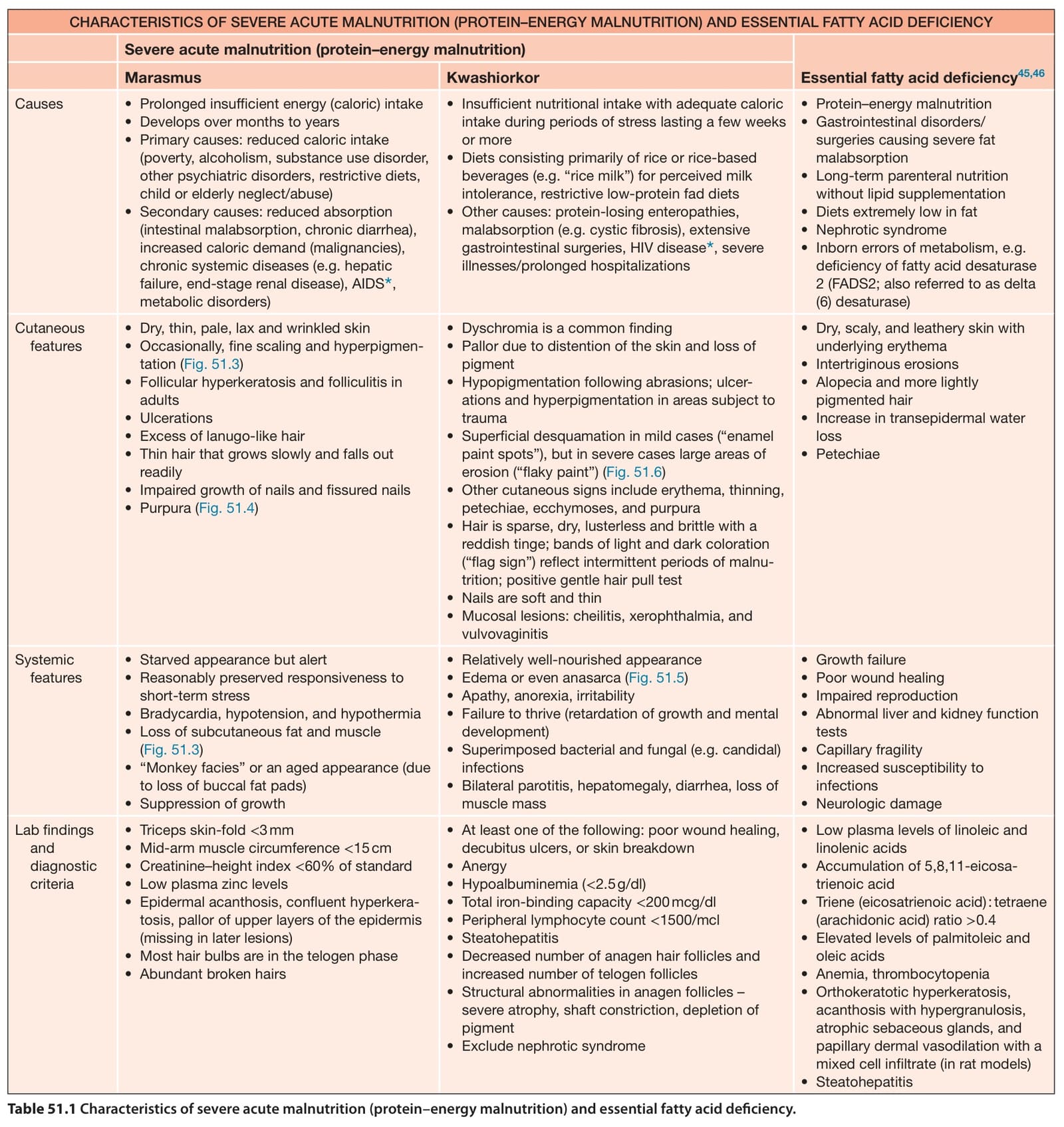
Table 51.1 Characteristics of severe acute malnutrition (protein–energy malnutrition) and essential fatty acid deficiency.
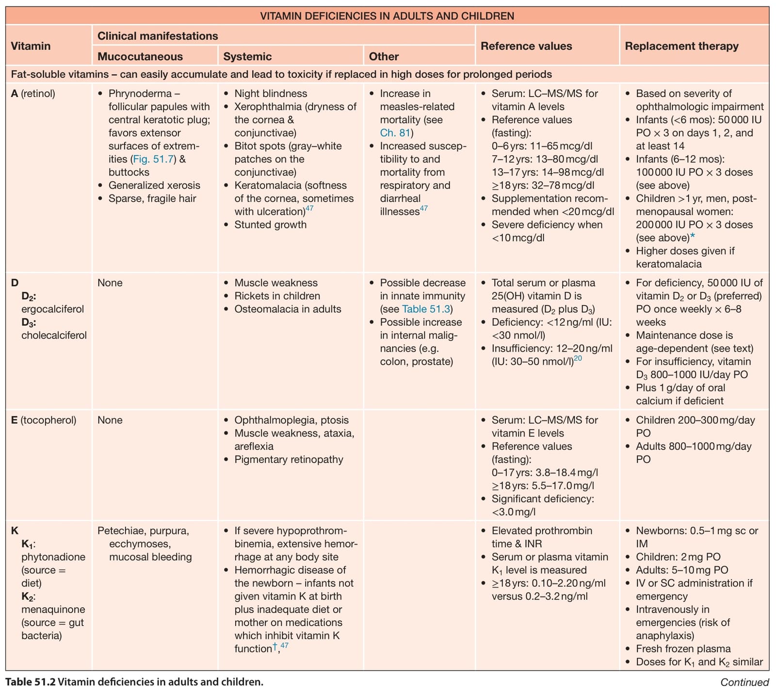
Table 51.2 Vitamin deficiencies in adults and children. Continued
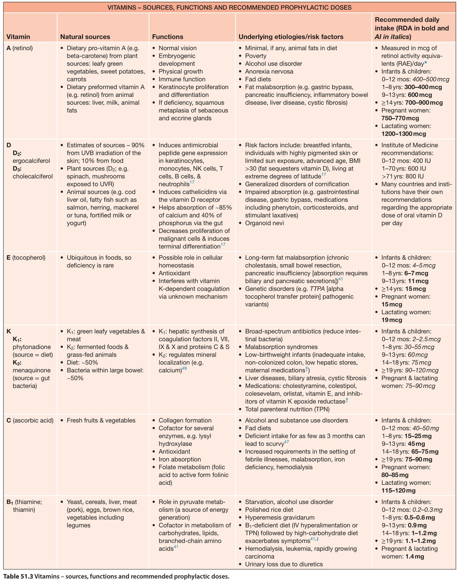
Table 51.3 Vitamins – sources, functions and recommended prophylactic doses.
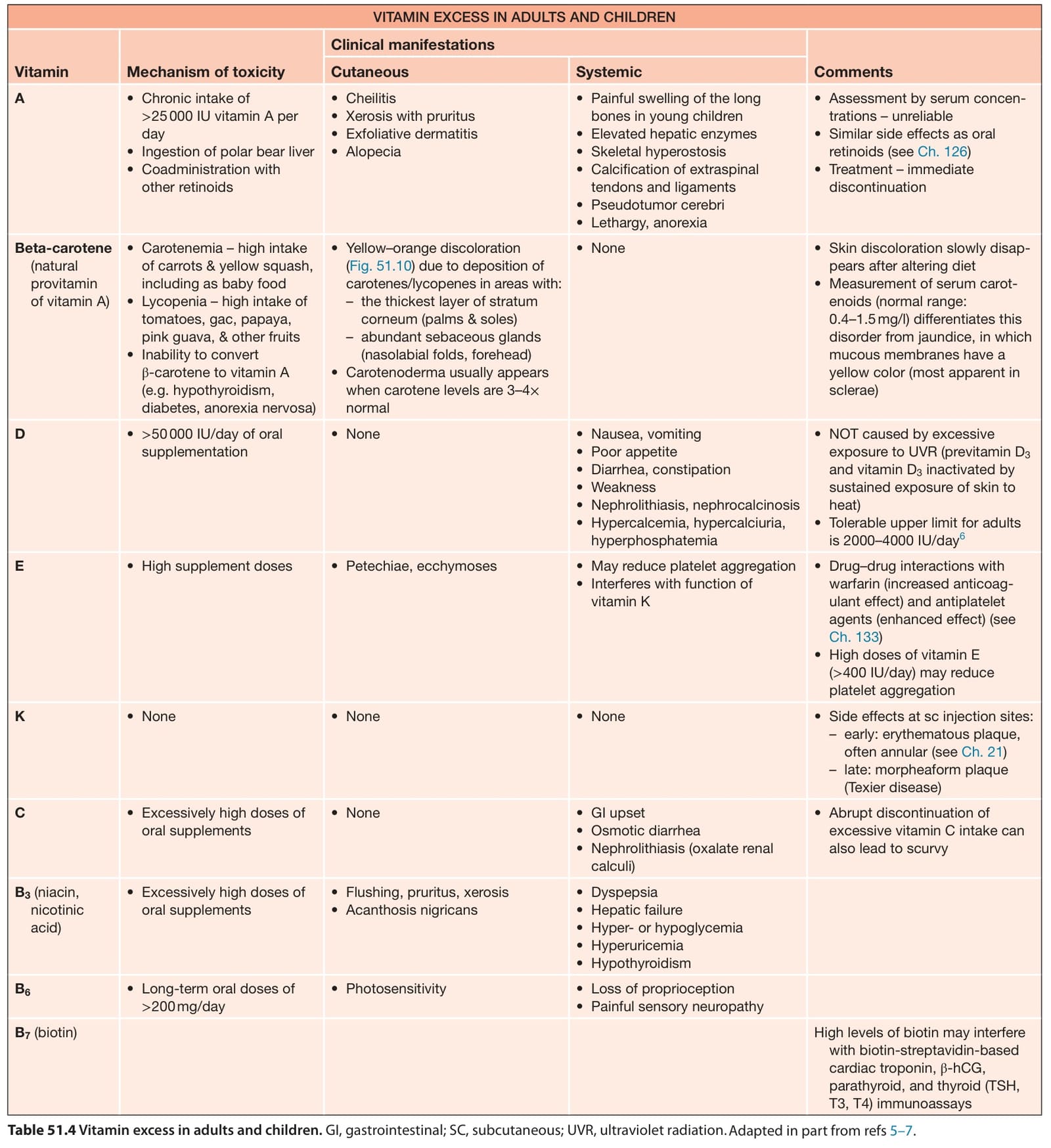
Table 51.4 Vitamin excess in adults and children. GI, gastrointestinal; SC, subcutaneous; UVR, ultraviolet radiation.Adapted in part from refs 5–7.