CLINICAL FEATURES
Acute GVHD
Acute GVHD presents in the skin as a morbilliform exanthem (Fig. 52.1), with an initial predilection for acral areas (e.g. dorsal hands and feet, palms, soles, forearms, ears), as well as the upper trunk. Lesions most commonly appear 4–6 weeks following HSCT despite prophylactic immunosuppressive therapy. Pruritus is variable and a folliculocentric pattern may be observed. Patients may be thrombocytopenic, which can give the eruption a hemorrhagic appearance. The gastrointestinal tract (nausea, voluminous diarrhea, abdominal pain) and the liver (cholestasis > transaminitis) are the two other primary organ systems affected by aGVHD. Together with body surface area of skin involvement, these manifestations are used for staging and grading of disease severity (Table 52.2). Stage IV acute skin disease consists of generalized involvement with bullae formation which may lead to skin sloughing resembling toxic epidermal necrolysis and portends a very low likelihood of survival. Fortunately, this degree of severity is nowadays rarely seen.
Chronic GVHD
In contrast to aGVHD, cGVHD may manifest in nearly any organ system (Fig. 52.2). Skin and mucosal involvement are exceedingly common, but there is remarkable variability in both presentation and severity. Other common sites of involvement include the eyes (keratoconjunctivitis sicca, blepharitis, corneal erosions), salivary glands (sicca syndrome), and lungs (bronchiolitis obliterans); less commonly, the esophagus (strictures, webs), liver, and pancreas (exocrine insufficiency) are affected.
Grade I aGVHD represents stage 1 or 2 cutaneous aGVHD while grade II aGVHD represents stage 3 cutaneous aGVHD; neither grade I nor II have significant liver or gut involvement. Grades III and IV aGVHD require internal involvement. BSA, body surface area.
In the past, differentiation of aGVHD and cGVHD was linked to time of onset following HSCT (≤ or >100 days); however, this is a somewhat arbitrary distinction and newer HSCT regimens have altered the onset of classic aGVHD and cGVHD manifestations. As a result, acute features may first present after day 100 (delayed acute GVHD). In addition, features of aGVHD can be seen in patients who also meet the diagnostic criteria for cGVHD and is referred to as overlap cGVHD. The NIH Consensus Project proposed a classification of cGVHD using organ-specific criteria. “Diagnostic” criteria, i.e. those skin manifestations sufficient to make a clinical diagnosis of cGVHD, are provided in Table 52.3. “Distinctive” cGVHD criteria (e.g. papulosquamous lesions, vitiligo, alopecia) require exclusion of other possible etiologies.
The most characteristic manifestation of epidermal cGVHD involvement is reticulate pink to violaceous papules and plaques with overlying scale, which in some patients resembles lichen planus (LP; Fig. 52.3A). Lesions often initially involve the dorsal aspects of the hands and feet, the forearms and trunk, but they can become widespread. In the past, the term “lichenoid” was used as a broad term which encompassed this presentation as well as all other non-sclerotic
cGVHD skin eruptions. In light of the greater appreciation for the breadth of clinical presentations of cutaneous cGVHD and to utilize more precise diagnostic terminology, the term “lichenoid” should be reserved for histologic description rather than as designation of a GVHD phenotype. In addition to LP-like lesions, psoriasiform plaques, keratosis pilaris-like follicular erythema, a subacute cutaneous LE-like eruption, or poikiloderma may develop (Table 52.4). Epidermal cGVHD involvement often leaves a remarkable patterned hyperpigmentation which is often reticulated and resolves very slowly over several months.
Similar to the use of “lichenoid”, the single designation “sclerodermoid” cGVHD is inadequate to describe the spectrum of sclerotic skin and subcutaneous tissue presentations. Sclerotic involvement includes superficial lichen sclerosus-like plaques, localized or widespread morphea-like involvement, and subcutaneous rippling of the skin with joint contractures resembling eosinophilic fasciitis. Lichen sclerosuslike involvement commonly presents as shiny, wrinkled, gray–white plaques on the upper back (Fig. 52.3B); there may be associated follicular plugging. Morphea-like (morpheaform) areas of sclerosis may occur anywhere on the body, but frequently localize to sites of previous
skin injury (e.g. port placement sites) as well as areas of friction (e.g. the waistband) (Fig. 52.3C–E). In young children, the diaper area can be the initial area of involvement. In contrast to systemic sclerosis, cGVHD is rarely associated with sclerodactyly, pinched facies, or Raynaud phenomenon. Deep-seated fibrosis of the subcutaneous tissue and fascia may present with insidious loss of joint range-ofmotion or as nonspecific myalgias and cramping. Subtle rippling of the overlying skin may be present with a firm, nodular texture noted with deep palpation (Fig. 52.4). Linear depressions within sites of fascial involvement (groove sign) demarcate the path of vascular structures
or fascial bundles. Longstanding fibrosis may result in skin ulceration, particularly of the legs and frictional surfaces, and the proliferation of benign angiomatous nodules (Fig. 52.3F).
Vitiligo can develop within skin affected by cGVHD as well as uninvolved skin. It occurs in 1%–2% of patients who have had an HSCT and ~5% of patients with cGVHD. The presence of vitiligo should alert the clinician to examine for additional evidence of cGVHD (Fig. 52.5A,B).
Mucosal surfaces are frequently affected by cGVHD (Fig. 52.6). Oral involvement resembles LP with lacy white plaques on the non-attached mucosa, ulceration, and gingivitis. Cheilitis is also a common manifestation (see Figs. 72.15 & 72.16 F) and the development of mucoceles is an additional clinical finding. Genital involvement, ranging from
A Vitiligo with patches of amelanosis; note the nail dystrophy. B Scarring alopecia and vitiligo in the setting of extensive sclerotic cGVHD. C Nail thinning, splitting, and dorsal pterygium formation.
erythema and fissuring to labial resorption and fusion, can occur in as many as 50% of female patients. Symptoms of burning, pruritus, and dyspareunia are common. Scarring of the vagina may lead to short-ening and narrowing of the vaginal canal and risk of hematocolpos in menstruating women. Male genital involvement is less common, but includes balanoposthitis and lichen planus-like lesions, and phimosis may develop in some patients.
Nail changes in cGVHD range from thin, brittle nails with distal splitting to dorsal pterygium and anonychia (Fig. 52.5C). In children, nail dystrophy, particularly dorsal pyterygium, has been associated with pulmonary cGVHD. Additional cutaneous findings include facial milia, alopecia areata, angiomatous nodules, scarring alopecia, and calcinosis cutis. The latter three entities are seen in patients with sclerotic disease and calcinosis cutis may contribute to decreased range of motion.
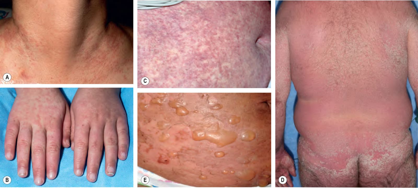
Fig. 52.1 Clinical spectrum of acute cutaneous graft-versus-host disease.AStage I – discrete and coalescing small pink papules on the upper chest and neck of a woman 6 weeks following allogeneic bone marrow transplant. B, CStage II – pink macules and papules on the dorsal aspect of the hands that are becoming confluent 4 weeks post allogeneic bone marrow transplant, and widespread pink–violet macules and papules on the abdomen in a liver transplant recipient. DStage III – diffuse erythema with desquamation, but without bullae formation. EStage IV – widespread bullae and epidermal necrosis in a patient who had received a donor lymphocyte infusion (DLI) following allogeneic bone marrow transplant; note the resemblance to toxic epidermal necrolysis. C, Courtesy Julie V. Schaffer, MD.
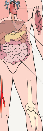
Fig. 52.2 Protean multi-organ manifestations of chronic graft-versus-host disease. The most common manifestations are in bold.
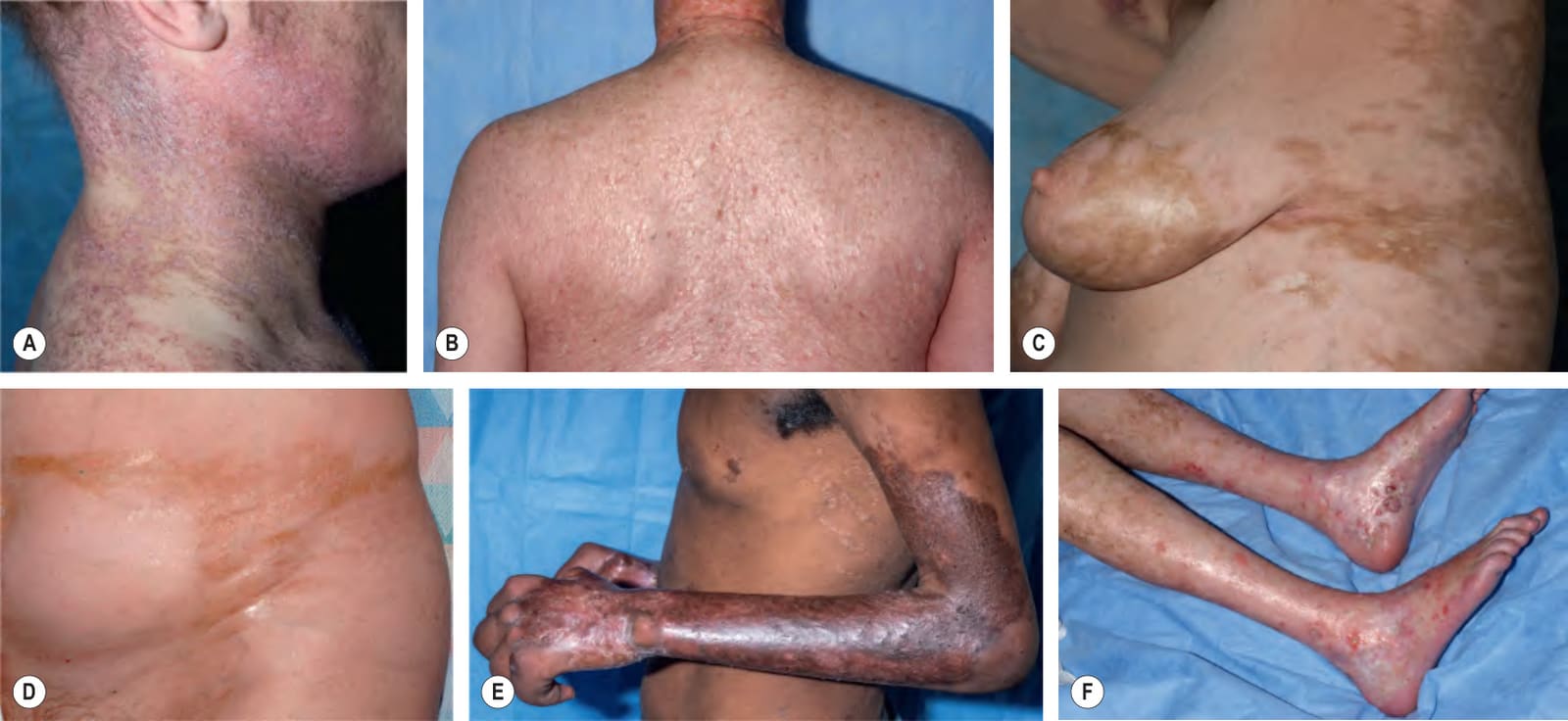
Fig. 52.3 Clinical spectrum of chronic cutaneous graft-versus-host disease.A Lichen planus-like: thin pink–violet papules and plaques with scale, admixed with postinflammatory hyperpigmentation. B Lichen sclerosus-like: multiple gray–white thin plaques with obvious wrinkling are present on the mid back. C, D Morphea-like: shiny, hyperpigmented, sclerotic plaques extending from the breasts to the lateral torso and in the girdle area; the latter is a common site of involvement for this form of cGVHD. E Morphea-like: sclerotic hyperpigmented plaque on the upper arm, with shiny bound-down skin of the forearm and dorsal hand; note the joint contractures. F Scleroderma-like: the skin is shiny and bound-down with dyspigmentation, hair loss, multiple erosions and ulceration; angiomatous nodules and marked reduction in range of motion of the ankles are also present.
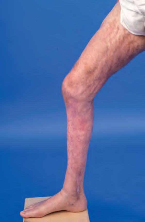
Fig. 52.4 Eosinophilic fasciitis-like presentation of chronic cutaneous graft-versus- host disease. A rippled appearance and irregular nodular texture to the skin is indicative of involvement of subcutaneous tissues. Especially in the earlier, edematous phase, the presence of hypereosinophilia may be a clue to the diagnosis.
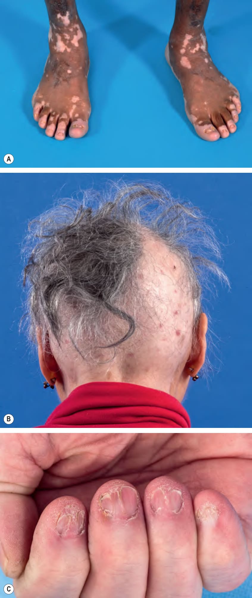
Fig. 52.5 Additional cutaneous findings in chronic graft-versus-host disease.
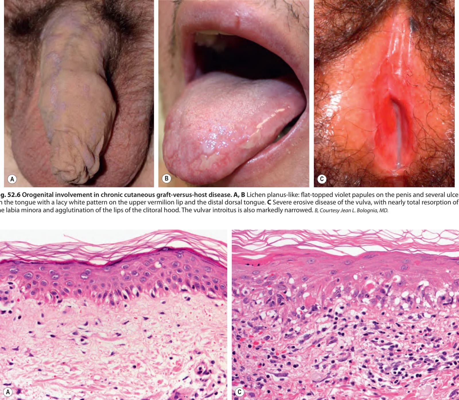
Fig. 52.6 Orogenital involvement in chronic cutaneous graft-versus-host disease.A, B Lichen planus-like: flat-topped violet papules on the penis and several ulcers on the tongue with a lacy white pattern on the upper vermilion lip and the distal dorsal tongue. C Severe erosive disease of the vulva, with nearly total resorption of the labia minora and agglutination of the lips of the clitoral hood. The vulvar introitus is also markedly narrowed. B, Courtesy Jean L. Bolognia, MD.
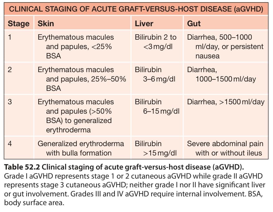
Table 52.2 Clinical staging of acute graft-versus-host disease (aGVHD).
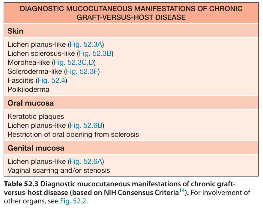
Table 52.3 Diagnostic mucocutaneous manifestations of chronic graft- versus-host disease (based on NIH Consensus Criteria). For involvement of other organs, see Fig. 52.2.
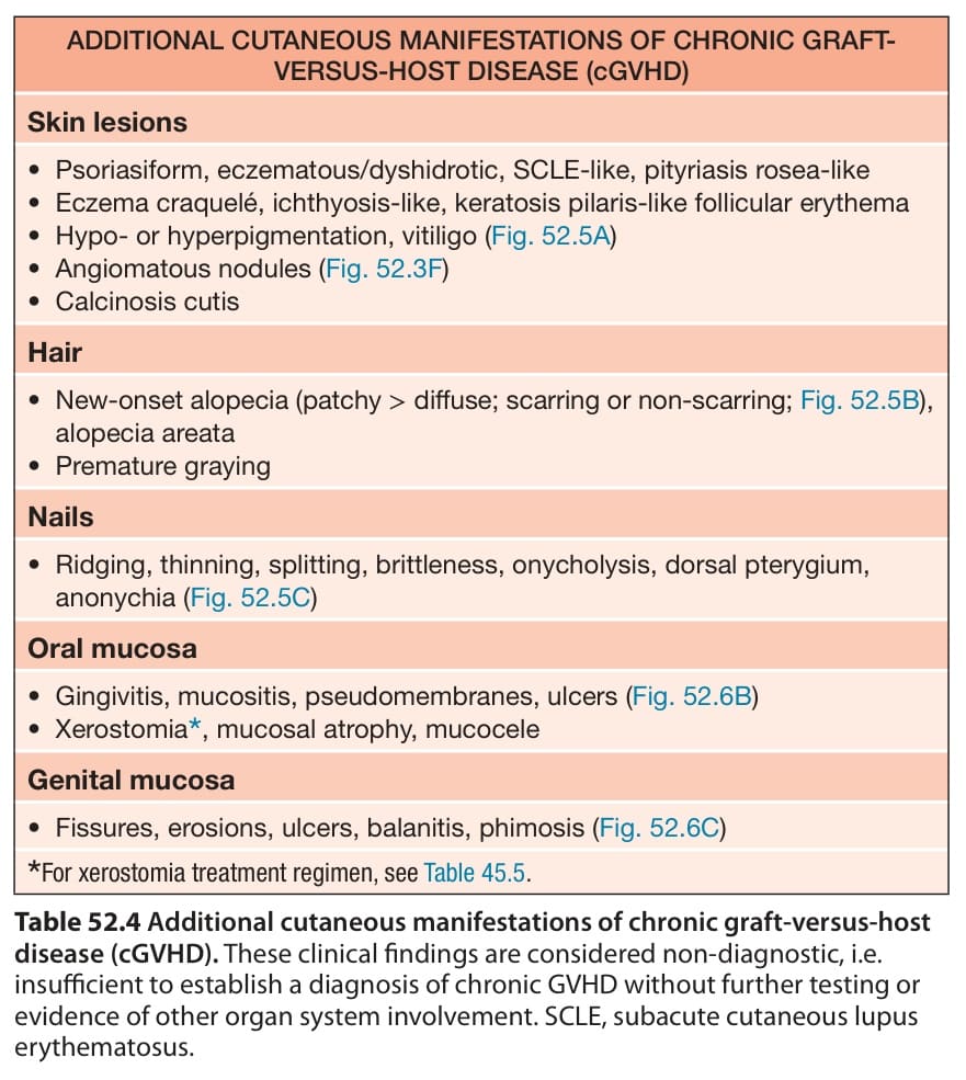
Table 52.4 Additional cutaneous manifestations of chronic graft-versus-host disease (cGVHD). These clinical findings are considered non-diagnostic, i.e. insufficient to establish a diagnosis of chronic GVHD without further testing or evidence of other organ system involvement. SCLE, subacute cutaneous lupus erythematosus.