PATHOLOGY
Histologically, aGVHD is characterized by widespread keratinocyte necrosis, basal layer hydropic degeneration, and a band-like lymphohistiocytic infiltrate in the upper dermis (Fig. 52.7). However, skin biopsies obtained shortly after disease onset may display more subtle findings, e.g. only focal vacuolar change of basal keratinocytes, and histologic features of aGVHD may be limited to the hair follicle. In addition, these histologic features overlap with those of viral exanthems and drug eruptions, making a definitive diagnosis difficult to render in some patients. With increasing severity of aGVHD, basilar vacuoles can fuse to form clefts and microvesicles and even complete separation of epidermis from dermis.
The histology of cGVHD reflects the variable skin manifestations of the condition. cGVHD with epidermal involvement may resemble aGVHD, but with a more robust lichenoid infiltrate and additional features of epidermal orthohyperkeratosis, hypergranulosis, and variable (usually mild) acanthosis. The sclerotic subtypes of cGVHD may have concurrent epidermal features or may only demonstrate sclerosis of the dermis (Fig. 52.8), subcutaneous tissue, and/or fascia. Thickening of subcutaneous fat septae indicates a deep-seated sclerosis and magnetic resonance imaging may be helpful to identify fascial involvement (Fig. 52.9).
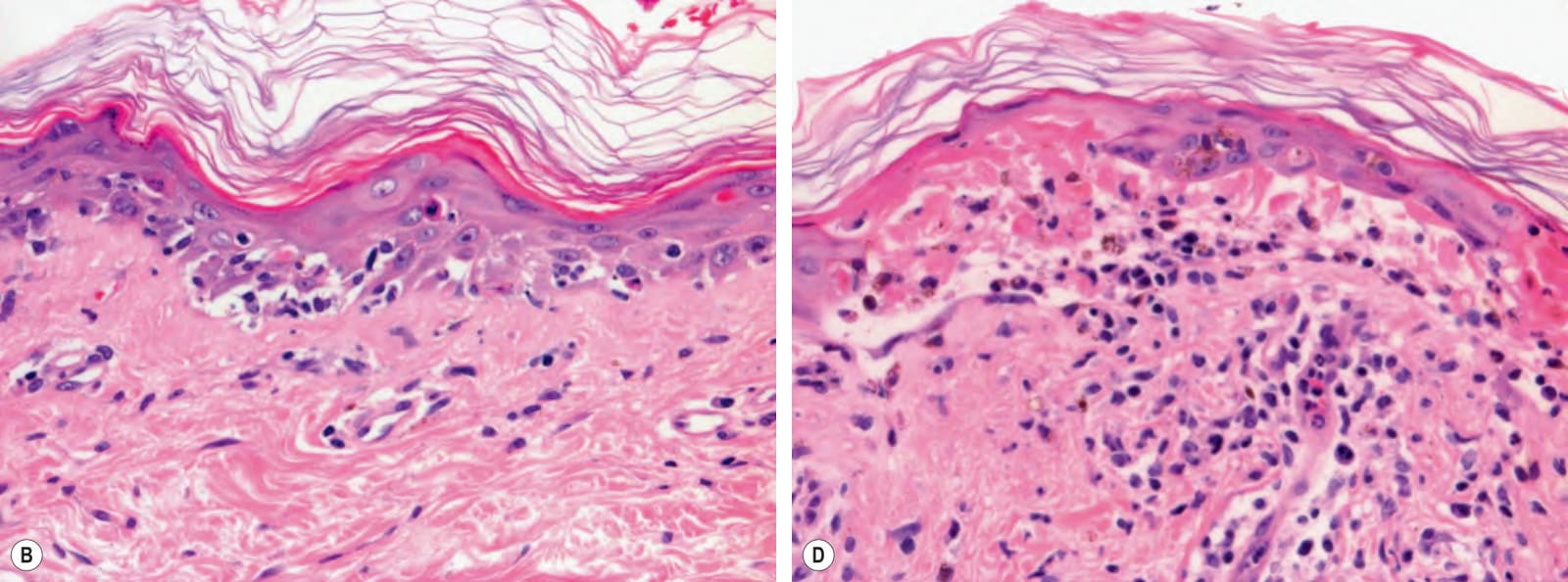
Fig. 52.7 Acute cutaneous graft-versus-host disease – histopathologic grades.A Grade I: focal vacuolar changes of the basal layer of the epidermis and sparse lymphocytic infiltrate. B Grade II: necrosis of keratinocytes and more obvious vacuolar degeneration of the basal layer. C Grade III: apoptosis of keratinocytes and blurring of the dermal–epidermal junction due to marked vacuolar degeneration and a lichenoid infiltrate of lymphocytes. D Grade IV: full-thickness necrosis of the epidermis with separation of the epidermis from the dermis. Of note, grade I changes are rather nonspecific and are also seen with autoimmune disorders showing the histopathologic pattern of an interface dermatitis (e.g. lupus erythematosus). Courtesy Lorenzo Cerroni, MD.
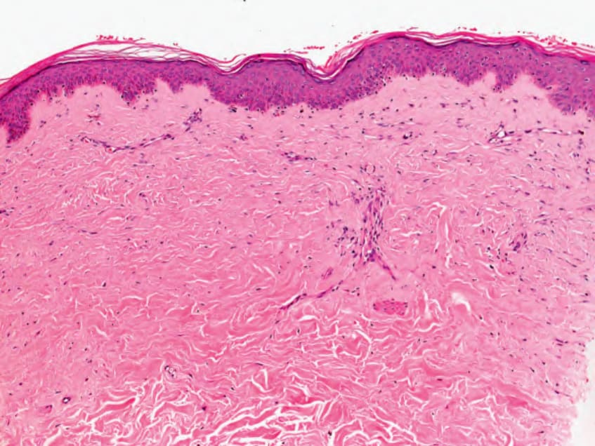
Fig. 52.8 Chronic cutaneous graft-versus-host disease (morphea-like) – histologic features. There is sclerosis of the dermis with thickening of collagen bundles. These same histologic features can be seen in morphea and systemic sclerosis (scleroderma). Pigmentary incontinence, with a few melanophages within the papillary dermis can also be seen in this form of chronic GVHD. Courtesy Lorenzo Cerroni, MD.
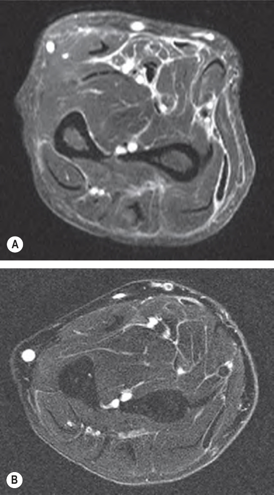
Fig. 52.9 Magnetic resonance imaging of the eosinophilic fasciitis-like presentation of chronic cutaneous graft-versus- host disease.A Prior to treatment (systemic corticosteroids), with a lace-like white pattern representing edema of the fascia. B After treatment, with marked diminution of edema. The round white circles represent blood vessels. Courtesy Dennis Cooper, MD.