病理學(PATHOLOGY)
組織學上,aGVHD 的特徵為廣泛的角質形成細胞壞死、基底層水腫變性(hydropic degeneration),以及上部真皮呈帶狀的淋巴組織球性浸潤(圖 52.7)。然而,疾病發生後不久取得的皮膚切片可能顯示更細微的發現,例如僅有基底角質形成細胞的局灶性空泡變化,且 aGVHD 的組織學特徵可能局限於毛囊。此外,這些組織學特徵與病毒疹及藥物疹者重疊,使得在部分病人難以做出確定診斷。隨著 aGVHD 嚴重度增加,基底部空泡可融合形成裂隙與微水疱,甚至表皮與真皮的完全分離。
cGVHD 的組織學反映了此病況多變的皮膚表現。具表皮侵犯的 cGVHD 可能類似 aGVHD,但具有更明顯的苔癬樣浸潤,並有表皮正角化過度(orthohyperkeratosis)、顆粒層增厚(hypergranulosis)與程度不一(通常為輕度)的棘皮症等額外特徵。cGVHD 的硬化性亞型可能同時具有表皮特徵,或僅顯示真皮(圖 52.8)、皮下組織及/或筋膜的硬化。皮下脂肪間隔的增厚指示深部硬化,而磁振造影可能有助於辨識筋膜侵犯(圖 52.9)。
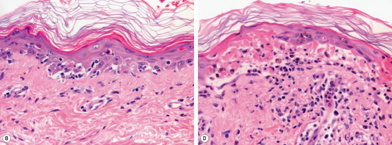
圖 52-7:急性皮膚移植物抗宿主病——組織病理學分級。A 第 I 級:表皮基底層的局灶性空泡變化與稀疏的淋巴球浸潤。B 第 II 級:角質形成細胞壞死與更明顯的基底層空泡變性。C 第 III 級:角質形成細胞凋亡,以及因顯著空泡變性與淋巴球苔癬樣浸潤所致的真皮–表皮交界模糊。D 第 IV 級:表皮全層壞死並伴表皮與真皮分離。值得注意的是,第 I 級的變化頗為非特異性,亦見於呈現介面性皮膚炎(interface dermatitis)組織病理型態的自體免疫疾患(例如紅斑性狼瘡)。承蒙 Lorenzo Cerroni, MD 提供。
Fig. 52.7 Acute cutaneous graft-versus-host disease – histopathologic grades.A Grade I: focal vacuolar changes of the basal layer of the epidermis and sparse lymphocytic infiltrate. B Grade II: necrosis of keratinocytes and more obvious vacuolar degeneration of the basal layer. C Grade III: apoptosis of keratinocytes and blurring of the dermal–epidermal junction due to marked vacuolar degeneration and a lichenoid infiltrate of lymphocytes. D Grade IV: full-thickness necrosis of the epidermis with separation of the epidermis from the dermis. Of note, grade I changes are rather nonspecific and are also seen with autoimmune disorders showing the histopathologic pattern of an interface dermatitis (e.g. lupus erythematosus). Courtesy Lorenzo Cerroni, MD.
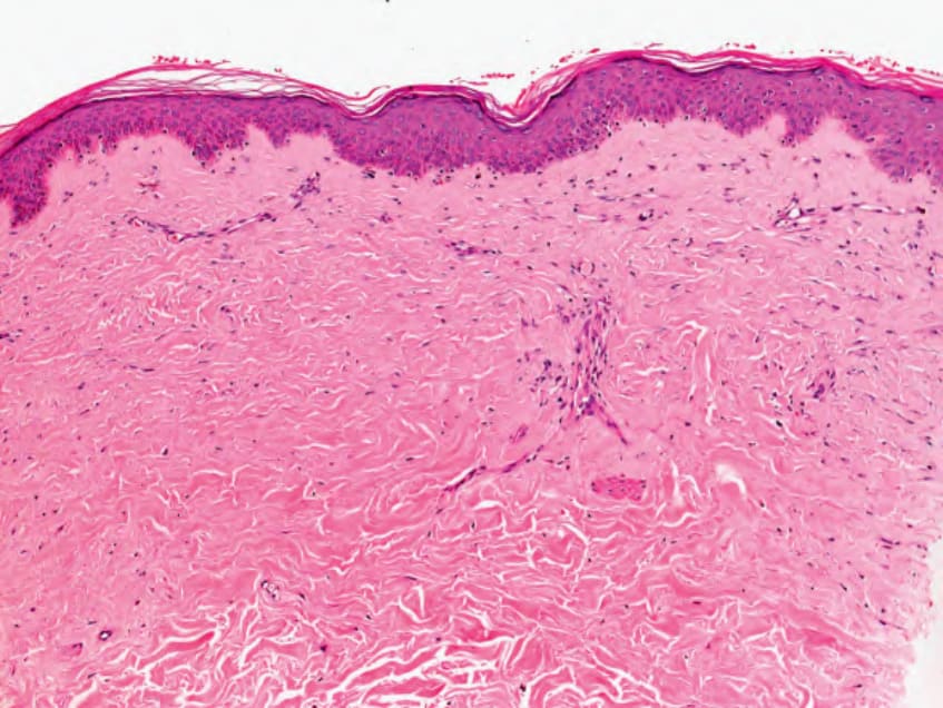
圖 52-8:慢性皮膚移植物抗宿主病(類局限性硬皮症)——組織學特徵。可見真皮硬化並伴膠原束增厚。這些相同的組織學特徵亦可見於局限性硬皮症與全身性硬皮症(scleroderma)。色素失禁(pigmentary incontinence)併乳頭層真皮內少數噬黑色素細胞,亦可見於此型慢性 GVHD。承蒙 Lorenzo Cerroni, MD 提供。
Fig. 52.8 Chronic cutaneous graft-versus-host disease (morphea-like) – histologic features. There is sclerosis of the dermis with thickening of collagen bundles. These same histologic features can be seen in morphea and systemic sclerosis (scleroderma). Pigmentary incontinence, with a few melanophages within the papillary dermis can also be seen in this form of chronic GVHD. Courtesy Lorenzo Cerroni, MD.
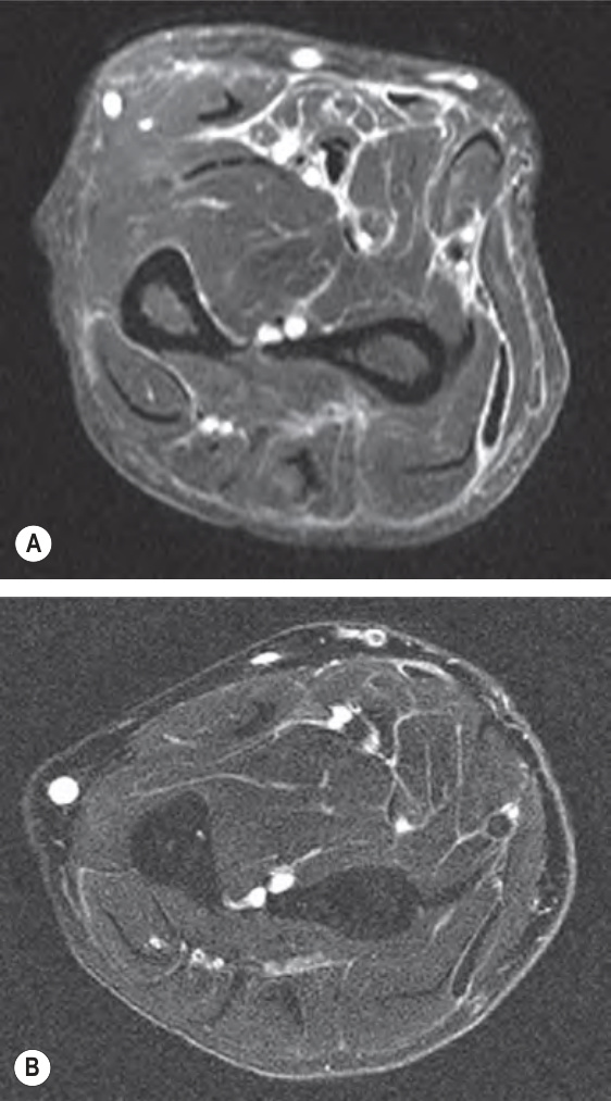
圖 52-9:慢性皮膚移植物抗宿主病之類嗜酸性球性筋膜炎表現的磁振造影。A 治療(全身性皮質類固醇)前,呈蕾絲狀白色圖案,代表筋膜的水腫。B 治療後,水腫顯著減少。圓形白色圈代表血管。承蒙 Dennis Cooper, MD 提供。
Fig. 52.9 Magnetic resonance imaging of the eosinophilic fasciitis-like presentation of chronic cutaneous graft-versus- host disease.A Prior to treatment (systemic corticosteroids), with a lace-like white pattern representing edema of the fascia. B After treatment, with marked diminution of edema. The round white circles represent blood vessels. Courtesy Dennis Cooper, MD.