TREATMENT
Acute GVHD
Limited cutaneous aGVHD (without other organ involvement) may initially be managed with topical corticosteroids while patients with more extensive skin involvement or internal manifestations require systemic treatment. First-line therapy consists of adding oral prednisone or IV methylprednisolone (0.5–1 mg/kg twice daily) to ongoing prophylactic GVHD therapy (usually a systemic calcineurin inhibitor plus either methotrexate or mycophenolate mofetil). This treatment approach results in disease control in ~50% of patients. Recently, the JAK inhibitor ruxolitinib was FDA-approved for steroid-refractory aGVHD and represents the preferred second-line therapy. Increasingly it is being used as a steroidsparing agent earlier after the diagnosis of aGVHD. Third-line therapies include other immunomodulatory agents such as TNF antagonists, but they are associated with an increased risk of infections. Unfortunately, until the recent introduction of ruxolitinib, patients who did not respond well to corticosteroid therapy were at high risk of mortality.
Chronic GVHD
Chronic GVHD continues to be a major therapeutic challenge, but the consistent observation that post-transplant cyclophosphamide has reduced its incidence without an increase in relapse or opportunistic infections is encouraging. With respect to cutaneous cGVHD, skindirected therapies can play an important adjunctive role. For an LP-like eruption or pruritus, medium- to high-potency topical corticosteroids and topical calcineurin inhibitors (pimecrolimus, tacrolimus) are effective. Of note, use of topical tacrolimus under occlusion may lead
to toxic drug levels in patients on concurrent systemic tacrolimus. Various forms of phototherapy have shown some benefit in small series, however controlled trial data are needed. The potential for increased skin cutaneous malignancies in the setting of concurrent immunosuppressive therapy or prophylactic voriconazole must also be weighed against the benefit of UV therapy.
As with aGVHD, first-line systemic therapy of cGVHD is systemic corticosteroids, but while the dose is less (maximum dose of prednisone is 1 mg/kg/day), the duration of treatment is often prolonged. This extended corticosteroid therapy is a major cause of non-relapse morbidity and mortality, especially from opportunistic infections. Approximately 50% of cGVHD patients will not respond adequately to corticosteroid therapy, and better treatment for this group remains a critical unmet need. Ibrutinib is FDA-approved for steroid-refractory cGVHD, but since the approval of ruxolitinib for the same indication, the use of ibrutinib has declined. For patients who have failed ≥2 lines of therapy, belumosudil, a well-tolerated oral inhibitor of rho-associated coil kinase 2 (ROCK2), has recently been FDA-approved given a response rate of >70%. There are also ongoing trials of the use of axatilimab, an anti- CSF-1R antibody, for cGVHD28a. Other options include extracorporeal photophoresis, sirolimus, imatinib mesylate, and rituxumab.
Patients with mucocutaneous cGVHD should undergo ocular, oral, and genital examinations. Site-specific therapies, including scleral contact lenses, oral rinses (e.g. dexamethasone elixir), and vaginal dilators can prove very helpful. Referral to physical therapy is strongly recommended in patients with sclerotic involvement to improve any decreased joint range-of-motion.
Long term, the importance of sun protection should be emphasized, as patients are typically prescribed one or more photosensitizing agents, including trimethoprim–sulfamethoxazole, levofloxacin, and voriconazole. In addition, the latter drug can increase the risk of developing cutaneous malignancies. Patients, especially those with significant photodamage and/or a previous history of cutaneous carcinomas, should be educated regarding skin cancer risk in the setting of immunosuppression, and they should receive regular surveillance for skin cancer. Ideally, patients with chronic mucocutaneous GVHD should be managed in a coordinated multidisciplinary manner by specialists in ophthalmology, gynecology, oral medicine, physiatry, and dermatology in order to optimize care.
Additional figures and tables on Risk factors associated with the development of graft-versus-host disease, and Comparison of clinical and laboratory features of sclerotic-type chronic GVHD and systemic sclerosis, available in our eBook (see inside front cover for access code).
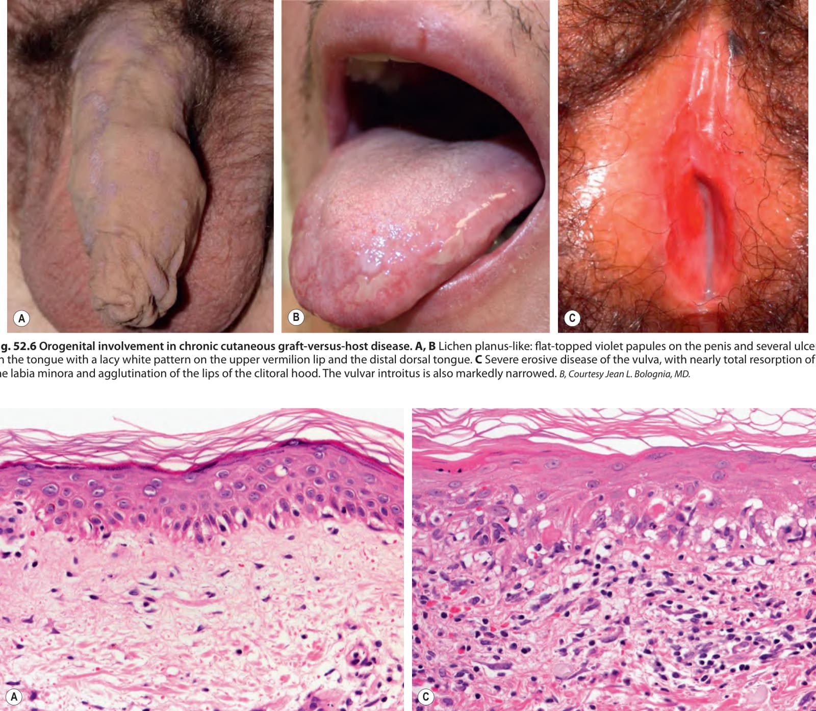
Fig. 52.6 Orogenital involvement in chronic cutaneous graft-versus-host disease.A, B Lichen planus-like: flat-topped violet papules on the penis and several ulcers on the tongue with a lacy white pattern on the upper vermilion lip and the distal dorsal tongue. C Severe erosive disease of the vulva, with nearly total resorption of the labia minora and agglutination of the lips of the clitoral hood. The vulvar introitus is also markedly narrowed. B, Courtesy Jean L. Bolognia, MD.
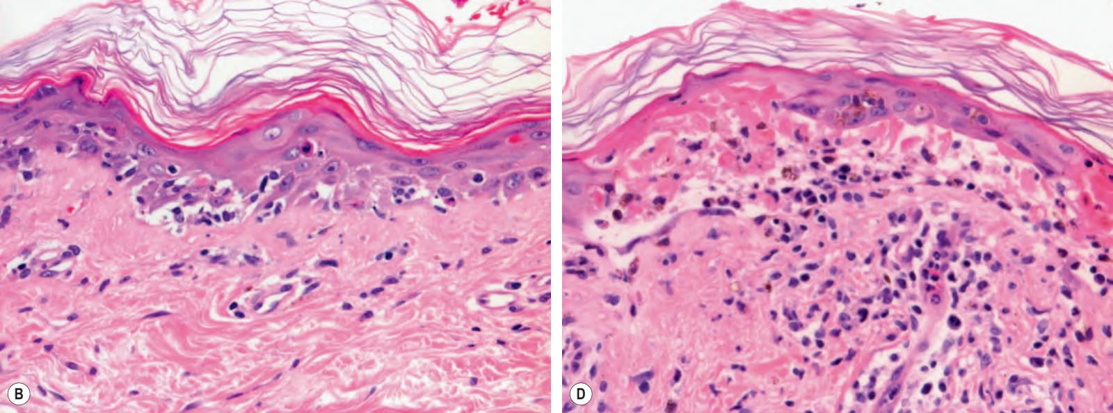
Fig. 52.7 Acute cutaneous graft-versus-host disease – histopathologic grades.A Grade I: focal vacuolar changes of the basal layer of the epidermis and sparse lymphocytic infiltrate. B Grade II: necrosis of keratinocytes and more obvious vacuolar degeneration of the basal layer. C Grade III: apoptosis of keratinocytes and blurring of the dermal–epidermal junction due to marked vacuolar degeneration and a lichenoid infiltrate of lymphocytes. D Grade IV: full-thickness necrosis of the epidermis with separation of the epidermis from the dermis. Of note, grade I changes are rather nonspecific and are also seen with autoimmune disorders showing the histopathologic pattern of an interface dermatitis (e.g. lupus erythematosus). Courtesy Lorenzo Cerroni, MD.
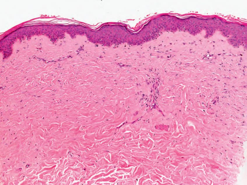
Fig. 52.8 Chronic cutaneous graft-versus-host disease (morphea-like) – histologic features. There is sclerosis of the dermis with thickening of collagen bundles. These same histologic features can be seen in morphea and systemic sclerosis (scleroderma). Pigmentary incontinence, with a few melanophages within the papillary dermis can also be seen in this form of chronic GVHD. Courtesy Lorenzo Cerroni, MD.
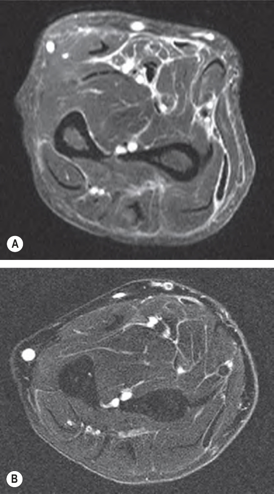
Fig. 52.9 Magnetic resonance imaging of the eosinophilic fasciitis-like presentation of chronic cutaneous graft-versus- host disease.A Prior to treatment (systemic corticosteroids), with a lace-like white pattern representing edema of the fascia. B After treatment, with marked diminution of edema. The round white circles represent blood vessels. Courtesy Dennis Cooper, MD.
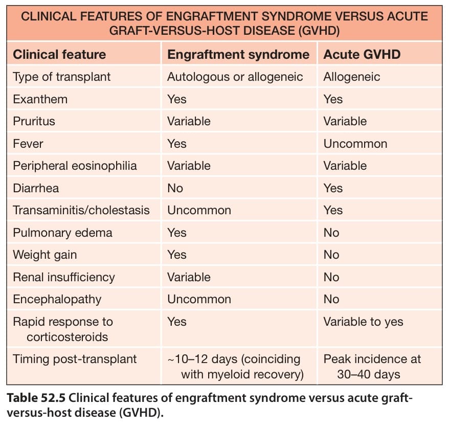
Table 52.5 Clinical features of engraftment syndrome versus acute graftversus-host disease (GVHD).