CHROMOSOMAL DISORDERS
The above sections discussed disease-causing mutations that affect a single gene. However, other disorders can result from larger aberrations at the chromosomal level, and these anomalies may produce severe phenotypes due to the involvement of multiple genes and regulatory regions. For example, the majority of spontaneous abortuses exhibit chromosomal anomalies.
Chromosomal anomalies can affect the number or structure of chromosomal segments or the entire chromosome. Numeric chromosomal abnormalities may involve the whole genome (polyploidy) or just one pair of homologs (aneuploidy). The forms of polyploidy observed in humans are triploidy with three complete sets of chromosomes (3n), and tetraploidy with four chromosome complements (4n). However, unless mosaic, polyploidy results in a spontaneous abortion or death soon after birth. Aneuploidy refers to extra or missing chromosome(s), such as trisomies with three copies of a single chromosome or
monosomies with only one copy. Trisomies for a whole chromosome that are compatible with extrauterine life (outside the setting of mosaicism) involve chromosomes 21 (Down syndrome), 18 (Edwards syndrome), 13 (Patau syndrome), and 23 (XXY, Klinefelter syndrome; XYY). Monosomy for the X chromosome (Turner syndrome) can also occur. Other trisomies and monosomies may be observed in mosaic or partial (e.g. due to translocations) forms.
Structural abnormalities involve breakage of chromosomes. They are balanced if there is no net gain or loss of genetic material, or unbalanced when they are accompanied by additional or missing genetic information. The stability of the resulting rearranged chromosome through cell division will depend on the presence of a centromere and two telomeres, the necessary elements for the correct segregation of chromosomes.
Balanced chromosomal abnormalities include some types of translocations and inversions. In a reciprocal translocation, two breaks in two non-homologous chromosomes occur and the chromosomal segments distal to the breaks are exchanged. The total number of chromosomes
remains the same. If the translocation breakpoint affects a gene or its regulatory sequence, or otherwise places a gene under the control of novel regulatory elements (e.g. the Philadelphia chromosome), it can result in an observable phenotype (e.g. chronic myelogenous leukemia). In addition, clinical manifestations related to reciprocal translocations can occur in the offspring of the carriers. Depending on the combination of parental chromosomes inherited, the offspring may have partial trisomy or monosomy for the chromosomal regions involved in the translocation, i.e. an unbalanced chromosomal anomaly.
Inversions are balanced rearrangements in which two breaks occur in the same chromosome and the intervening segment is inverted before the chromosome is reconstituted. In a pericentric inversion, the inverted segment includes the centromere, whereas in a paracentric inversion, the two breaks occur in the same chromosome arm, without involvement of the centromere.
Numerical and structural chromosomal aberrations can be detected using karyotypes, which allow visualization of banding patterns in stained metaphase chromosomes (Fig. 54.4). Fluorescence in situ hybridization (FISH) can identify much smaller changes in the chromosomal constitution that are not visible with conventional banding techniques but requires the use of preselected probes (see Table 3.5 and Fig. 3.8). More recently, advances in microarray technology have enabled the use of array-based comparative genomic hybridization (CGH) for detection of copy number variations (CNVs) at a considerably higher level of resolution than can be accomplished via karyotypes. Conventional CGH identifies chromosomal gains and losses and all CNVs in a single hybridization procedure. While this method has improved resolution (5–10 Mb) compared to chromosomal banding, it still requires metaphase chromosomal preparations and is unable to identify balanced chromosomal rearrangements. However, array-based CGH (aCGH) that incorporates DNA microarray technologies has an improved resolution of ~100 Kb. Currently, single nucleotide polymorphism (SNP) oligonucleotide microarray analysis (SOMA), which combines SNP data with the detection of CNVs, yields the highest resolution (~6 Kb) and detects both visible and submicroscopic imbalances, including cryptic insertions/deletions in apparently balanced chromosomes. It also provides SNP genotype information for additional studies, such as assessment of uniparental disomy (UPD) and zygosity.
Classic examples of skin conditions associated with chromosomal anomalies are linear nevoid hypo- or hyperpigmentation (including “hypomelanosis of Ito” and “linear and whorled nevoid hypomelanosis”), where streaks and swirls of hypo- or hyperpigmentation following the lines of Blaschko reflect mosaicism. In a subset of such patients, especially those with associated extracutaneous manifestations, a mosaic chromosomal anomaly can be identified in affected skin via culture of lesional fibroblasts. Other genodermatoses are occasionally caused by chromosomal abnormalities. For example, hypohidrotic ectodermal dysplasia can result from X;autosome translocations or genomic deletions involving EDA.
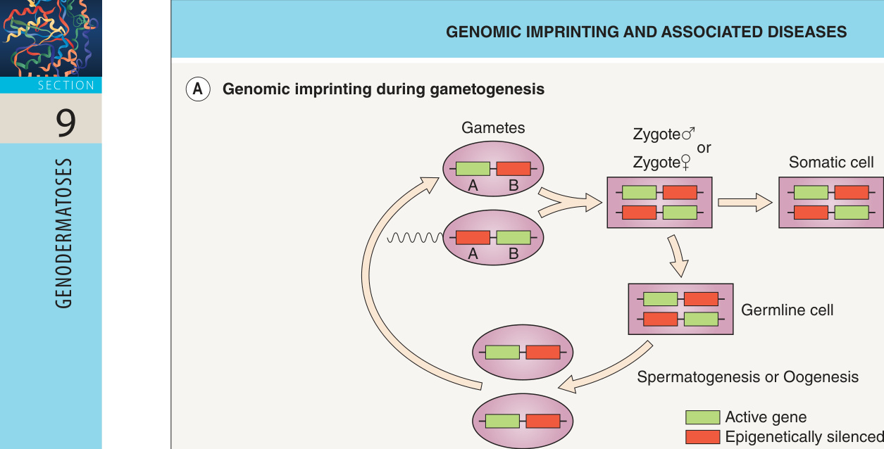
Fig. 54.3 Genomic imprinting and associated diseases. A Genomic imprinting occurs during gametogenesis. Genes A and B are epigenetically silenced in male and female gametes, respectively. Somatic cells in males and females maintain the imprinting pattern present in the gametes. In the germline, on the contrary, the imprinting is erased and the corresponding sex-specific imprint established. B Prader–Willi and Angelman syndromes represent diseases shaped by genomic imprinting.
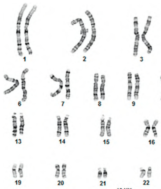
Fig. 54.4 Normal female karyotype. Metaphase chromosomes are photographed, cut out and placed in pairs from largest to smallest, including 22 autosomal pairs and 2 sex chromosomes.
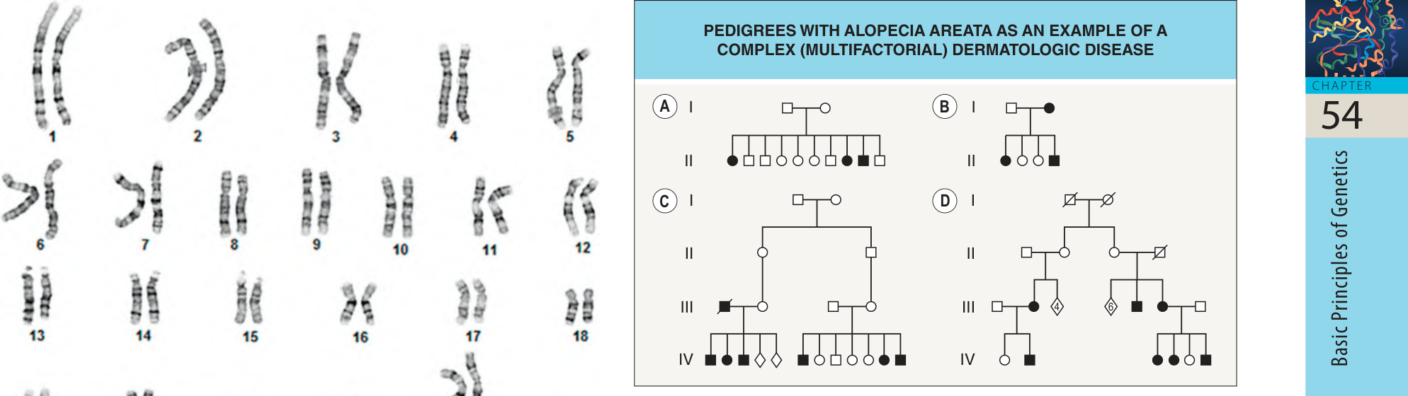
Fig. 54.5 Pedigrees with alopecia areata as an example of a complex (multi- factorial) dermatologic disease. Although some of the pedigrees individually can mimic Mendelian patterns of inheritance (A, B), others show a more complex pattern (C, D). In some cases, the disease can be inherited through two unrelated branches of the family (C). A single mode of inheritance cannot be established for the disease as a group. The key for symbols commonly used for pedigree drawing is indicated at the bottom of Fig. 54.1.