CUTANEOUS MOSAICISM
The current medical definition of a mosaic is: the coexistence of at least two genotypes in an individual derived from a single zygote, by the time
of birth, and which gives rise to a disease phenotype (see Ch. 62). As a uniquely accessible organ, the skin provides an excellent opportunity to visualize and study mosaicism. Patterns on the skin due to mosaicism include the lines of Blaschko, phylloid (see below), segmental with quadrilateral shapes, and non-segmental with circular shapes (see Fig. 62.1). Mosaicism for activating mutations affecting RAS-MAPK signaling or G protein α-subunits have been found to underlie a variety of birthmarks, including congenital melanocytic, epidermal, and sebaceous nevi as well as port-wine birthmarks and dermal melanocytosis. A single heterozygous mutation in a multipotent progenitor cell can lead to multiple types of birthmarks involving different skin lineages, such as nevus spilus (melanocytic) plus nevus sebaceus in phakomatosis pigmentokeratotica (HRAS) or port-wine birthmarks (vascular) plus dermal melanocytosis in phakomatosis pigmentovascularis (GNAQ or GNA11). Extracutaneous manifestations may occur if the mutation involves other organs such as the brain, eye, or bones.
Mosaic forms of autosomal dominant genodermatoses include: (1) a heterozygous postzygotic mutation occurring on a wild-type (“normal”) background (type 1); and (2) a postzygotic “second hit” leading to loss-of-heterozygosity in the setting of a heterozygous constitutional mutation (type 2). The latter manifests clinically as a superimposed area of earlier onset or more severe disease than the background of generalized disease, e.g. linear porokeratosis in a patient with disseminated superficial actinic porokeratosis. Examples of type 1 mosaicism
A Hyperpigmented macules and dermal atrophy with fat “herniation” on the posterior thigh. B Raspberry-like papillomas on the lower lip. C Ectrodactyly.
that have been confirmed on a molecular level include epidermolytic (K1 or K10) and non-epidermolytic (fibroblast growth factor receptor 3 [FGFR3]) epidermal nevi; constitutional mutations in these genes produce epidermolytic ichthyosis and craniosynostosis syndromes associated with severe acanthosis nigricans (Fig. 55.8), respectively. Both type 1 and type 2 mosaicism have been documented for Darier disease (ATP2A2) and Hailey–Hailey disease (ATP2C1) (Fig. 55.9), while type 2 mosaicism has been confirmed for several genodermatoses due to a defective tumor suppressor gene, such as NF1, basal cell nevus syndrome (PTCH1), and PTEN hamartoma tumor syndrome.
All women and girls are functional mosaics due to the random, irreversible, stably inherited inactivation of one of the two X chromosomes that occurs in each cell during early embryonic development (lyonization). However, 15% of genes on the human X chromosome escape lyonization and 10% are variably inactivated. In women heterozygous for X-linked disorders, functional mosaicism can result in skin lesions that follow the lines of Blaschko or have other mosaic distribution patterns. This occurs in female patients with X-linked dominant, male-lethal disorders such as incontinentia pigmenti (IKBKG/NEMO), CHILD (congenital hemidysplasia with ichthyosiform nevus and limb defects) syndrome (NSDHL), Conradi–Hünermann–Happle syndrome (EBP), and Goltz syndrome (PORCN; see Fig. 55.7) as well as in female “carriers” of X-linked dermatoses such as hypohidrotic ectodermal dysplasia (EDA; Fig. 55.10) and ichthyosis follicularis–atrichia–photophobia (IFAP) syndrome (MBTPS2; Fig. 55.11). The gene encoding steroid sulfatase escapes lyonization, explaining why skin lesions in a mosaic pattern are not observed in female carriers of X-linked recessive ichthyosis.
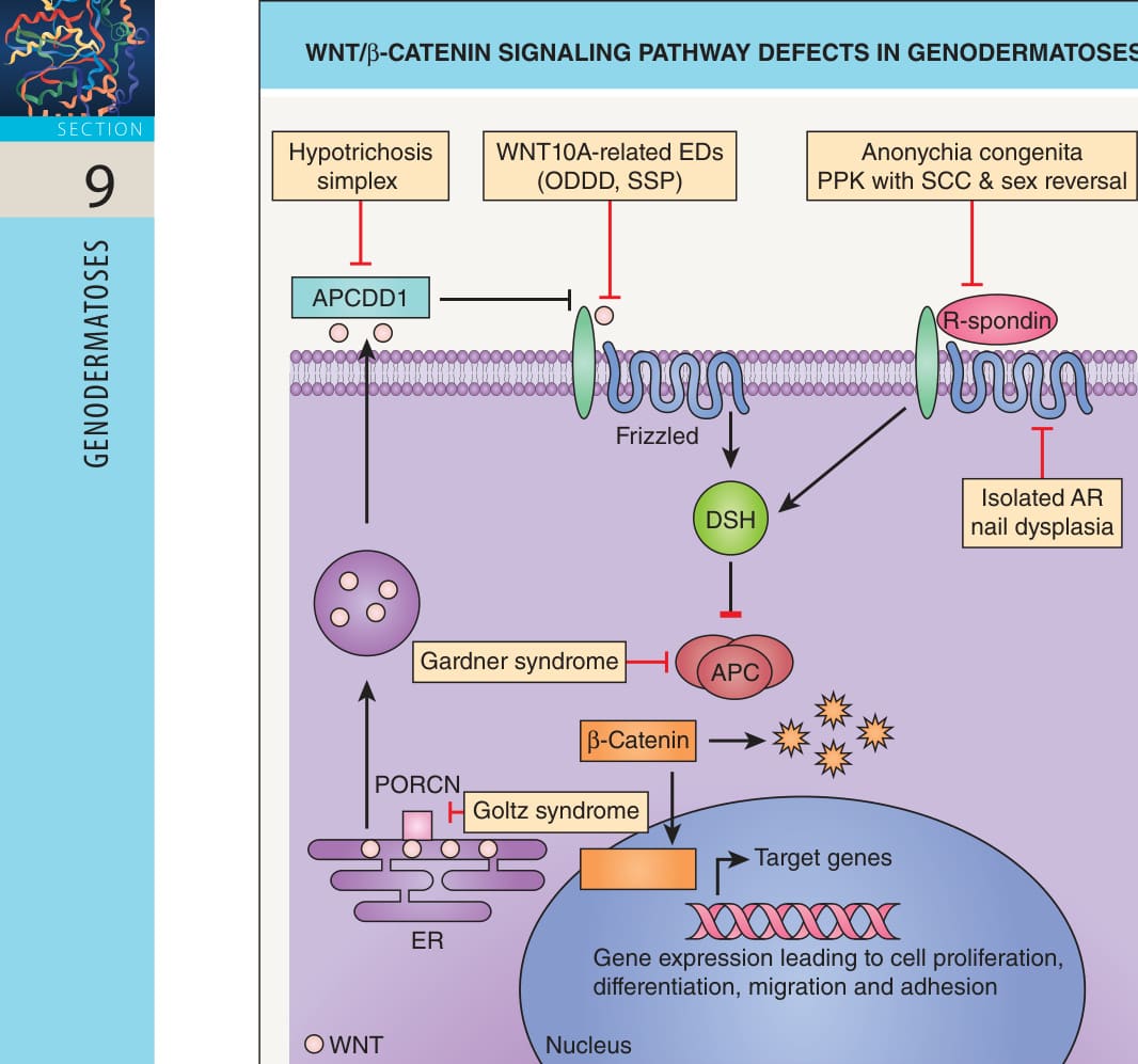
Fig. 55.6 WNT/β-catenin signaling pathway defects in genodermatoses. The porcupine homolog (PORCN) O-acyltransferase in the endoplasmic reticulum (ER) palmitoylates WNT proteins, facilitating their secretion. Extracellular WNT binds to the frizzled transmembrane receptor, which activates dishevelled (DSH), a cytosolic protein that inhibits the “destruction complex” (containing the adenomatous polyposis coli [APC] tumor suppressor protein) that mediates proteasomal degradation of β-catenin. As a result, β-catenin accumulates and is translocated to the nucleus, where it induces the transcription of genes that lead to cell proliferation, differentiation, migration, and adhesion. APC downregulated 1 (APCDD1) can inhibit WNT signaling. R-spondins are other secreted proteins that bind to frizzled and activate the WNT/β-catenin pathway. AR, autosomal recessive; EDs, ectodermal dysplasias; ODDD, odonto-onycho-dermal dysplasia; PPK, palmoplantar keratoderma; SCC, squamous cell carcinoma; SSP, Schöpf–Schulz–Passarge syndrome.
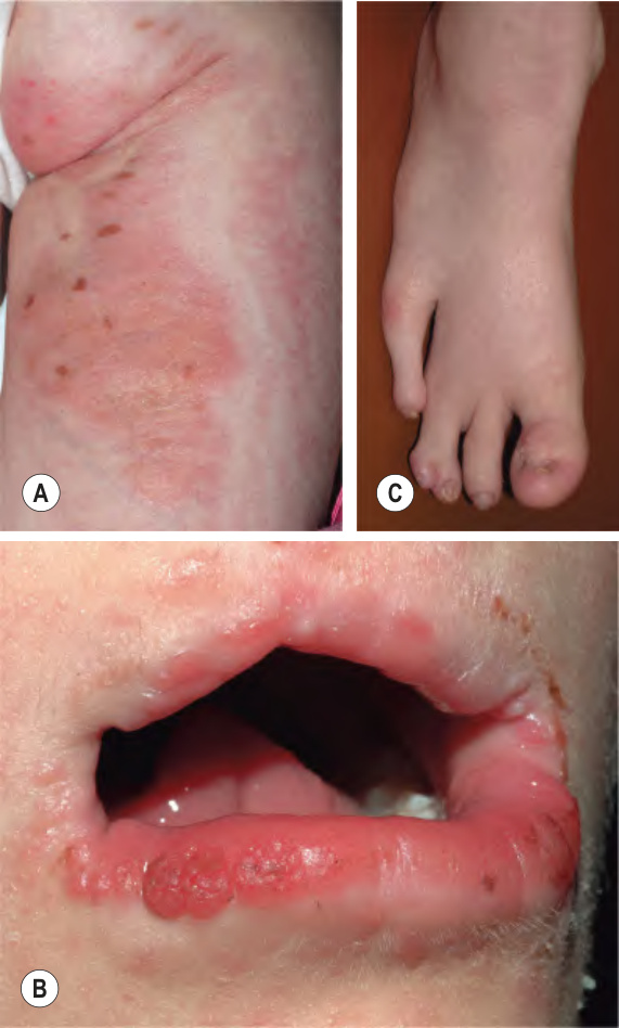
Fig. 55.7 Goltz syndrome (focal dermal hypoplasia).
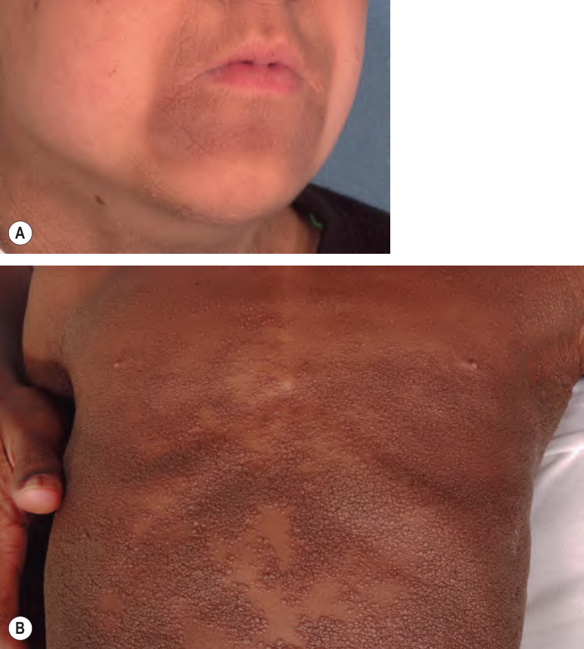
Fig. 55.8 Skin disease due to fibroblast growth factor receptor 3 (FGFR3) defects.
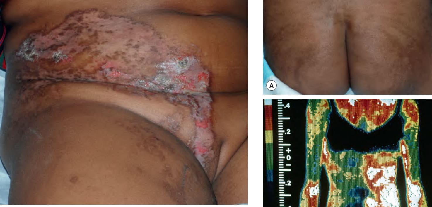
Fig. 55.9 Type 2 segmental Hailey–Hailey disease. This 7-year-old girl had a history of recurrent blistering on the right abdomen, groin, and thigh since infancy.
Fig. 55.10 Female “carriers” of X-linked hypohidrotic ectodermal dysplasia.
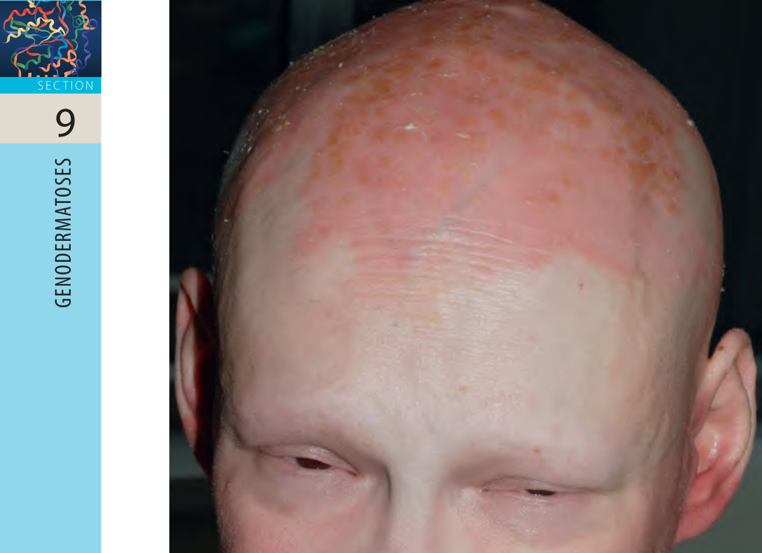
Fig. 55.11 Ichthyosis follicularis with atrichia and photophobia (IFAP)