KERATIN INTERMEDIATE FILAMENTS
Keratin intermediate filaments provide resilience to keratinocytes and represent the largest group of intermediate filament proteins (Table 56.1). Compared to microfilaments and microtubules, the other two major classes of filaments, intermediate filaments lack intrinsic polarity. The current classification system includes 54 human keratin genes which can be divided into three categories: (1) epithelial keratin genes; (2) hair keratin genes; and (3) keratin pseudogenes. Based upon their biochemical properties, keratins are classified as either type I (KRT9–KRT28, KRT31–KRT40) or type II (KRT1–KRT8, KRT71– KRT86) (Table 56.2). Type I and type II keratins form obligatory heteropolymers, i.e. pairs composed of one keratin from each group. These heteropolymers represent the basic building blocks of epithelial inter-mediate filaments (Fig. 56.4).
Similar to other intermediate filament proteins, keratins have a structure composed of domains. The central α-helical rod domain consists of 310 amino acid residues and is divided into four segments (1 A, 1B, 2 A, 2B), which are interrupted by three non-helical segments of variable lengths, termed linkers (Fig. 56.5). The rod domain is composed of seven-residue amino acid sequence repeats (a-b-c-d-ef-g)n termed “heptad repeats”, where positions “a” and “d” represent hydrophobic residues that are considered crucial for stabilization of the heterodimer. In the middle of the 2B domain, the heptad pattern is interrupted, giving rise to the “stutter”. This helical segment is highly conserved among intermediate filaments and does not participate in the formation of the coiled-coil dimer (see Fig. 56.4).
The beginning and end of the α-helical rod domain, referred to as the helix initiation and helix termination motifs, are highly conserved within different keratins and play a pivotal role in keratin intermediate filament assembly (e.g. filament elongation). These helix boundary peptides represent genetic “hot spots” for mutations in many of the hereditary keratin disorders (see Fig. 56.5). The severity and other phenotypic features of genodermatoses caused by keratin defects often correlate with the position of the underlying mutation, with more severe disease resulting from mutations in the helix initiation and termination motifs (see Table 56.4).
The head and tail regions that flank the rod domain are subdivided into extreme end (E), variable (V), and (in type II keratins) homologous (H) domains. Epithelial keratins possess glycine- and serine-rich head and tail domains, whereas these regions in hair keratins have a high content of cysteine and proline. Variations in the head and tail domains account for much of the diversity among different keratin proteins, suggesting that these domains play an important role in cell typespecific functions such as interactions with cytosolic proteins.
Keratins and Signaling
Keratins are markers of keratinocyte differentiation and are required to maintain epidermal integrity. However, keratins may also influence other basic cell functions, such as cell cycle progression, metabolic activity, apoptosis, and migration. The mechanisms by which keratins regulate these processes are not well understood, but accumulating evidence suggests that intermediate filaments directly interact with several cell signaling pathways including TNF receptor 2-induced apoptosis, Src-mediated migration, and Akt/mTOR-related growth. Interestingly, deletion of the entire murine type II keratin gene cluster, which completely prevented the assembly of keratin intermediate filaments, resulted in an early embryonic lethal phenotype with growth retardation and defects in yolk sac hematopoiesis and vasculogenesis. This underscores the importance of keratins in cellular signaling and differentiation of non-epithelial cell lineages.
Hair Keratins
The mature hair is the differentiation product of trichocytes and is found within the central core of the hair follicle. The anagen (growing) follicle is a complex structure consisting of eight distinct, concentrically arranged cell layers (see Fig. 68.5). The innermost compartment is the hair shaft, which is composed of a medulla, cortex, and one-layered cuticle; the latter serves as a protective coat for the hair shaft. The innermost living cell layer of the hair follicle is the inner root sheath (IRS). It surrounds the growing hair shaft and consists of the IRS cuticle as well as Huxley and Henle layers. The outermost compartment is the outer root sheath (ORS), which is continuous
Intermediate filament assembly takes place in several stages and begins with the heterodimerization of one type I and one type II keratin protein in a coiled-coil fashion. Two heterodimers then associate to form a tetramer. Lateral aggregation of tetramers yields higher-order polymers which eventually make up the filament network of the keratinocyte. Courtesy Julie V. Schaffer, MD.
with the interfollicular epidermis. The companion layer is located between the IRS and the ORS.
The medulla contains a mixture of epithelial keratins (KRT17, KRT75) and hair keratins (KRT33, KRT34, KRT36, KRT37, KRT81), whereas the cortex contains type I hair keratins (KRT31–KRT38) and type II hair keratins (KRT81, KRT83, KRT85, and KRT86) (Fig. 56.6, see Fig. 68.6). The cuticle expresses hair keratins KRT32 and KRT35 (type I) and their partners KRT82 and KRT85 (type II). KRT71, KRT74, and KRT73 can be detected in the three IRS layers. The epithelial keratins KRT5 and KRT14 are found throughout the full thickness of the ORS, while expression of KRT6, KRT16, and KRT17 is limited to the isthmus and the lower ORS. Additional keratins expressed in the ORS are KRT15 and KRT19.
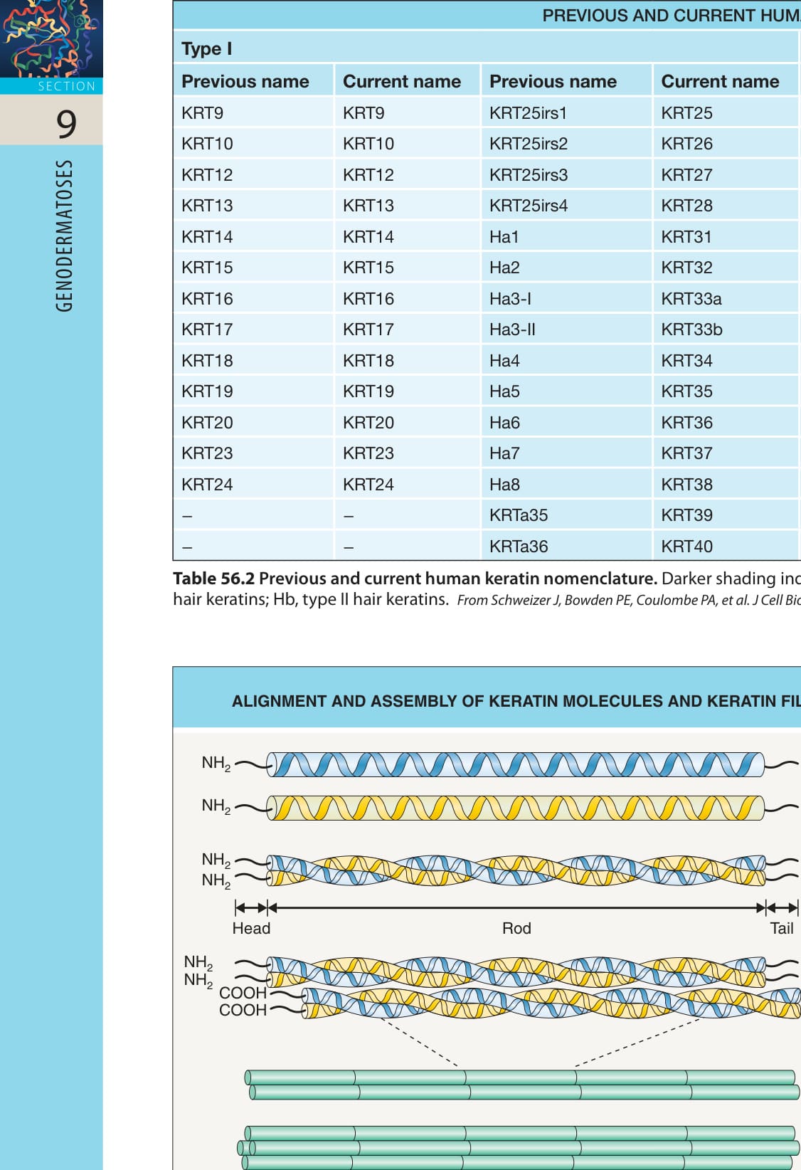
Fig. 56.4 Alignment and assembly of keratin molecules and keratin filament packing.
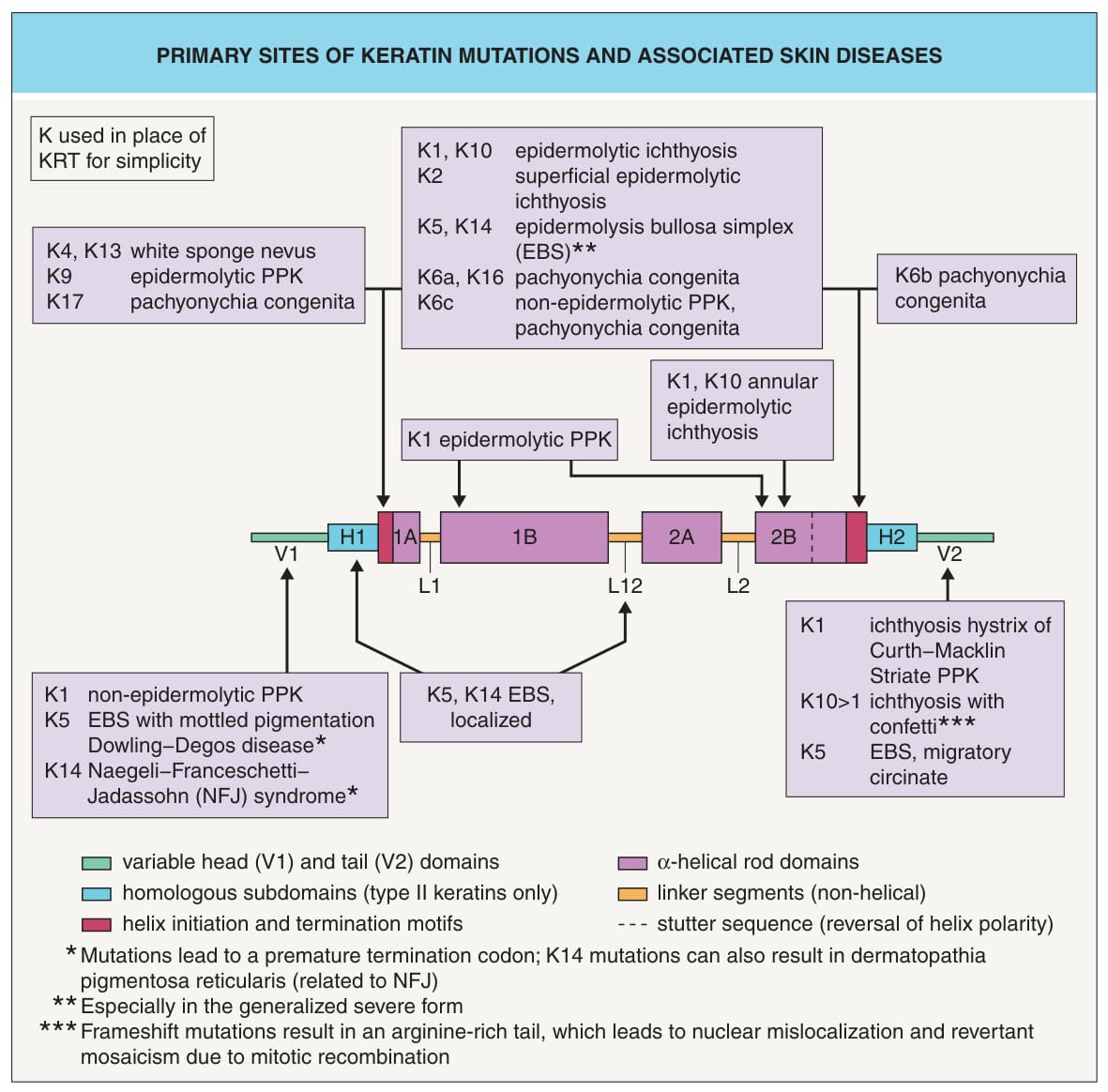
Fig. 56.5 Primary sites of keratin mutations and associated skin diseases. Over 90% of pathogenic alterations in keratins are missense mutations. Genotype–phenotype correlations are particularly well established for EBS. In the severe form of EBS, many patients have a “hot spot” mutation in a highly conserved arginine (Arg125) located within the helix initiation motif of KRT14. The corresponding arginine (Arg156) in KRT10 is also a mutational “hot spot” in epidermolytic ichthyosis. In the intermediate EBS subtype, mutations are more centrally located in the rod domains of KRT5 and KRT14. In contrast, mutations in the localized form of EBS are often outside the rod domain, e. g. in the L12 linker motifs of KRT5 and KRT14 or the H1 homologous subdomain of KRT5. Mutations in the variable head (V1) and tail (V2) domains of KRT1 can lead to non-epidermolytic PPK and ichthyosis hystrix Curth–Macklin. Rather than interfering with keratin intermediate filament assembly, mutations in these domains may cause intracellular maldistribution of loricrin. K or KRT, keratin; PPK, palmoplantar keratoderma. Courtesy Julie V. Schaffer, MD.
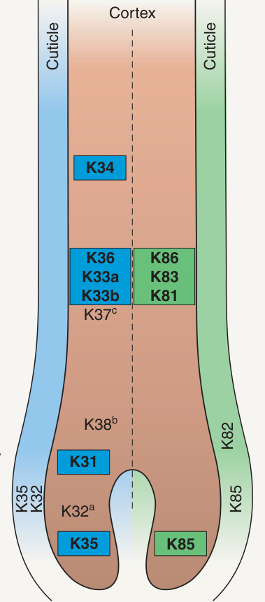
Fig. 56.6 Complex pattern of hair keratin expression in the human anagen hair shaft. Major type I hair keratins are in blue, and major type II hair keratins are in green. Minor hair keratins are in pink. aThis protein is weakly expressed at this site. bTo date, expression of this protein has only been detected in single cortex cells. cTo date, this protein has only been detected in vellus hairs. Autosomal dominant monilethrix is caused by mutations in KRT81, KRT83, and KRT86. Redrawn from Langbein L, et al. The catalog of human hair keratins. J Biol Chem 2001;276:35123–32.
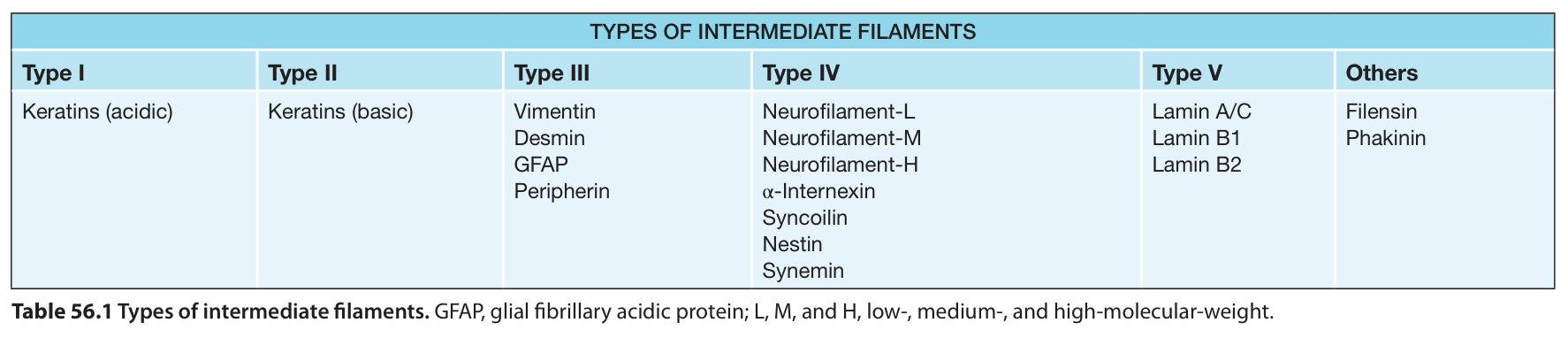
Table 56.1 Types of intermediate filaments. GFAP, glial fibrillary acidic protein; L, M, and H, low-, medium-, and high-molecular-weight.
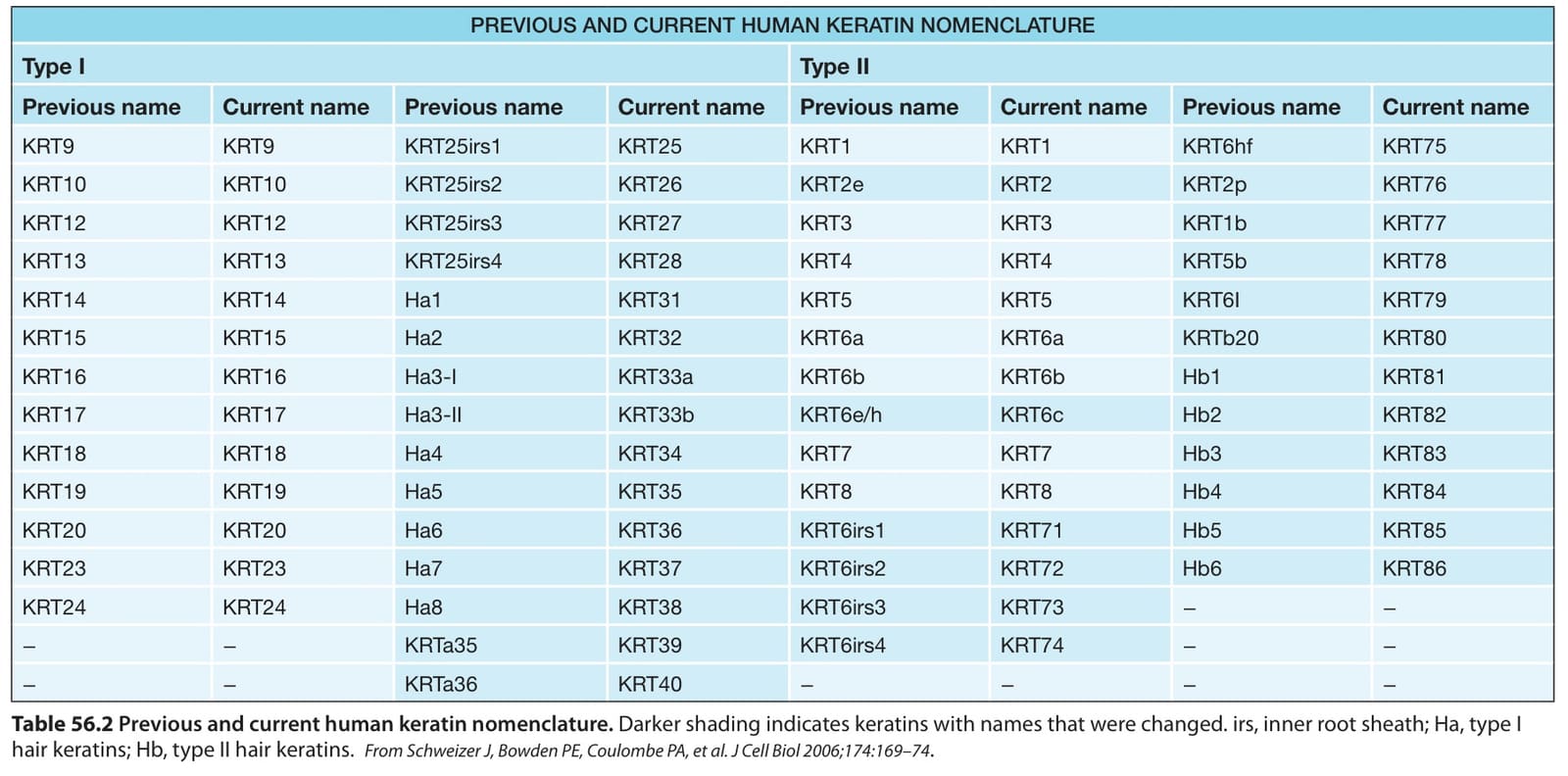
Table 56.2 Previous and current human keratin nomenclature. Darker shading indicates keratins with names that were changed. irs, inner root sheath; Ha, type I hair keratins; Hb, type II hair keratins.From Schweizer J, Bowden PE, Coulombe PA, et al. J Cell Biol 2006;174:169–74
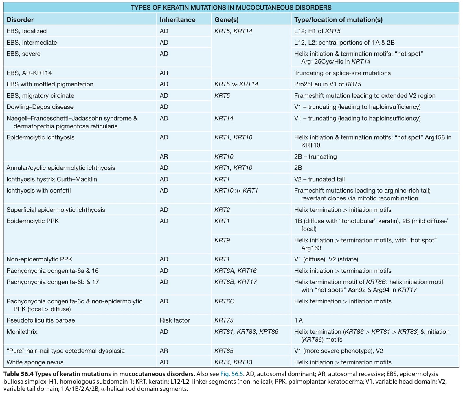
Table 56.4 Types of keratin mutations in mucocutaneous disorders. Also see Fig. 56.5. AD, autosomal dominant; AR, autosomal recessive; EBS, epidermolysis bullosa simplex; H1, homologous subdomain 1; KRT, keratin; L12/L2, linker segments (non-helical); PPK, palmoplantar keratoderma; V1, variable head domain; V2, variable tail domain; 1 A/1B/2 A/2B, α-helical rod domain segments.