KERATINOCYTE ADHESION
Desmosomes
Desmosomes are multi-protein complexes that function as cell–cell adhesion structures (junctions) in epidermal cells (Fig. 56.8). They form within areas of specialized plasma membrane, termed lipid raft domains, that are enriched with sphingolipids and cholesterol. Desmosomes also provide attachment sites for the keratin intermediate filament cytoskeleton of keratinocytes. Consequently, these junctions are critical components of a supracellular keratin network that traverses the interfollicular epidermis and epithelia of cutaneous adnexae, such as hair follicles.
Although the biochemical composition of desmosomes varies from tissue to tissue, the core of the desmosome is formed mainly by transmembrane glycoproteins that belong to the desmoglein (Dsg) and desmocollin (Dsc) subfamilies of Ca+-dependent cell adhesion proteins (cadherins). Heterophilic and homophilic interactions between Dsg and Dsc proteins are required to establish cell–cell coupling (see Ch. 29). Another protein, termed Perp, is also crucial for this process. The transmembrane proteins are linked to the keratin intermediate filament network via a complex of several proteins, including desmoplakin, plakoglobin, one of several plakophilin (PKP) isoforms, and additional accessory proteins (see Fig. 29.3B).
To complicate matters, several desmosomal components are encoded by multigene families. In humans, for example, there are four DSG genes, three DSC genes, and three PKP genes. In the epidermis, several Dsg and Dsc isoforms can be present in the same cell and even in the same desmosome. The specific composition of the desmosome is thought to affect its adhesive properties. Similar to the epidermis, a differential expression pattern of desmosomal proteins is observed in the living layers of the hair follicle.
Regulating adhesiveness of junctions via changes in the protein composition or through protein modifications is also a crucial prerequisite for regulating cell migration, cell sorting, and the formation of the proper tissue histoarchitecture during embryonic development. An example of differential regulation of desmosomes during tissue/ organ development is the formation of adnexae (e.g. hair follicles) and mammary glands. Regardless of the type of adnexal structure that is being formed, the initial steps are very similar. First, keratinocytes from the basal cell layer change their polarity and downregulate desmosomal proteins. Next, these cells migrate into the underlying dermal tissue. Eventually, the cells begin to differentiate in order to form the various cell layers that constitute the new adnexal structure. During this process, desmosomal adhesion is re-established.
The molecular pathways that control differential expression of desmosomal genes during such developmental processes are not well under-stood. However, based on work focusing on the regulation of classical cadherins (e.g. E-cadherin), it is likely that major epidermal signaling pathways, such as the Wnt and NF-κB, are involved (see Figs. 55.6 and 130.4). Of note, the phenomenon of local proliferation of keratinocytes followed by invasion into dermal tissue also occurs in cutaneous wound healing and the initial stages of cancer progression. It is therefore likely that the same gene regulatory pathways that govern adnexal development also control these biological processes.
Desmosomes act as signaling centers that affect the basic characteristics of epithelial cells. For example, induction of epidermal Dsg1 expression is thought to counteract epidermal growth factor (EGF) signaling, triggering cell cycle withdrawal, expression of differentiation-associated proteins, and delamination of keratinocytes from the basal layer. Desmosomal proteins are also involved in nuclear signaling. Plakoglobin (also known as junction plakoglobin) is associated with both desmosomes (see Fig. 56.8) and adherens junctions (see below) and is involved in relaying signals to the nucleus. Plakoglobin belongs to the armadillo protein family, the prototype of which is β-catenin. In addition to binding desmosomal cadherins (Dsg, Dsg) at the plasma membrane, plakoglobin forms complexes with Tcf/ Lef transcription factors to control gene expression, thereby influencing cell proliferation, migration, and apoptosis. Plakophilins can also affect cytoplasmic signaling pathways, and at least two isoforms (PKP1, PKP2) have nuclear functions.
Type 1 transmembrane glycoproteins from the cadherin family (desmogleins and desmocollins) are linked to intermediate filaments via a complex that contains catenins (plakoglobin, plakophilins) and plakin proteins (desmoplakin). Both homophilic and heterophilic interactions have been postulated to participate in establishing desmosomal adhesion. Examples of inherited skin diseases affecting the components of desmosomes are shown. AR, autosomal recessive; PPK, palmoplantar keratoderma.
What happens when desmosomes do not function
Tissues that are subjected to mechanical stress, such as the skin and its adnexae, mucous membranes (e.g. oral cavity), and the heart, are often affected by desmosomal defects. The clinical symptoms of desmosomal diseases typically reflect tissue fragility, with mucocutaneous blistering and cardiomyopathy representing major manifestations. In fact, skin and/or heart diseases have been linked to dysfunction of at least 12 desmosomal proteins, including Dsc1–3, Dsg1–4, desmoplakin, plakoglobin, plakophilin 1–2, and corneodesmosin (see Fig. 56.8). These disorders include genodermatoses, autoantibody-mediated diseases such as pemphigus vulgaris (PV) and pemphigus foliaceus (PF), and bacterial toxin-induced staphylococcal scalded skin syndrome.
The first mucocutaneous disorders linked to a desmosomal defect were PV and PF (see Ch. 29). In these diseases, autoantibodies bind to Dsg and induce epidermal and/or mucous membrane blistering via acantholysis, a process thought to be caused by loss of Dsg. In contrast to the blisters observed in patients with epidermolysis bullosa simplex due to keratin mutations, PV and PF blisters are caused by cell–cell separation, not cytolysis. Nevertheless, both groups of diseases illustrate the point that desmosomes and the associated keratin intermediate filaments must function properly to maintain tissue integrity.
The decreased function of desmosomal proteins may also lead to abnormal distribution of gap junction proteins, contributing to the development of cardiomyopathy in patients with mutations in desmosomal genes. These findings support the emerging picture that desmosomes are part of a highly dynamic network of cellular junctions that interact with each other and the keratinocyte cytoskeleton in signal transduction and regulation of cell properties such as mechanical stress resistance, proliferation, migration, and differentiation.
Adherens Junctions
Adherens junctions consist of classical cadherins, in particular, E-and P-cadherin, as well as a complex of cytoplasmic plaque proteins (α-catenin, β-catenin, γ-catenin [also known as plakoglobin]) that connect the transmembrane proteins to the actin microfilament cytoskeleton (see Fig. 29.3A). Note that the armadillo protein plakoglobin can bind to both desmosomal and classical cadherins (see above).
P-cadherin is expressed primarily in the basal layer of mouse epidermis, whereas E-cadherin is expressed throughout the interfollicular epidermis. Genetically engineered loss of P-cadherin in mice has little effect on the interfollicular epidermis, whereas loss of E-cadherin can lead to severe defects in the homeostasis and function of the epidermis. Interestingly, it appears that loss of E-cadherin can affect tight junctions and thus the skin barrier function of the epidermis. Based on an analysis of genetically engineered mice, it has become evident that normal expression of the plaque proteins α-catenin, β-catenin, and plakoglobin is also required for normal epidermal function.
Similar to desmosomal cadherins, classical cadherins are differentially regulated during the development of adnexae within the skin. For example, E-cadherin is downregulated in keratinocytes that invade the underlying dermis. A failure to downregulate cadherin-based adhesion suppresses invasive growth of keratinocytes and thus formation of adnexal structures.
As indicated above, armadillo proteins (e.g. β-catenin, plakoglobin) are involved in signaling from cell junctions to the nucleus. While β-catenin is a downstream effector of the classical (canonical) Wnt pathway, plakoglobin appears to be able to signal either via Wnt components or independently. Interestingly plakoglobin also counteracts classical Wnt signaling.
Tight Junctions
Tight junctions (zonula occludens) “seal” the intercellular space, thus preventing the free diffusion of macromolecules. The seal formed by tight junctions in the granular layer is required for maintaining the water barrier function of the epidermis (see Fig. 124.1). Together, tight junctions, adherens junctions, and desmosomes form the apical junctional complex in polarized epithelial cells.
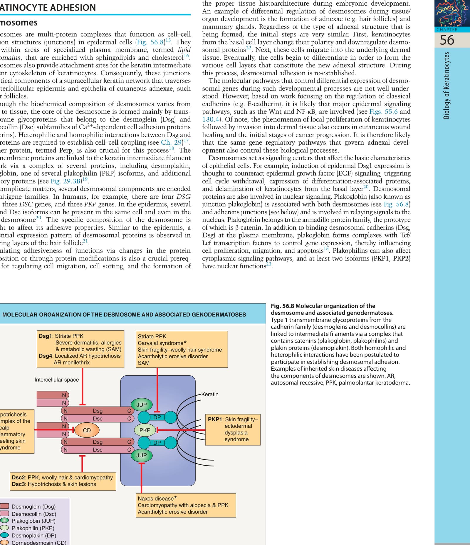
Fig. 56.8 Molecular organization of the desmosome and associated genodermatoses.
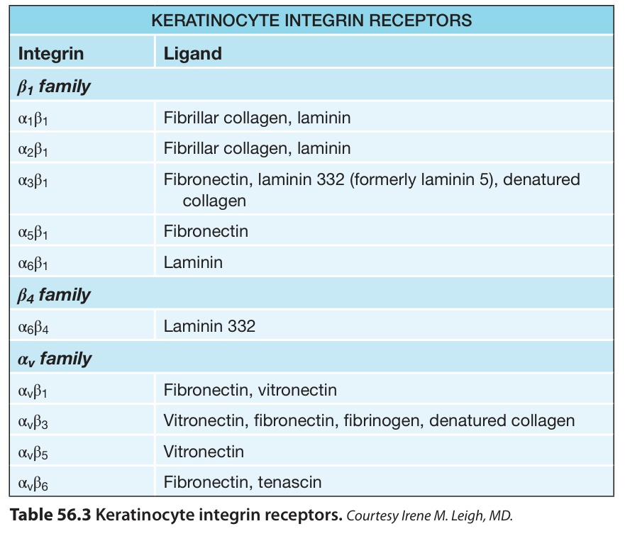
Table 56.3 Keratinocyte integrin receptors.Courtesy Irene M. Leigh, MD.