RELATED DISEASES
Keratin Disorders
Mutations in keratin genes cause a variety of disorders of the skin and other epithelia, usually with an autosomal dominant pattern of inheritance. The underlying mutations typically act in a dominant-negative fashion, interfering with normal intermediate filament assembly. This causes aggregation of disorganized keratin bundles, which often leads to clinically evident cell fragility. Mutations in the helix initiation and termination motifs are generally associated with relatively severe disease phenotypes, whereas mutations affecting other keratin domains usually cause milder disease (see Fig. 56.5). The location of mutations and genetic “hot spots” in skin disorders caused by mutations in keratin genes are presented in Table 56.4.
Epidermolysis bullosa simplex
Epidermolysis bullosa (EB) comprises a group of heritable skin fragility disorders characterized by skin and sometimes mucosal blistering following minor mechanical trauma (see Ch. 32). Mutations in genes encoding desmosomal components can lead to several disorders associated with skin fragility (see Fig. 56.8). Genotype–phenotype correlations are well established for EB simplex due to KRT5 and KRT14 mutations (see Table 56.4). The clinical severity is related to the location of the mutations and the degree to which they perturb keratin
structure. Interestingly, hyperpigmentation is a major feature of Dowling–Degos disease, Naegeli–Franceschetti–Jadassohn syndrome, and dermatopathia pigmentosa reticularis (disorders that do not feature prominent blistering; see Ch. 67) as well as EBS with mottled pigmentation, demonstrating that mutations in keratin genes expressed in the basal epidermis can lead to pigmentary changes in addition to skin fragility (see Fig. 56.5).
Epidermolytic ichthyosis and palmoplantar keratoderma
Epidermolytic ichthyosis (EI) is a disorder of keratinization caused by mutations in KRT1 or KRT10 (see Ch. 57). It is usually inherited in an autosomal dominant fashion due to heterozygous missense mutations (typically involving the helix initiation and termination motifs) that lead to clumping of keratin filaments in the suprabasal layers of the epidermis and subsequent cytolysis. EI associated with PPK is mainly due to KRT1 mutations, while EI without palmoplantar involvement is primarily due to KRT10 mutations. KRT1 mutations (outside of the critical helix initiation and termination regions) have also been identified in non-epidermolytic and epidermolytic variants of isolated PPK, which is more commonly caused by mutations in KRT9. A recessive form of EI characterized by a complete absence of the KRT10 protein has also been described. Somatic mutations in KRT1 or KRT10 that occur during embryogenesis can present as an epidermolytic epidermal nevus
(see Ch. 62), representing a mosaic form of EI. Vertical transmission of the mutation is possible if the gonads are involved, causing generalized disease in affected offspring. The types of mutations in keratin disorders are outlined in Table 56.4.
White sponge nevus
White sponge nevus is characterized by white plaques involving the oral mucosa, with occasional involvement of other mucosal surfaces such as the esophagus, vagina, rectum, and nasal cavity. The plaques may wax and wane over time, and suprabasal cytolysis and keratin clumping are observed histologically. Mutations in the genes encoding KRT4 and KRT13, which are specifically expressed in mucosal keratinocytes, cause this condition.
Gastrointestinal disorders
KRT8 and KRT18 are the major keratins that are expressed in gastrointestinal epithelia, including the liver, pancreas, and gut. Mutations in these simple keratins are typically located within the head and tail domains and do not involve the highly conserved helix boundary regions. KRT8 and KRT18 mutations are considered as risk factors for developing liver and gastrointestinal disorders (e.g. cirrhosis, inflammatory bowel disease) with additional genetic and environmental alterations likely required for disease development. The
mutation-associated predisposition to tissue injury is likely related to mechanical and non-mechanical keratin functions, including maintenance of cell integrity and protection from oxidative injury and apoptosis.
Filaggrin Deficiency Disorders
Filaggrin is a component of the cornified cell envelope and is responsible for aggregating keratins. It represents the processed product of profilaggrin, which is cleaved into individual filaggrin polypeptides by caspase
14. Loss-of-function mutations in filaggrin underlie ichthyosis vulgaris (Fig. 56.9), a semidominant condition with incomplete penetrance (~90% in homozygotes and ~60% in heterozygotes; see Ch. 57). Whereas patients with a heterozygous filaggrin mutation display mild scaling or no phenotype, those with homozygous or compound heterozygous mutations have more severe ichthyosis vulgaris with dry, scaly skin and a substantially altered cutaneous barrier.
In addition to causing ichthyosis vulgaris, loss-of-function mutations in filaggrin also predispose to atopic dermatitis (see Ch. 12). Approximately 20%–50% of patients with atopic dermatitis have at least one filaggrin null allele. Furthermore, individuals who carry filaggrin mutations and develop atopic dermatitis are also predisposed to the subsequent onset of asthma, suggesting that epicutaneous sensitization and inflammation of the skin in the context of an abnormal epidermal barrier may play a role in the development of airway hyperreactivity.
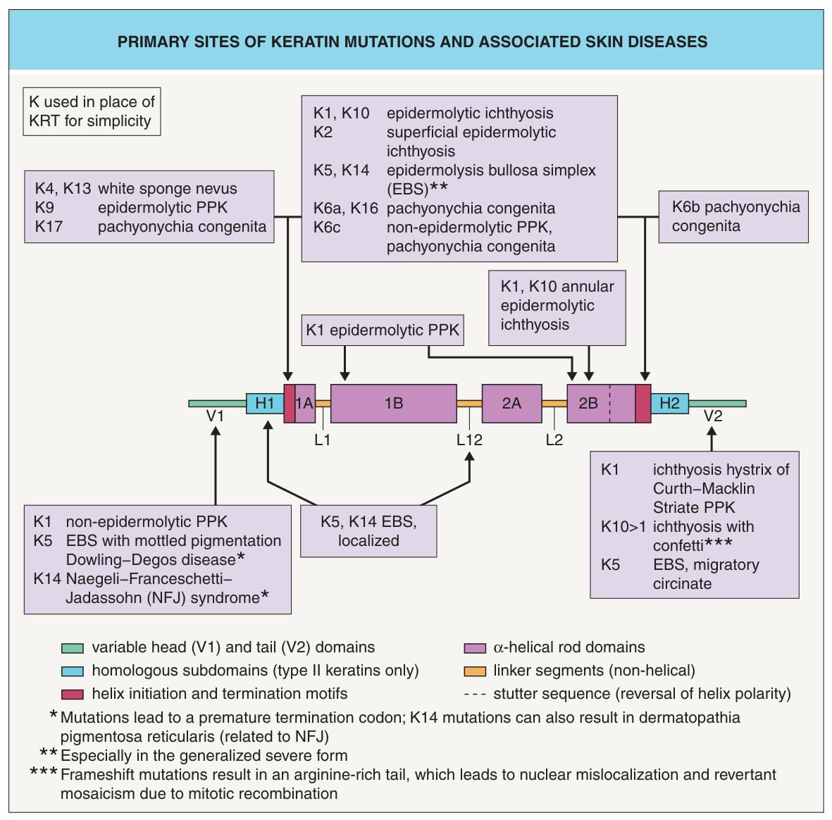
Fig. 56.5 Primary sites of keratin mutations and associated skin diseases. Over 90% of pathogenic alterations in keratins are missense mutations. Genotype–phenotype correlations are particularly well established for EBS. In the severe form of EBS, many patients have a “hot spot” mutation in a highly conserved arginine (Arg125) located within the helix initiation motif of KRT14. The corresponding arginine (Arg156) in KRT10 is also a mutational “hot spot” in epidermolytic ichthyosis. In the intermediate EBS subtype, mutations are more centrally located in the rod domains of KRT5 and KRT14. In contrast, mutations in the localized form of EBS are often outside the rod domain, e. g. in the L12 linker motifs of KRT5 and KRT14 or the H1 homologous subdomain of KRT5. Mutations in the variable head (V1) and tail (V2) domains of KRT1 can lead to non-epidermolytic PPK and ichthyosis hystrix Curth–Macklin. Rather than interfering with keratin intermediate filament assembly, mutations in these domains may cause intracellular maldistribution of loricrin. K or KRT, keratin; PPK, palmoplantar keratoderma. Courtesy Julie V. Schaffer, MD.
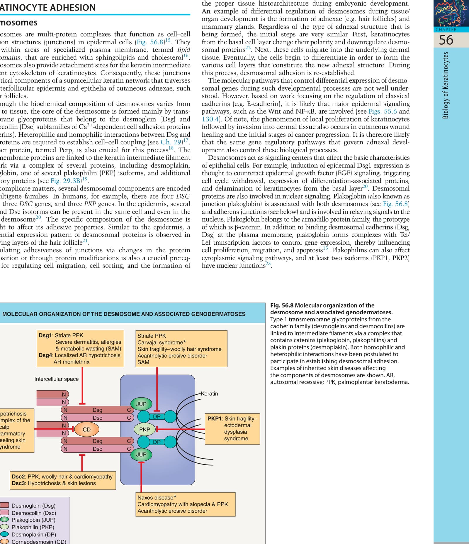
Fig. 56.8 Molecular organization of the desmosome and associated genodermatoses.
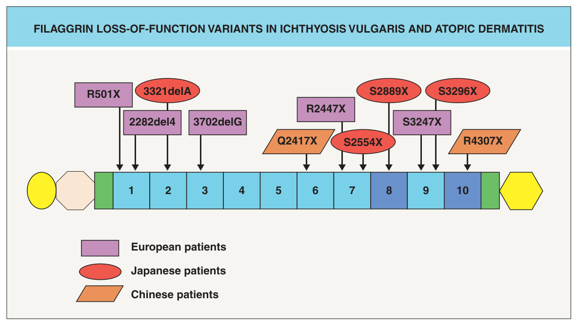
Fig. 56.9 Filaggrin loss-of-function variants in ichthyosis vulgaris and atopic dermatitis. The filaggrin protein consists of several domains: an S100 Ca+-binding domain (yellow oval), a B-domain (beige octagon), two imperfect filaggrin repeats (green rectangles), 10 filaggrin repeats (blue numbered rectangles; some individuals have two copies of repeat 8 and/or 10), and a C-terminal domain (yellow hexagon). Mutations in filaggrin that have been identified in patients with ichthyosis vulgaris and atopic dermatitis are indicated.
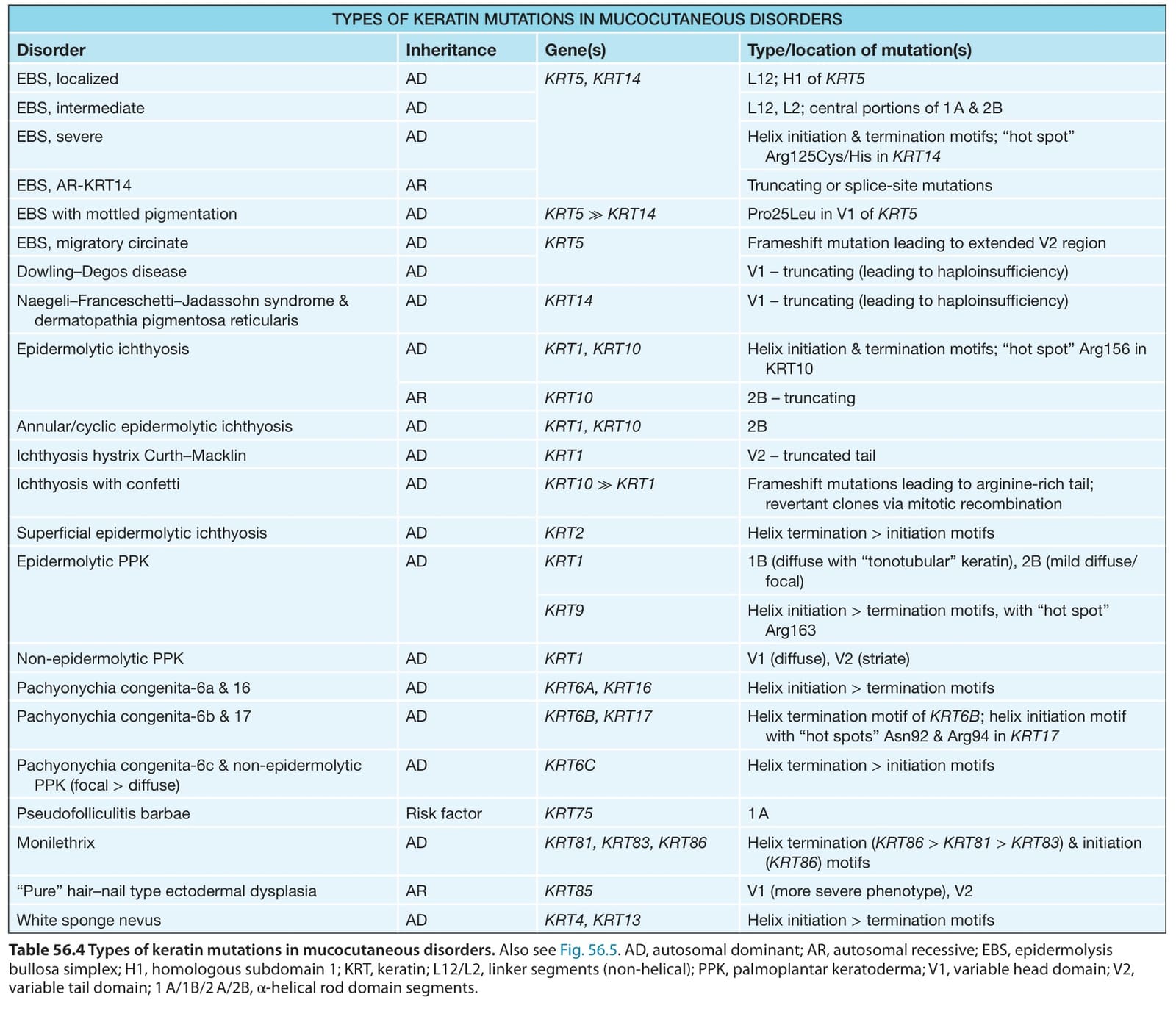
Table 56.4 Types of keratin mutations in mucocutaneous disorders. Also see Fig. 56.5. AD, autosomal dominant; AR, autosomal recessive; EBS, epidermolysis bullosa simplex; H1, homologous subdomain 1; KRT, keratin; L12/L2, linker segments (non-helical); PPK, palmoplantar keratoderma; V1, variable head domain; V2, variable tail domain; 1 A/1B/2 A/2B, α-helical rod domain segments.