CONCLUSION
We have made great progress in understanding the structure and function of the epidermis. Another challenge is to better elucidate the gene regulatory mechanisms and cell signaling pathways required for the development and maintenance of the skin. Identification of epigenetic factors and modifier genes will help to explain phenotypic variations and establish more precise genotype–phenotype correlations. This knowledge will form the foundation for the development of new therapeutic strategies for acquired and inherited skin diseases.
Additional table on Mouse models for human skin diseases due to abnormalities in structural and desmosomal proteins, available in our eBook (see inside front cover for access code).
-
Watt FM, Estrach S, Ambler CA. Epidermal Notch neity and dynamics of keratinocyte differentiation. Curr Opin Cell Biol. 2020;67:92–98.2. Brandner JM, Zorn-Kruppa M, Yoshida T, et al. Epidermal
-
Garrod D, Chidgey M. Desmosome structure, signalling: differentiation, cancer and adhesion. Curr Opin Cell Biol. 2008;20:171–179.11. Bikle DD, Xie Z, Tu CL. Calcium regulation of keratinocyte tight junctions in health and disease. Tissue Barriers. 2015;3:e974451.3. Kalinin AE, Kajava AV, Steinert PM. Epithelial barrier composition and function. Biochim Biophys Acta. 2008;1778:572–587.20. Green KJ, Jaiganesh A, Broussard JA. Desmosomes:
differentiation. Expert Rev Endocrinol Metab. 2012;7:461–472.12. Denning MF. Epidermal keratinocytes: regulation of essential contributors to an integrated intercellular junction network. F1000Res. 2019;8:F1000 Faculty Rev-2150.21. Kurzen H, Moll I, Moll R, et al. Compositionally different multiple cell phenotypes by multiple protein kinase C isoforms. Int J Biochem Cell Biol. 2004;36:1141–1146.13. Tu CL, Oda Y, Komuves L, Bikle DD. The role of the function: assembly and structural features of the cornified cell envelope. Bioessays. 2002;24:789–800.4. Samuelov L, Sprecher E. Peeling off the genetics of desmosomes in the various compartments of the human hair follicle. Differentiation. 1998;63:295–304.22. Nanba D, Hieda Y, Nakanishi Y. Remodeling of desmo- calcium-sensing receptor in epidermal differentiation. Cell Calcium. 2004;35:265–273.14. Koster MI, Roop DR. Mechanisms regulating epithelial atopic dermatitis-like congenital disorders. J Allergy Clin Immunol. 2014;134:808–815.5. Schweizer J, Bowden PE, Coulombe PA, et al. New somal and hemidesmosomal adhesion systems during early morphogenesis of mouse pelage hair follicles. J Invest Dermatol. 2000;114:171–177.23. Bass-Zubek AE, Godsel LM, Delmar M, Green KJ.
stratification. Annu Rev Cell Dev Biol. 2007;23:93–113.15. Schmidt A, Koch PJ. Desmosomes: just cell adhesion or is consensus nomenclature for mammalian keratins. J Cell Biol. 2006;174:169–174.6. Steinert PM. Structure, function, and dynamics of there more? Cell Adh Migr. 2007;1:28–32.16. Zimmer SE, Kowalczyk AP. The desmosome as a model keratin intermediate filaments. J Invest Dermatol. 1993;100:729–734.7. Gu LH, Coulombe PA. Keratin function in skin epithelia: a
Plakophilins: multifunctional scaffolds for adhesion and signaling. Curr Opin Cell Biol. 2009;21:708–716.24. Broussard JA, Getsios S, Green KJ. Desmosome for lipid raft driven membrane domain organization. Biochim Biophys Acta Biomembr. 2020;1862:183329.17. Chitaev NA, Troyanovsky SM. Direct Ca2+-dependent broadening palette with surprising shades. Curr Opin Cell Biol. 2007;19:13–23.8. Koster MI, Roop DR. Sorting out the p63 signaling regulation and signaling in disease. Cell Tissue Res. 2015;360:501–512.25. Rübsam M, Broussard JA, Wickström SA, et al. Adherens heterophilic interaction between desmosomal cadherins, desmoglein and desmocollin, contributes to cell-cell adhesion. J Cell Biol. 1997;138:193–201.18. Ihrie RA, Marques MR, Nguyen BT, et al. Perp is a network. J Invest Dermatol. 2008;128:1617–1619.9. Fisher ML, Balinth S, Mills AA. p63-related signaling at a junctions and desmosomes coordinate mechanics and signaling to orchestrate tissue morphogenesis and function: an evolutionary perspective. Cold Spring Harb Perspect Biol. 2018;10:a029207.
p63-regulated gene essential for epithelial integrity. Cell. 2005;120:843–856.
glance. J Cell Sci. 2020:133. jcs. 228015.
The generation of mouse models has significantly increased our under-standing of skin biology and the pathophysiology of genodermatoses. Animals harboring mutational “hot spots” that have been identified in humans are important for testing novel disease-targeted treatments and can help to bring new technologies closer to clinical use. For example, a reporter mouse model was developed to assess topical formulations that deliver short interfering RNAs (siRNAs) into the skin to silence dominant-negative mutant alleles.
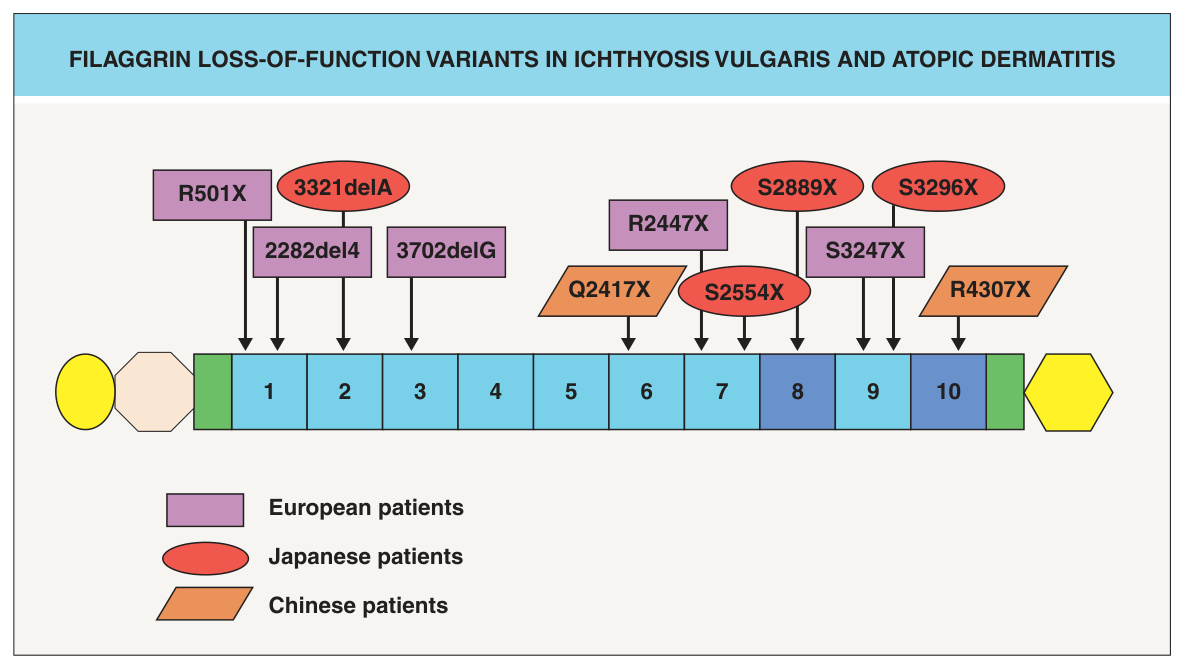
Fig. 56.9 Filaggrin loss-of-function variants in ichthyosis vulgaris and atopic dermatitis. The filaggrin protein consists of several domains: an S100 Ca+-binding domain (yellow oval), a B-domain (beige octagon), two imperfect filaggrin repeats (green rectangles), 10 filaggrin repeats (blue numbered rectangles; some individuals have two copies of repeat 8 and/or 10), and a C-terminal domain (yellow hexagon). Mutations in filaggrin that have been identified in patients with ichthyosis vulgaris and atopic dermatitis are indicated.