結論(CONCLUSION)
我們在理解表皮的結構與功能方面已有長足進展。另一項挑戰是更進一步闡明皮膚發育與維持所需的基因調控機轉與細胞訊息傳遞路徑。鑑定表觀遺傳因子(epigenetic factors)與修飾基因(modifier genes)將有助於解釋表現型的變異,並建立更精確的基因型—表現型相關性。此知識將構成為後天性與遺傳性皮膚疾病開發新治療策略的基礎。
關於因結構性與胞橋小體蛋白異常所致人類皮膚疾病之小鼠模式的補充表格,可於本書電子書版本中取得(存取碼見封面內頁)。
參考文獻(References,原文照錄)
-
Watt FM, Estrach S, Ambler CA. Epidermal Notch neity and dynamics of keratinocyte differentiation. Curr Opin Cell Biol. 2020;67:92–98.2. Brandner JM, Zorn-Kruppa M, Yoshida T, et al. Epidermal
-
Garrod D, Chidgey M. Desmosome structure, signalling: differentiation, cancer and adhesion. Curr Opin Cell Biol. 2008;20:171–179.11. Bikle DD, Xie Z, Tu CL. Calcium regulation of keratinocyte tight junctions in health and disease. Tissue Barriers. 2015;3:e974451.3. Kalinin AE, Kajava AV, Steinert PM. Epithelial barrier composition and function. Biochim Biophys Acta. 2008;1778:572–587.20. Green KJ, Jaiganesh A, Broussard JA. Desmosomes:
differentiation. Expert Rev Endocrinol Metab. 2012;7:461–472.12. Denning MF. Epidermal keratinocytes: regulation of essential contributors to an integrated intercellular junction network. F1000Res. 2019;8:F1000 Faculty Rev-2150.21. Kurzen H, Moll I, Moll R, et al. Compositionally different multiple cell phenotypes by multiple protein kinase C isoforms. Int J Biochem Cell Biol. 2004;36:1141–1146.13. Tu CL, Oda Y, Komuves L, Bikle DD. The role of the function: assembly and structural features of the cornified cell envelope. Bioessays. 2002;24:789–800.4. Samuelov L, Sprecher E. Peeling off the genetics of desmosomes in the various compartments of the human hair follicle. Differentiation. 1998;63:295–304.22. Nanba D, Hieda Y, Nakanishi Y. Remodeling of desmo- calcium-sensing receptor in epidermal differentiation. Cell Calcium. 2004;35:265–273.14. Koster MI, Roop DR. Mechanisms regulating epithelial atopic dermatitis-like congenital disorders. J Allergy Clin Immunol. 2014;134:808–815.5. Schweizer J, Bowden PE, Coulombe PA, et al. New somal and hemidesmosomal adhesion systems during early morphogenesis of mouse pelage hair follicles. J Invest Dermatol. 2000;114:171–177.23. Bass-Zubek AE, Godsel LM, Delmar M, Green KJ.
stratification. Annu Rev Cell Dev Biol. 2007;23:93–113.15. Schmidt A, Koch PJ. Desmosomes: just cell adhesion or is consensus nomenclature for mammalian keratins. J Cell Biol. 2006;174:169–174.6. Steinert PM. Structure, function, and dynamics of there more? Cell Adh Migr. 2007;1:28–32.16. Zimmer SE, Kowalczyk AP. The desmosome as a model keratin intermediate filaments. J Invest Dermatol. 1993;100:729–734.7. Gu LH, Coulombe PA. Keratin function in skin epithelia: a
Plakophilins: multifunctional scaffolds for adhesion and signaling. Curr Opin Cell Biol. 2009;21:708–716.24. Broussard JA, Getsios S, Green KJ. Desmosome for lipid raft driven membrane domain organization. Biochim Biophys Acta Biomembr. 2020;1862:183329.17. Chitaev NA, Troyanovsky SM. Direct Ca2+-dependent broadening palette with surprising shades. Curr Opin Cell Biol. 2007;19:13–23.8. Koster MI, Roop DR. Sorting out the p63 signaling regulation and signaling in disease. Cell Tissue Res. 2015;360:501–512.25. Rübsam M, Broussard JA, Wickström SA, et al. Adherens heterophilic interaction between desmosomal cadherins, desmoglein and desmocollin, contributes to cell-cell adhesion. J Cell Biol. 1997;138:193–201.18. Ihrie RA, Marques MR, Nguyen BT, et al. Perp is a network. J Invest Dermatol. 2008;128:1617–1619.9. Fisher ML, Balinth S, Mills AA. p63-related signaling at a junctions and desmosomes coordinate mechanics and signaling to orchestrate tissue morphogenesis and function: an evolutionary perspective. Cold Spring Harb Perspect Biol. 2018;10:a029207.
p63-regulated gene essential for epithelial integrity. Cell. 2005;120:843–856.
glance. J Cell Sci. 2020:133. jcs. 228015.
小鼠模式(Mouse models)
小鼠模式的建立已顯著增進我們對皮膚生物學以及遺傳性皮膚病病理生理學的理解。帶有已在人類中被鑑定出之突變「熱點(hot spots)」的動物,對於測試新型疾病標的治療十分重要,並有助於使新技術更接近臨床應用。舉例而言,曾開發出一種報導基因(reporter)小鼠模式,用以評估將短干擾 RNA(short interfering RNAs, siRNAs)遞送至皮膚以靜默顯性負向突變等位基因的外用製劑。
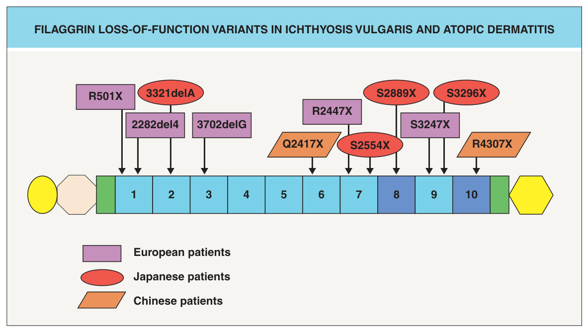
圖 56-9:尋常性魚鱗癬與異位性皮膚炎中的 filaggrin 功能喪失型變異。Filaggrin 蛋白由數個區域組成:一個 S100 Ca+ 結合區域(黃色橢圓)、一個 B 區域(米色八角形)、兩個不完全的 filaggrin 重複序列(綠色長方形)、10 個 filaggrin 重複序列(藍色編號長方形;部分個體帶有兩份重複序列 8 及/或 10)以及一個 C 端區域(黃色六角形)。圖中標示了在尋常性魚鱗癬與異位性皮膚炎病人中已被鑑定出的 filaggrin 突變。