INTRODUCTION
Ichthyoses and erythrokeratodermas are disorders of cornification in which abnormal differentiation and desquamation of the epidermis result in a defective cutaneous barrier. Ichthyoses represent a large clinically and etiologically heterogeneous group of conditions that feature generalized scaling of the skin (Tables 57.1 and 57.2). Erythrokeratodermas are characterized by circumscribed areas of erythema and hyperkeratosis without obvious scaling. Willan introduced the term “ichthyosis”, derived from the Greek root “ichthy” meaning fish, in his textbook of dermatology in 1808. Since then, nomenclature and nosology of ichthyoses have continuously evolved, including a variety of descriptive names, eponyms, and synonyms.
Milestones in recognizing distinct entities include the description of harlequin ichthyosis in the nineteenth century, separation of the bullous and non-bullous types of ichthyosis by Brocq (who also coined the name “congenital ichthyosiform erythroderma”) in the early 20th century, and differentiation between autosomal dominant “epidermolytic hyperkeratosis” and autosomal recessive “lamellar ichthyosis” by Frost and Van Scott in 1966. Alibert mentioned the autosomal dominant inheritance of ichthyosis vulgaris as early as 1806. X-linked ichthyosis, despite several earlier reports, was only fully recognized in Wells and Kerr’s study in 1966, followed by the identification of steroid sulfatase deficiency in the 1970s6. Advances in the molecular etiology and biology of ichthyoses and erythrokeratodermas have provided tools to categorize these disorders of cornification, at least in part, based on their underlying genetic defects. In 2009, an international group of clinical and research experts developed a revised nomenclature and classification system for ichthyoses and other disorders of cornification, incorporating molecular causes as well as functional aspects of disease pathogenesis.
Establishing the correct diagnosis in a patient with ichthyosis is a prerequisite for making prognostic predictions, planning therapy, and offering genetic counseling. This may pose a considerable challenge, as these disorders are uncommon and have overlapping clinical spectra. However, a systematic approach utilizing clinical and laboratorybased clues in conjunction with genetic testing can aid in making the diagnosis (Fig. 57.1).
In general, it is helpful to determine whether an ichthyosis presented at birth (e.g. as a collodion baby) or later in life and whether manifestations are limited to the skin or are part of a multisystem disorder. The quality and distribution of the scale as well as the presence or absence of erythroderma, blistering, and abnormalities of cutaneous adnexa are other useful clinical features. A thorough family history is essential for recognizing the inheritance pattern, and examination of both parents (even in a seemingly sporadic case) may reveal valuable diagnostic hints such as an epidermal nevus representing a mosaic presentation of epidermolytic ichthyosis. Patients whose parents are clinically unaffected may have: (1) a recessive ichthyosis, especially in the setting of consanguinity and/or affected siblings; (2) a dominant ichthyosis due to a “new” mutation; or (3) for a male patient, an X-linked recessive ichthyosis where the mother may be an asymptomatic carrier but her male relatives could be affected. A few disorders are recognizable by characteristic histopathologic and ultrastructural features, such as epidermolytic ichthyosis and ichthyosis hystrix Curth–Macklin. Laboratory studies suggest the diagnosis in other conditions, including enzyme analysis in steroid sulfatase deficiency and observation of lipid vacuoles in circulating leukocytes in neutral lipid storage disease (see Fig. 57.1).
This strategy allows the clinician to identify some ichthyoses based on their key features. Genetic testing can confirm the diagnosis in ~90% of patients with ichthyosis, allowing for more precise genetic counseling and providing the basis for prenatal testing.
The latter can potentially utilize circulating fetal cells as well as samples of chorionic villi or amniotic fluid. However, despite advances in our understanding of the pathomechanisms underlying ichthyoses, effective therapies are available for only a subset of these conditions, and safety concerns may limit long-term use of treatments such as oral retinoids. Hopefully, ongoing research will lead to the development of targeted treatments that will be of greater benefit to ichthyosis patients. Patient organizations such as the Foundation for Ichthyosis and Related Skin Types (FIRST; www.firstskinfoundation.org) can provide valuable support to affected individuals and their families.
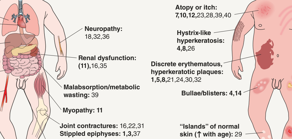
Fig. 57.1 Approach to the neonate or infant with signs of ichthyosis and diagnostic clues.
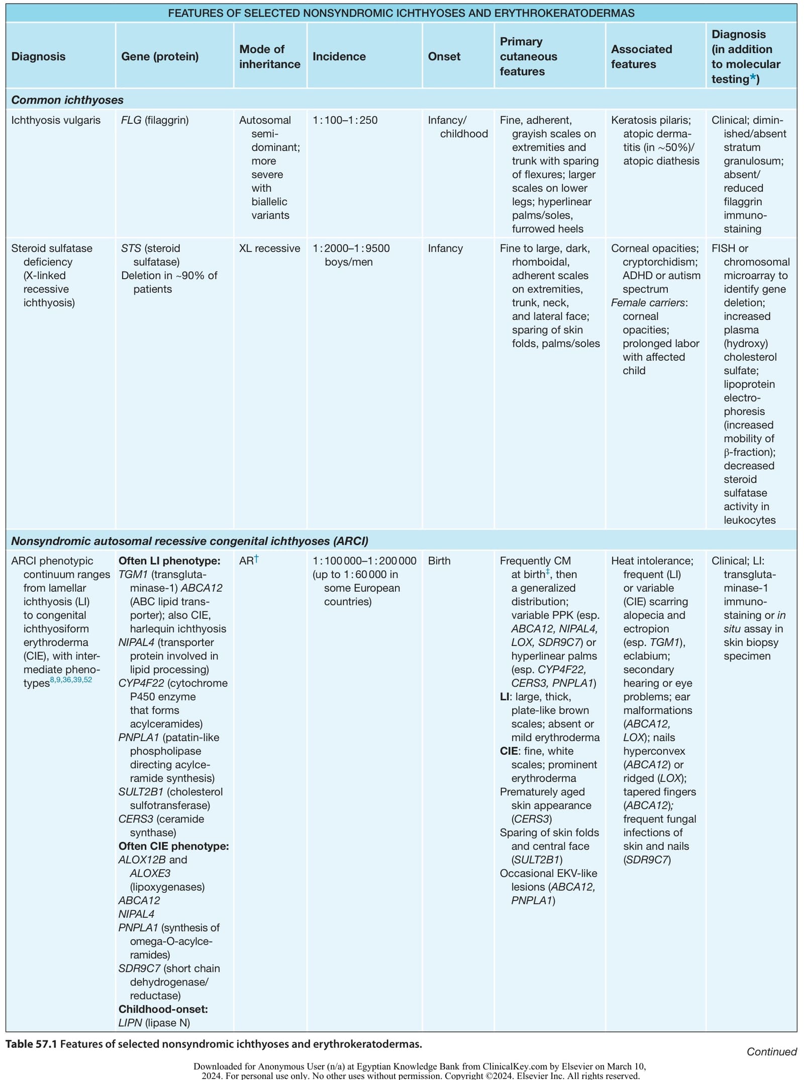
Table 57.1 Features of selected nonsyndromic ichthyoses and erythrokeratodermas. Continued
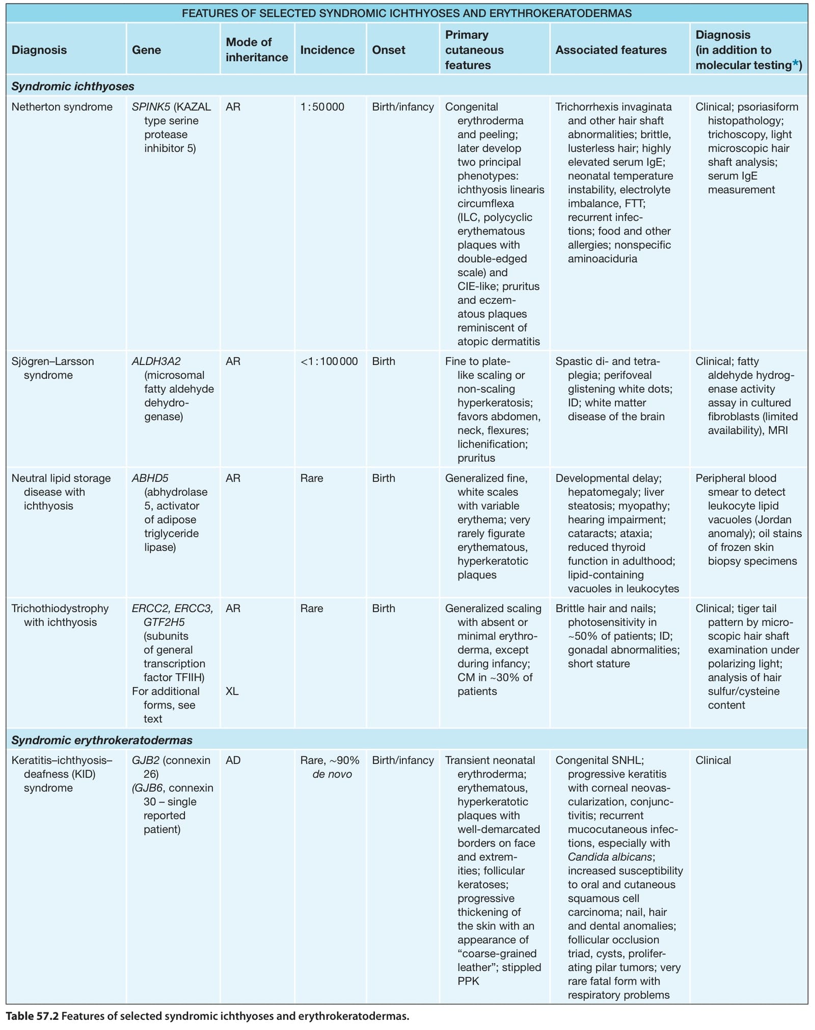
Table 57.2 Features of selected syndromic ichthyoses and erythrokeratodermas.