INTRODUCTION
Palmoplantar keratodermas (PPKs) represent a heterogeneous group of hereditary and acquired disorders of cornification characterized by prominent hyperkeratosis of the skin on the palms and soles. Inherited PPK sometimes represents a component of a syndromic phenotype with extracutaneous manifestations such as cardiomyopathy or deafness. Likewise, acquired PPK can be drug-induced or associated with cancer. Early diagnosis with determination of the specific subtype of PPK and recognition of associated features is therefore important (Table 58.1).
Several classification systems for PPKs have been proposed, but none of these satisfactorily incorporates clinical presentation, pathology, and molecular pathogenesis. Traditional nomenclature is non-uniform and includes many eponyms. In addition, PPK may represent a component of other genodermatoses such as ichthyoses, erythrokeratodermas, epidermolysis bullosa, and ectodermal dysplasias (Table 58.2).
A simple working classification divides PPKs into three major types based on the clinical pattern of involvement:
●diffuse PPK – involvement of the entire palmoplantar surface, usually but not always including the central palmar skin and instep (Fig. 58.1).
●focal PPK – localized areas of hyperkeratosis, with two major patterns: (1) the areata/nummular type – oval lesions, located mainly over pressure points; and (2) the striate type – linear hyperkeratotic lesions, most commonly extending from the palms to the volar surface of the fingers, overlying flexor tendons (Fig. 58.2; see Fig. 58.15A)
●punctate PPK – multiple small keratotic papules or pits (usually from removal of a keratotic plug) that are scattered or aggregated on the palms and soles (Fig. 58.3). Many hereditary forms of PPK do not develop until the first months or years of life. The onset and severity of disease expression depend in part on mechanical stress, e.g. exposure of the feet to friction and pressure. Some patients have diffuse PPK on the soles and focal PPK (e.g. striate) on the palms. In addition, focal involvement may evolve into a diffuse PPK over time, e.g. from infancy to adulthood. Some types of PPK don’t even manifest until adulthood, while others have spontaneous improvement and recurrences over time. In addition, substantial variability can often be observed within the same family.
Further clinical classification depends upon whether there are associated cutaneous and extracutaneous features (see Table 58.1). Many PPKs demonstrate transgrediens, which refers to extension of the hyperkeratosis onto the dorsal aspects of the fingers, toes, hands, and feet as well as the flexor aspects of the wrists and heels, either diffusely or as callosities on pressure points (e.g. the knuckles). Confluent hyperkeratosis may also extend circumferentially around entire digits. In scarring PPKs, keratotic constriction bands (pseudoainhum) can develop around digits and may lead to autoamputation (Table 58.3). Additional associated findings include hyperhidrosis, maceration, blistering, malodor, pain, and fungal infections.
When evaluating a patient with a PPK, a thorough family history should be obtained and the entire skin surface examined as well as the mucous membranes, nails, and hair. Hearing and the ability to sweat should also be assessed. Exclusion of extracutaneous manifestations may require referral for cardiac, audiologic, ophthalmologic, or dental evaluation.
Hyperkeratosis of the entire palmar (A) and plantar (B) surface with sharp demarcation. A, Courtesy Julie V. Schaffer, MD; B, Courtesy Alfons Krol, MD, and Dawn Siegel, MD.
A skin biopsy for histologic examination may provide additional diagnostic clues (Table 58.4). The molecular defect underlying PPKs may involve epidermal proteases as well as proteins of the keratinocyte cytoskeleton, cornified envelope, desmosome (see Fig. 56.8), or gap junction (Fig. 58.4, Table 58.5); the known underlying genetic mutations are summarized in Table 58.6. Genetic analysis is sometimes helpful to establish the specific diagnosis, facilitate identification of affected family members, and enable prenatal diagnosis.
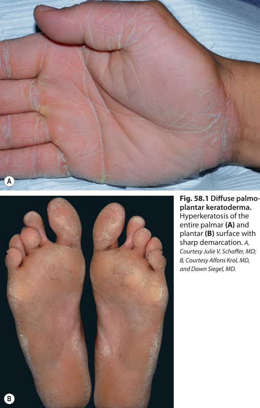
Fig. 58.1 Diffuse palmoplantar keratoderma.
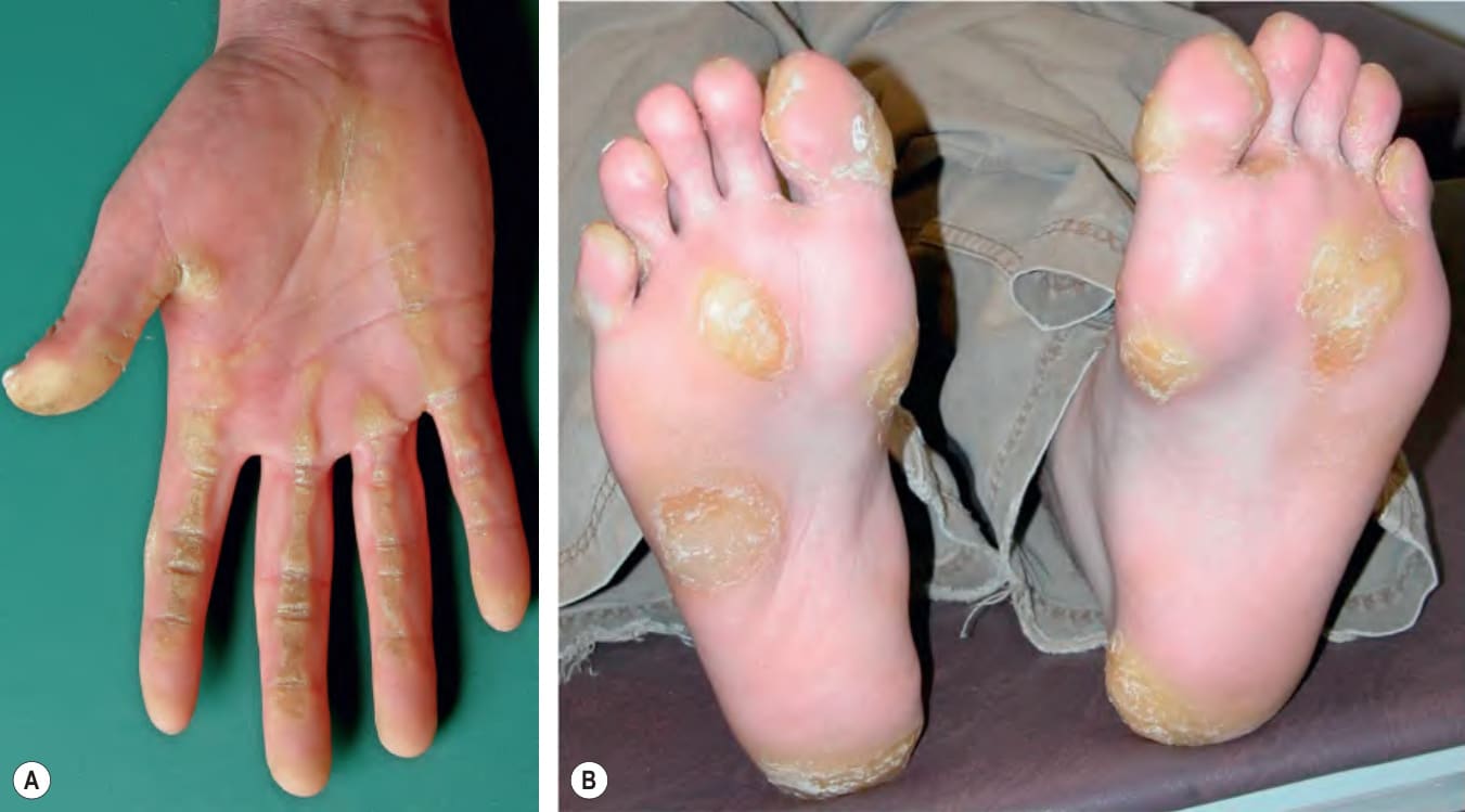
Fig. 58.2 Focal palmoplantar keratoderma. A Striate type with linear hyperkeratosis on the palm overlying flexor tendons. B Areata type on the soles. B, Courtesy Alfons Krol, MD, and Dawn Siegel, MD.
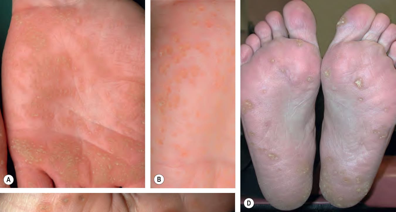
Fig. 58.3 Punctate palmoplantar keratoderma. Keratotic papules, some coalescing to form plaques, on the palms (A–C) and soles (D, E). C, Courtesy Kalman Watsky, MD.
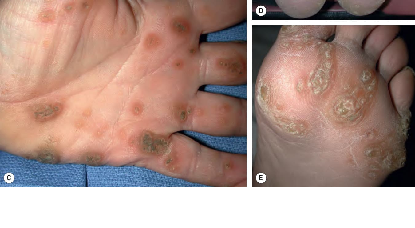
Fig. 58.4 Connexin channels and the gap junction plaque. Gap junctions are specialized transmembrane channels that connect the cytoplasm of neighboring cells, facilitating cell-to-cell communication. The status of the channels (i.e. open or closed) is controlled by mechanisms, including voltage, calcium concentration, pH, and phosphorylation. Each gap junction channel is composed of integral membrane proteins called connexins. The half-life of each connexin is short (i.e. hours), which results in a constant state of assembly and degradation of channels. Different connexins are expressed in a tissue- and differentiation-specific manner as well as in response to stimuli. Six connexin molecules oligomerize to form a connexon hemichannel with a central pore that has a maximum diameter of 2 nm. Connexons may be homomeric if all the participating molecules are of the same connexin species, or heteromeric if they differ. Connexons of adjacent cells dock in the intercellular gap to form a complete gap junction intercellular channel. Redrawn with permission from Richard G, Smith LE, Bailey RA, et al. Mutations in the human connexin gene GJB3 cause erythrokeratoderma variabilis. Nat Genet 1998;20:366–9.
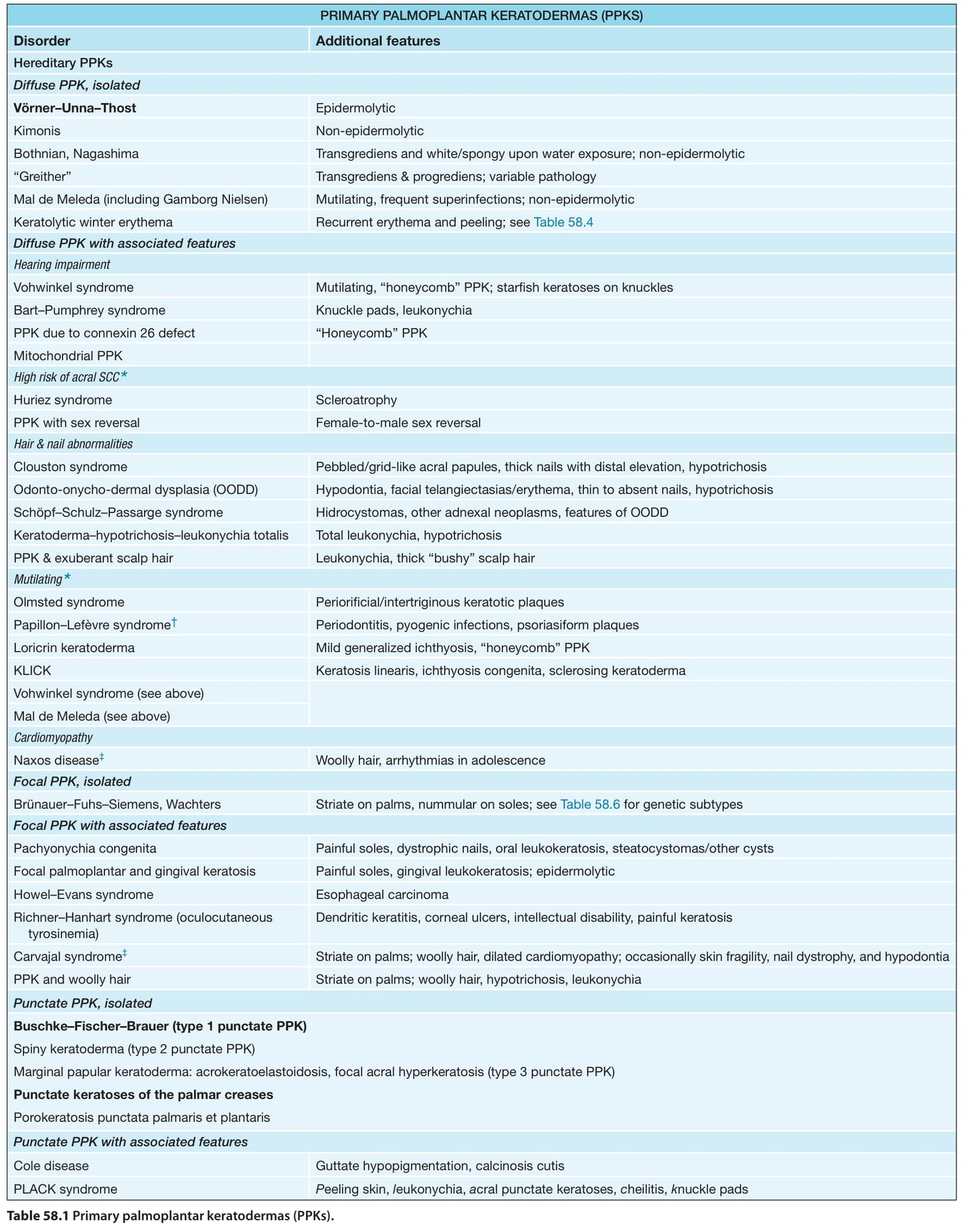
Table 58.1 Primary palmoplantar keratodermas (PPKs).
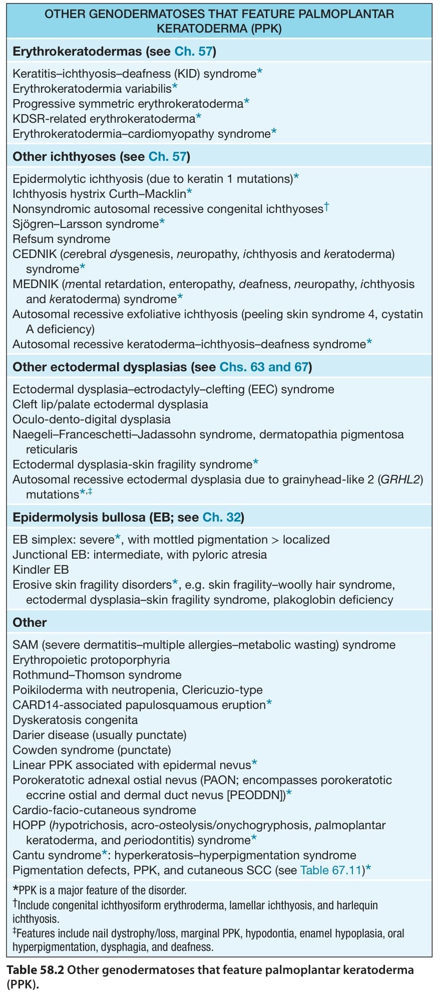
Table 58.2 Other genodermatoses that feature palmoplantar keratoderma (PPK).
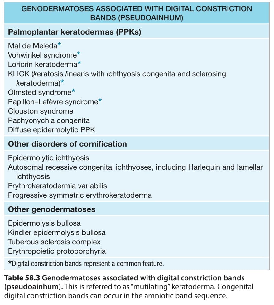
Table 58.3 Genodermatoses associated with digital constriction bands
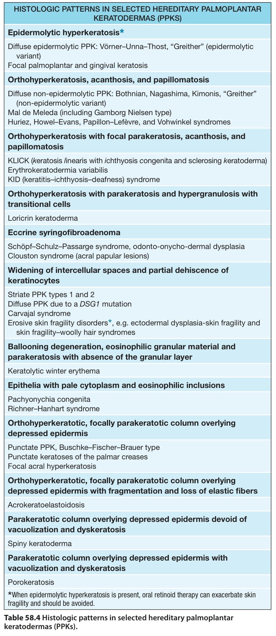
Table 58.4 Histologic patterns in selected hereditary palmoplantar keratodermas (PPKs).
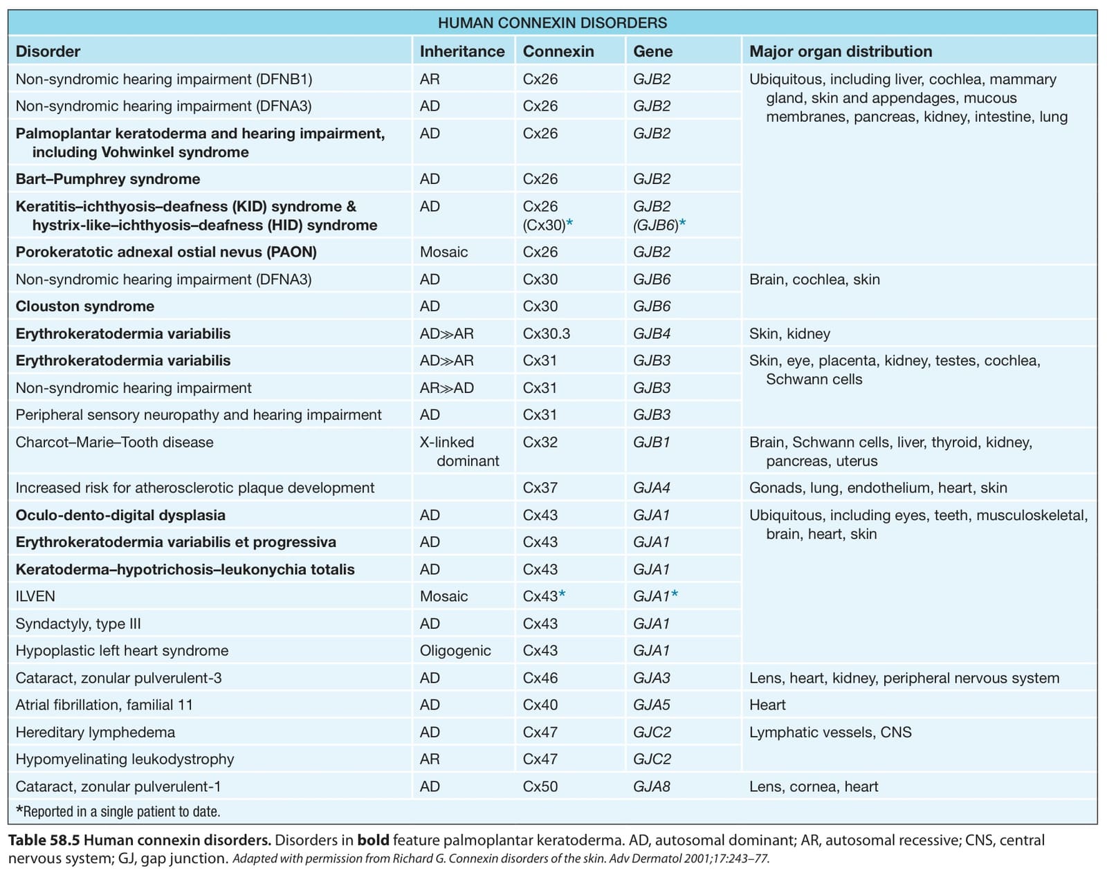
Table 58.5 Human connexin disorders. Disorders in bold feature palmoplantar keratoderma. AD, autosomal dominant; AR, autosomal recessive; CNS, central nervous system; GJ, gap junction.
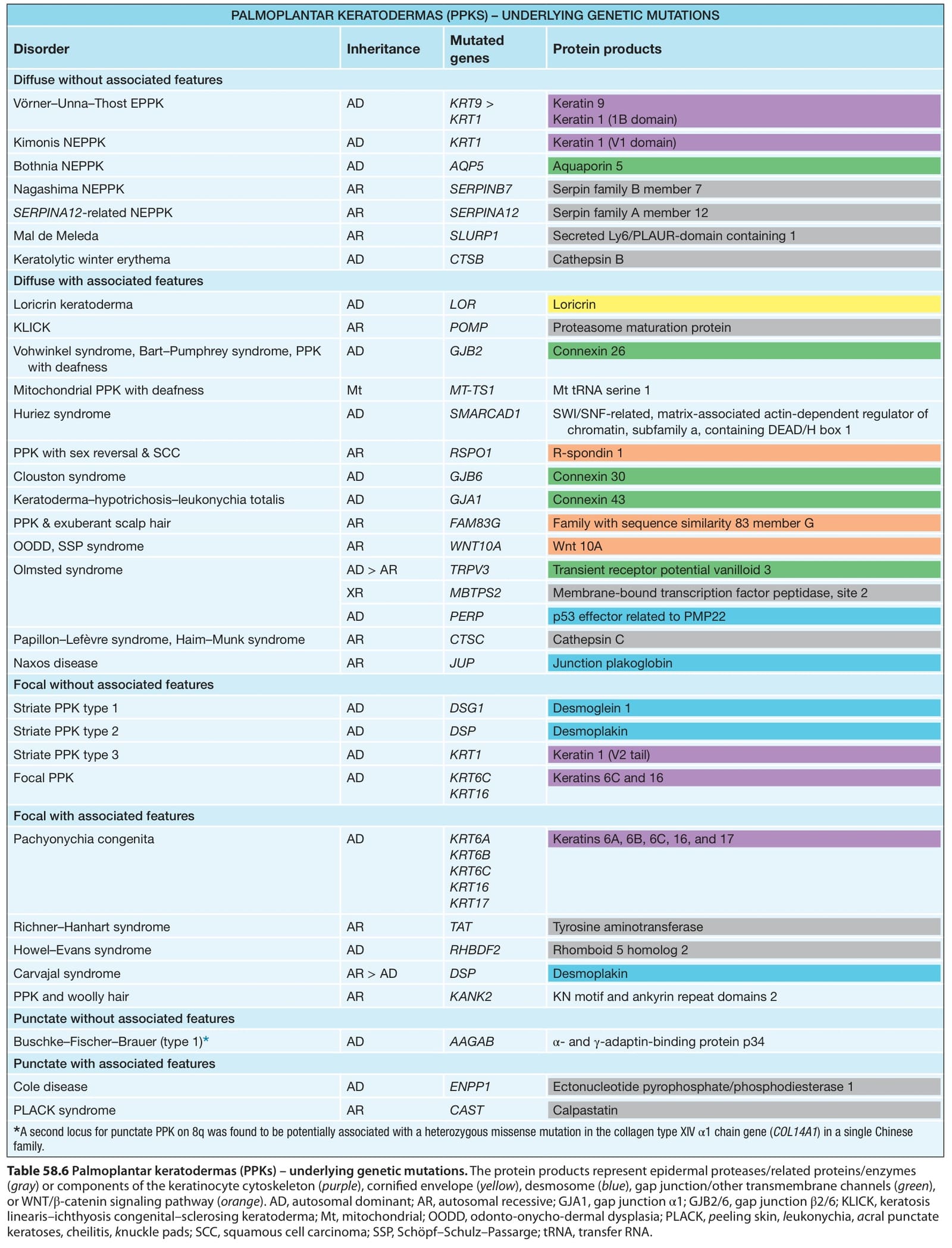
Table 58.6 Palmoplantar keratodermas (PPKs) – underlying genetic mutations. The protein products represent epidermal proteases/related proteins/enzymes (gray) or components of the keratinocyte cytoskeleton (purple), cornified envelope (yellow), desmosome (blue), gap junction/other transmembrane channels (green), or WNT/β-catenin signaling pathway (orange). AD, autosomal dominant; AR, autosomal recessive; GJA1, gap junction α1; GJB2/6, gap junction β2/6; KLICK, keratosis linearis–ichthyosis congenital–sclerosing keratoderma; Mt, mitochondrial; OODD, odonto-onycho-dermal dysplasia; PLACK, peeling skin, leukonychia, acral punctate keratoses, cheilitis, knuckle pads; SCC, squamous cell carcinoma; SSP, Schöpf–Schulz–Passarge; tRNA, transfer RNA.