引言(INTRODUCTION)
掌蹠角皮症(palmoplantar keratodermas, PPKs)代表一群異質性的遺傳性與後天性角化障礙(disorders of cornification),其特徵為手掌與足底皮膚顯著的過度角化(hyperkeratosis)。遺傳性 PPK 有時是某種症候群表型的一部分,伴隨皮膚外表現,例如心肌病變(cardiomyopathy)或耳聾。同樣地,後天性 PPK 可能為藥物誘發或與癌症相關。因此,早期診斷並判定 PPK 的特定亞型、辨識其相關特徵,十分重要(表 58.1)。
已有數種 PPK 的分類系統被提出,但沒有一種能令人滿意地整合臨床表現、病理學與分子致病機轉。傳統命名法並不統一,且包含許多冠名(eponyms)。此外,PPK 可能是其他遺傳性皮膚病的一部分,例如魚鱗癬(ichthyoses)、紅斑角皮症(erythrokeratodermas)、水皰性表皮鬆解症(epidermolysis bullosa)與外胚層發育不良(ectodermal dysplasias)(表 58.2)。
一個簡單實用的分類法依據侵犯的臨床型態將 PPKs 分為三大類:
● 瀰漫型 PPK(diffuse PPK)——整個掌蹠表面受侵犯,通常但不一定包括手掌中央皮膚與足弓(instep)(圖 58.1)。
● 局灶型 PPK(focal PPK)——局限區域的過度角化,有兩種主要型態:(1) 斑塊/錢幣狀型(areata/nummular type)——卵圓形病灶,主要位於壓力點上;以及 (2) 條紋型(striate type)——線狀過度角化病灶,最常從手掌延伸至手指的掌側面,位於屈肌腱之上(圖 58.2;見圖 58.15A)
● 點狀型 PPK(punctate PPK)——多發性的小型角化性丘疹或凹陷(通常是角化栓被移除後留下),散在或聚集於手掌與足底(圖 58.3)。許多遺傳型的 PPK 要到出生後最初數月或數年才會發生。疾病表現的發病時間與嚴重度部分取決於機械性壓力,例如足部承受的摩擦與壓力。有些病人在足底為瀰漫型 PPK,而在手掌為局灶型 PPK(例如條紋型)。此外,局灶性侵犯可能隨時間演變成瀰漫型 PPK,例如從嬰兒期到成年期。有些型態的 PPK 甚至要到成年期才顯現,而其他型態則隨時間出現自發性改善與復發。此外,在同一家族內常可觀察到相當大的變異性。
進一步的臨床分類則取決於是否有相關的皮膚與皮膚外特徵(見表 58.1)。許多 PPKs 表現出越界性(transgrediens),此詞指過度角化延伸至手指、腳趾、手部與足部的背側面,以及手腕與足跟的屈側面,可呈瀰漫性或呈壓力點上的胼胝(callosities,例如指節)。融合性的過度角化也可能環繞整個指趾呈環狀延伸。在瘢痕性 PPKs 中,角化性縮窄環(keratotic constriction bands, pseudoainhum)可在指趾周圍形成,並可能導致自截(autoamputation)(表 58.3)。其他相關發現包括多汗(hyperhidrosis)、浸潤糜爛(maceration)、水皰形成、惡臭、疼痛與黴菌感染。
在評估 PPK 病人時,應取得詳盡的家族史,並檢查整個皮膚表面以及黏膜、指甲與毛髮。也應評估聽力與流汗能力。要排除皮膚外表現,可能需要轉介進行心臟、聽力、眼科或牙科評估。
整個手掌(A)與足底(B)表面的過度角化,具清楚界限。A,由 Julie V. Schaffer, MD 提供;B,由 Alfons Krol, MD 與 Dawn Siegel, MD 提供。
皮膚切片進行組織學檢查可提供額外的診斷線索(表 58.4)。PPKs 潛在的分子缺陷可能涉及表皮蛋白酶(epidermal proteases),以及角質形成細胞細胞骨架(keratinocyte cytoskeleton)、角質化外套膜(cornified envelope)、胞橋小體(desmosome,見圖 56.8)或間隙連接(gap junction)的蛋白質(圖 58.4,表 58.5);已知的潛在基因突變總結於表 58.6。基因分析有時有助於確立特定診斷、促進受影響家族成員的辨識,並使產前診斷成為可能。
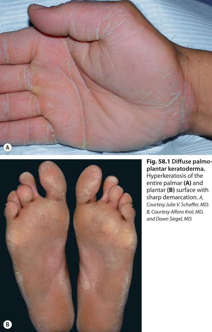
Fig. 58.1 Diffuse palmoplantar keratoderma.
圖 58-1:瀰漫型掌蹠角皮症。
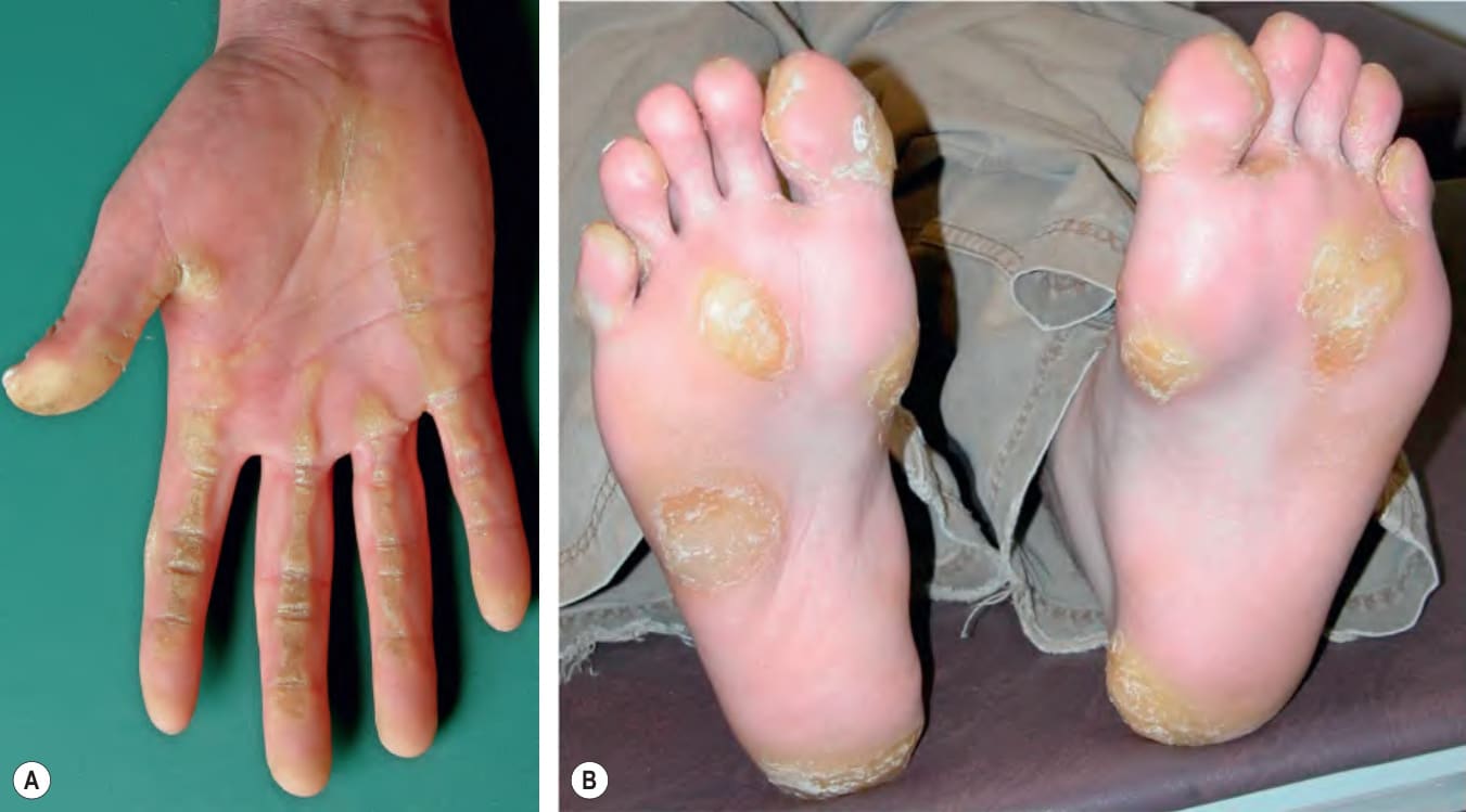
Fig. 58.2 Focal palmoplantar keratoderma. A Striate type with linear hyperkeratosis on the palm overlying flexor tendons. B Areata type on the soles. B, Courtesy Alfons Krol, MD, and Dawn Siegel, MD.
圖 58-2:局灶型掌蹠角皮症。A 條紋型,手掌上位於屈肌腱之上的線狀過度角化。B 足底的斑塊型。B,由 Alfons Krol, MD 與 Dawn Siegel, MD 提供。
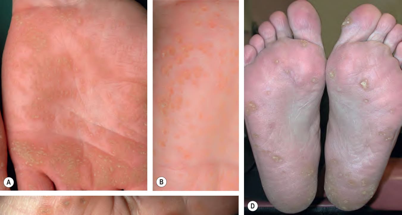
Fig. 58.3 Punctate palmoplantar keratoderma. Keratotic papules, some coalescing to form plaques, on the palms (A–C) and soles (D, E). C, Courtesy Kalman Watsky, MD.
圖 58-3:點狀型掌蹠角皮症。手掌(A–C)與足底(D、E)上的角化性丘疹,部分融合形成斑塊。C,由 Kalman Watsky, MD 提供。
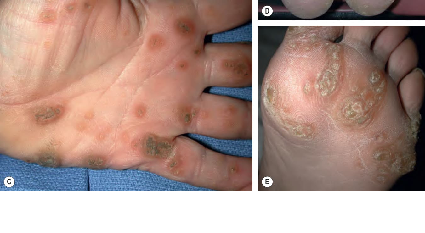
Fig. 58.4 Connexin channels and the gap junction plaque. Gap junctions are specialized transmembrane channels that connect the cytoplasm of neighboring cells, facilitating cell-to-cell communication. The status of the channels (i.e. open or closed) is controlled by mechanisms, including voltage, calcium concentration, pH, and phosphorylation. Each gap junction channel is composed of integral membrane proteins called connexins. The half-life of each connexin is short (i.e. hours), which results in a constant state of assembly and degradation of channels. Different connexins are expressed in a tissue- and differentiation-specific manner as well as in response to stimuli. Six connexin molecules oligomerize to form a connexon hemichannel with a central pore that has a maximum diameter of 2 nm. Connexons may be homomeric if all the participating molecules are of the same connexin species, or heteromeric if they differ. Connexons of adjacent cells dock in the intercellular gap to form a complete gap junction intercellular channel. Redrawn with permission from Richard G, Smith LE, Bailey RA, et al. Mutations in the human connexin gene GJB3 cause erythrokeratoderma variabilis. Nat Genet 1998;20:366–9.
圖 58-4:連接蛋白(connexin)通道與間隙連接斑塊。間隙連接(gap junctions)是連接相鄰細胞細胞質的特化跨膜通道,促進細胞間的溝通。通道的狀態(即開啟或關閉)由多種機制控制,包括電位、鈣濃度、pH 值與磷酸化。每個間隙連接通道由稱為連接蛋白(connexins)的膜整合蛋白組成。每個連接蛋白的半衰期很短(即數小時),造成通道處於持續組裝與降解的狀態。不同的連接蛋白以組織特異性與分化特異性的方式表現,也會因應刺激而表現。六個連接蛋白分子寡聚化形成一個連接子半通道(connexon hemichannel),具有最大直徑 2 nm 的中央孔道。若所有參與的分子屬於同一種連接蛋白,則連接子為同聚型(homomeric);若不同,則為異聚型(heteromeric)。相鄰細胞的連接子在細胞間隙中對接,形成完整的間隙連接細胞間通道。經授權重繪自 Richard G, Smith LE, Bailey RA, et al. Mutations in the human connexin gene GJB3 cause erythrokeratoderma variabilis. Nat Genet 1998;20:366–9。
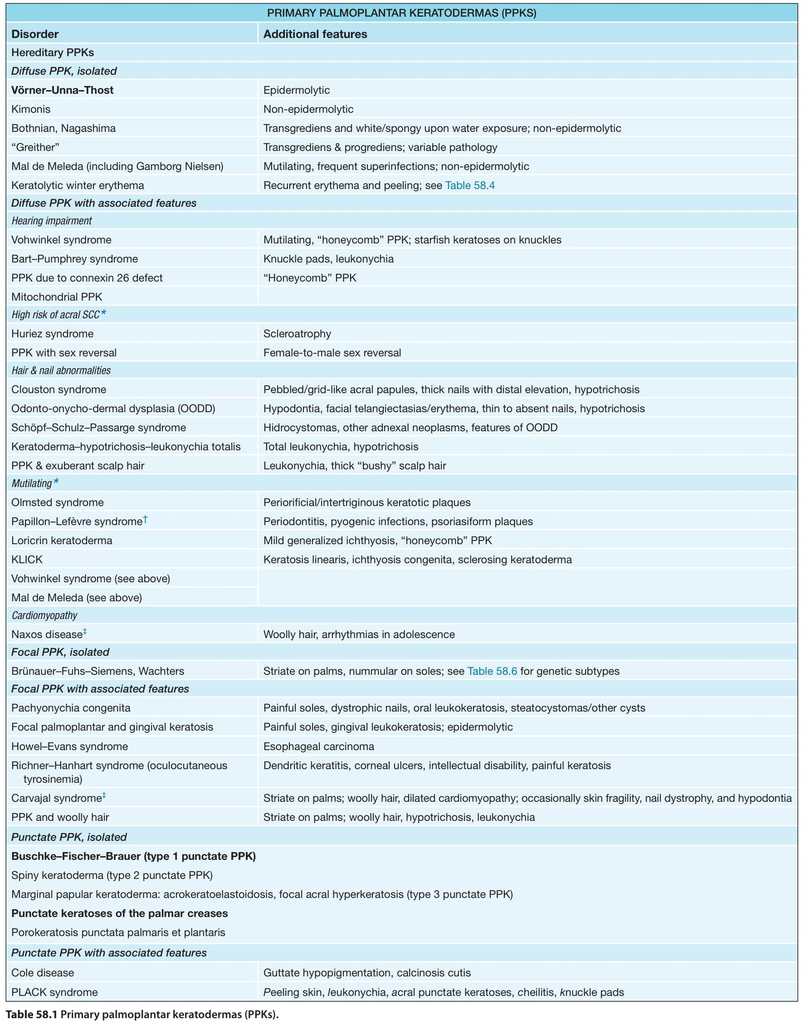
Table 58.1 Primary palmoplantar keratodermas (PPKs).
表 58-1:原發性掌蹠角皮症(PPKs)。
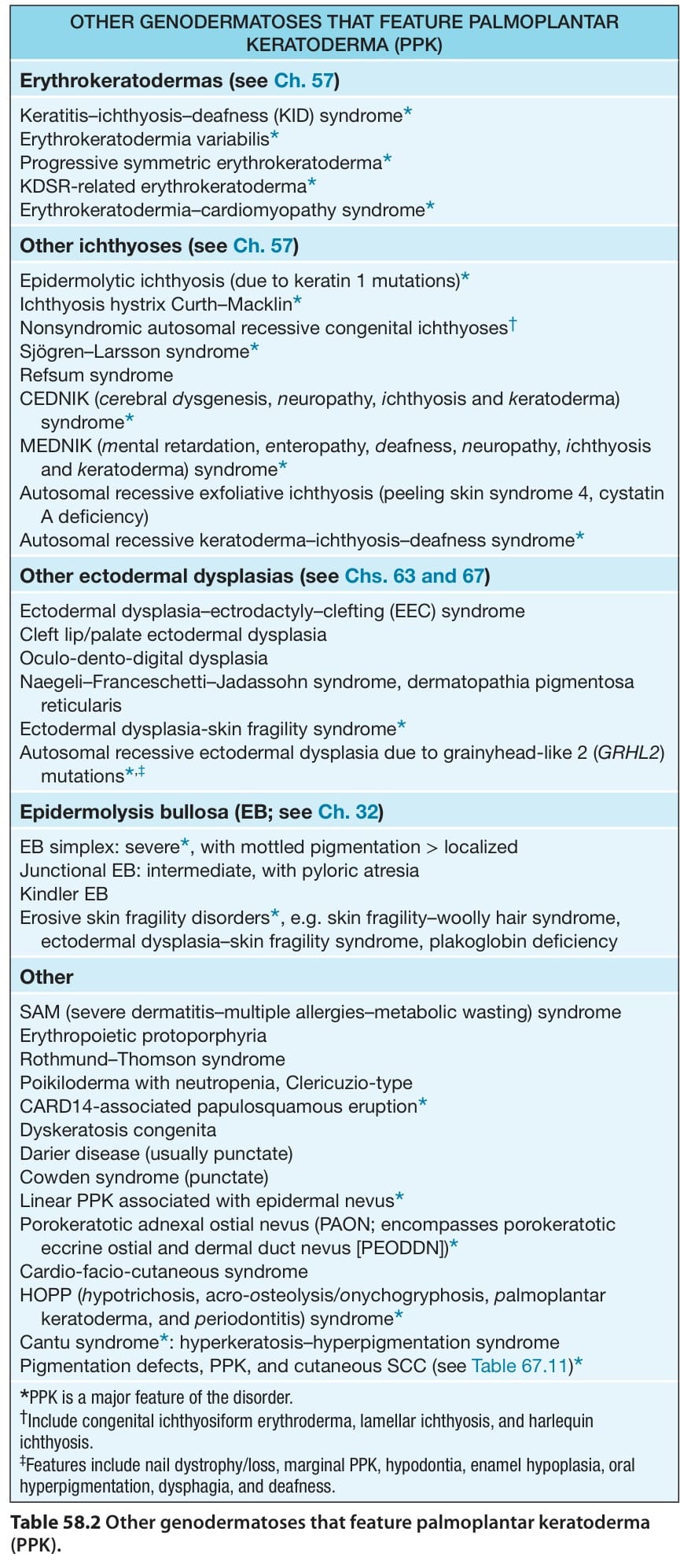
Table 58.2 Other genodermatoses that feature palmoplantar keratoderma (PPK).
表 58-2:其他以掌蹠角皮症(PPK)為特徵的遺傳性皮膚病。
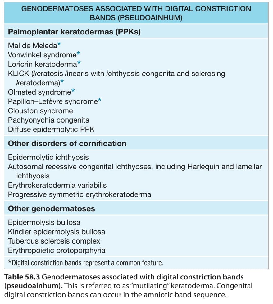
Table 58.3 Genodermatoses associated with digital constriction bands
表 58-3:與指趾縮窄環相關的遺傳性皮膚病
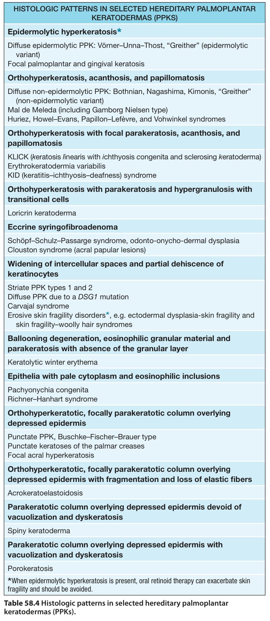
Table 58.4 Histologic patterns in selected hereditary palmoplantar keratodermas (PPKs).
表 58-4:選定的遺傳性掌蹠角皮症(PPKs)之組織學型態。
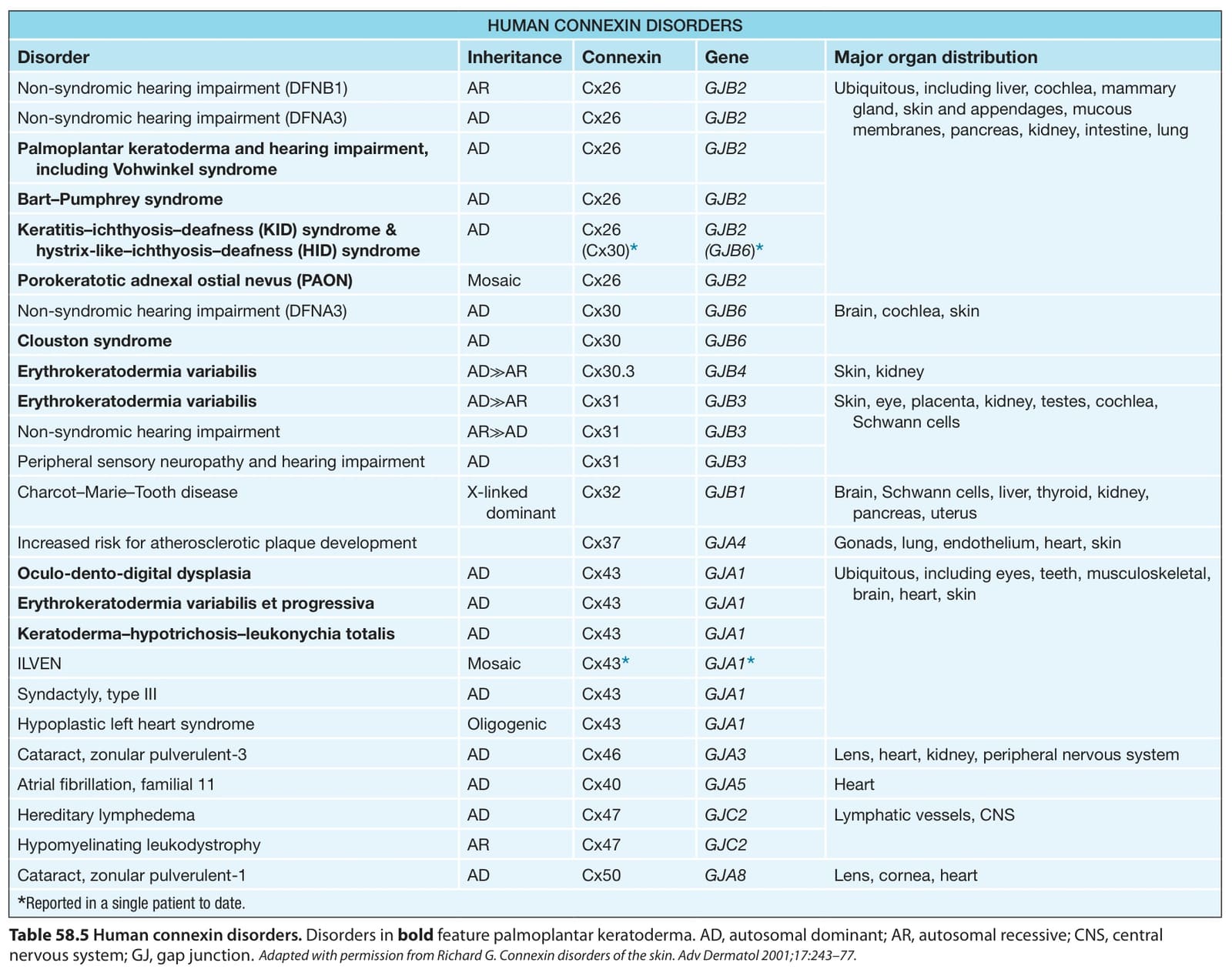
Table 58.5 Human connexin disorders. Disorders in bold feature palmoplantar keratoderma. AD, autosomal dominant; AR, autosomal recessive; CNS, central nervous system; GJ, gap junction.
表 58-5:人類連接蛋白(connexin)疾病。粗體字的疾病以掌蹠角皮症為特徵。AD,體染色體顯性;AR,體染色體隱性;CNS,中樞神經系統;GJ,間隙連接。
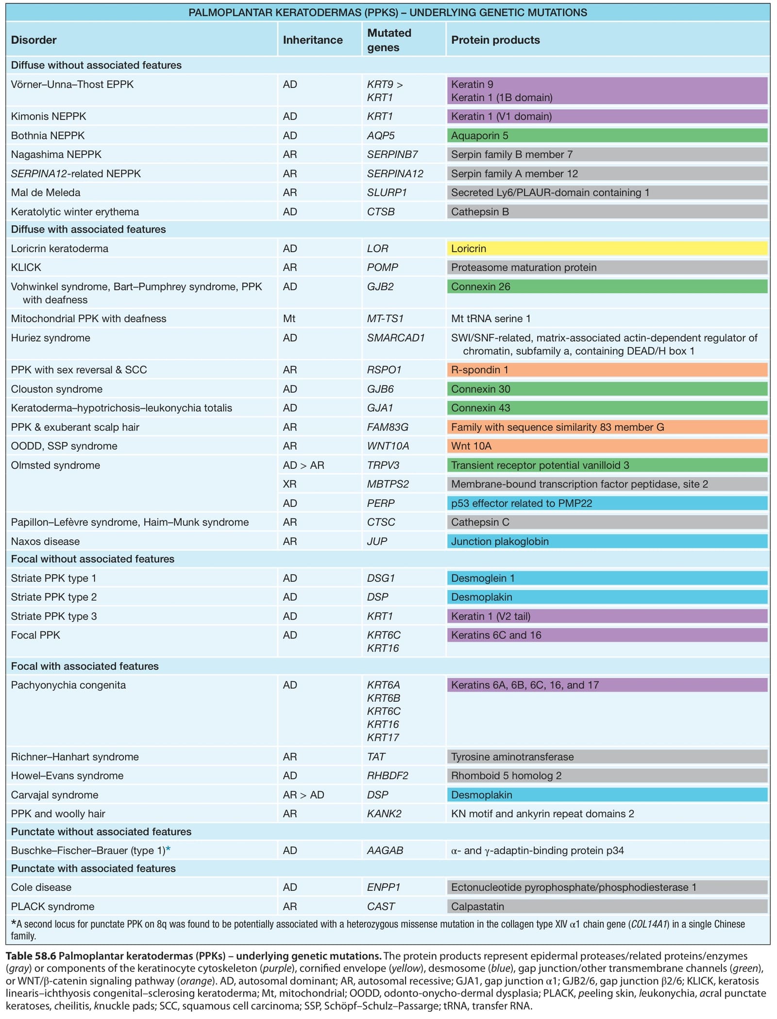
Table 58.6 Palmoplantar keratodermas (PPKs) – underlying genetic mutations. The protein products represent epidermal proteases/related proteins/enzymes (gray) or components of the keratinocyte cytoskeleton (purple), cornified envelope (yellow), desmosome (blue), gap junction/other transmembrane channels (green), or WNT/β-catenin signaling pathway (orange). AD, autosomal dominant; AR, autosomal recessive; GJA1, gap junction α1; GJB2/6, gap junction β2/6; KLICK, keratosis linearis–ichthyosis congenital–sclerosing keratoderma; Mt, mitochondrial; OODD, odonto-onycho-dermal dysplasia; PLACK, peeling skin, leukonychia, acral punctate keratoses, cheilitis, knuckle pads; SCC, squamous cell carcinoma; SSP, Schöpf–Schulz–Passarge; tRNA, transfer RNA.
表 58-6:掌蹠角皮症(PPKs)——潛在的基因突變。蛋白質產物代表表皮蛋白酶/相關蛋白/酵素(灰色),或角質形成細胞細胞骨架(紫色)、角質化外套膜(黃色)、胞橋小體(藍色)、間隙連接/其他跨膜通道(綠色)、或 WNT/β-catenin 訊息傳遞路徑(橘色)的組成成分。AD,體染色體顯性;AR,體染色體隱性;GJA1,間隙連接 α1;GJB2/6,間隙連接 β2/6;KLICK,keratosis linearis–ichthyosis congenital–sclerosing keratoderma;Mt,粒線體;OODD,odonto-onycho-dermal dysplasia;PLACK,peeling skin, leukonychia, acral punctate keratoses, cheilitis, knuckle pads;SCC,鱗狀細胞癌;SSP,Schöpf–Schulz–Passarge;tRNA,轉運 RNA。