ACQUIRED KERATODERMAS AND RELATED CONDITIONS
Keratoderma Climactericum
This common, acquired form of focal plantar hyperkeratosis favoring women >45 years of age was first described by Haxthausen in 1934. It is often associated with obesity, cold dry climates, and wearing backless shoes (e.g. sandals). Hyperkeratosis begins over pressure points on the heels, and the formation of fissures makes walking painful. If present, involvement of the hands is much milder. Endocrine dysfunction, vitamin A deficiency, contact dermatitis, and fungal infection were excluded in reported patients. Treatment options include keratolytic preparations containing 20%–40% urea or 12% ammonium lactate and, if severe, a low-dose oral retinoid. Similar changes described in young women following bilateral oophorectomy responded to estrogen replacement therapy.
Keratoderma and Cancer
Acquired diffuse PPK has been observed in association with bronchial carcinoma, and filiform PPK has been reported in patients with cancer of the breast, colon, and kidney. “Tripe palms” refers to velvety, thickened palmar skin with exaggerated ridges. It is often associated with acanthosis nigricans and may herald malignancies of the lung, stomach, or genitourinary tract. Bazex syndrome (acrokeratosis paraneoplastica) is discussed in Chapter 53. Esophageal carcinoma in association with focal PPK (Howel–Evans syndrome) as well as the increased risk of acral SCC in patients with Huriez syndrome and
PPK with sex reversal are discussed above. Acral melanoma and the epithelioma cuniculatum variant of SCC have also been reported in individuals with mutilating forms of PPK and (for acral melanoma in Asians) Nagashima-type PPK. In addition, an autosomal recessive syndrome featuring pigmentation defects, PPK, and cutaneous SCC can result from mutations in SASH1 (SAM and SH3 domain-containing 1; see Table 67.11). Mycosis fungoides represents another diagnostic consideration in patients with acquired PPK.
Arsenical keratoses present as small, corn-like areas of hyperkeratosis on the palms and soles (see Fig. 88.8). Over time, the lesions enlarge, thicken, and increase in number, spreading to the dorsal surfaces of the hands and feet. Ulceration often occurs when SCC develops. On histologic examination, epidermal changes vary from benign hyperplasia to moderate atypia or frank SCC. The latent period between ingestion of inorganic arsenic and onset of keratoses is 10 to 30 years or longer. Visceral malignancies, particularly of the lung and genitourinary tract, usually develop after the onset of skin tumors.
Other Etiologies of Acquired Keratoderma
Diffuse or focal palmoplantar hyperkeratosis can develop in association with hypothyroidism, often in the setting of myxedema. The PPK improves or resolves with thyroid replacement therapy.
With the increasing use of targeted therapies for the treatment of malignancies, in particular tyrosine kinase inhibitors, drug-induced palmoplantar hyperkeratosis also needs to be considered (see Table 21.16).
Other conditions that have been associated with keratoderma are listed in Table 58.1.
Aquagenic Palmoplantar Keratoderma
Synonyms: Transient reactive papulotranslucent acrokeratoderma Aquagenic syringeal acrokeratoderma Acquired aquagenic palmoplantar keratoderma Aquagenic wrinkling of the palms and soles Transient aquagenic keratoderma
Patients with aquagenic PPK develop thickening and white to translucent, “pebbly” changes on their palms shortly after immersion in water (e.g. within 3 minutes) (Fig. 58.19). Often, there is associated edema and burning pain. Involvement of the dorsal hands or soles is less frequently observed. These findings disappear shortly after drying the hands. Onset is typically during the second decade of life, with a predilection for girls and women. By dermoscopy, the papular lesions are at sites of dilated acrosyringeal ostia. Histologically, normal skin or dilated eccrine ostia and a mildly hyperkeratotic stratum corneum may be seen.
These exaggerated changes occur after water immersion. Courtesy Cindy E. Owen, MD.
Autosomal recessive or dominant inheritance of aquagenic PPK has been described, and an onset associated with the use of cyclooxygenase-2 inhibitors was reported. Aquagenic wrinkling of the palms can be observed in ~50% of patients with cystic fibrosis and ~10%–25% of heterozygous carriers of CFTR mutations. Aquagenic PPK should be differentiated from hereditary papulotranslucent acrokeratoderma, in which lesions are persistent once they appear, and other forms of PPK that are accentuated upon exposure to water, e.g. the Bothnian and Nagashima types of diffuse NEPPK (see above).
Improvement has been described with botulinum toxin injections, application of 20% aluminum chloride hexahydrate followed by urea cream, tap water iontophoresis, and oral acitretin.
Circumscribed Palmar or Plantar Hypokeratosis
This entity, which was first described in 2002, presents as a well-circumscribed, mildly erythematous, circular depression on the thenar or hypothenar region of the palm or less often the medial
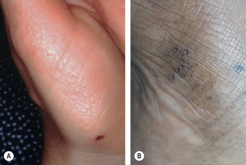
Fig. 58.18 Marginal papular keratoderma. A Acrokeratoelastoidosis presenting with multiple skin-colored to yellowish papules at the margin of the palmar skin. B Focal acral hyperkeratosis presenting with umbilicated keratotic papules at the margin of volar skin in an African-American woman. B, Courtesy Boni Elewski, MD.
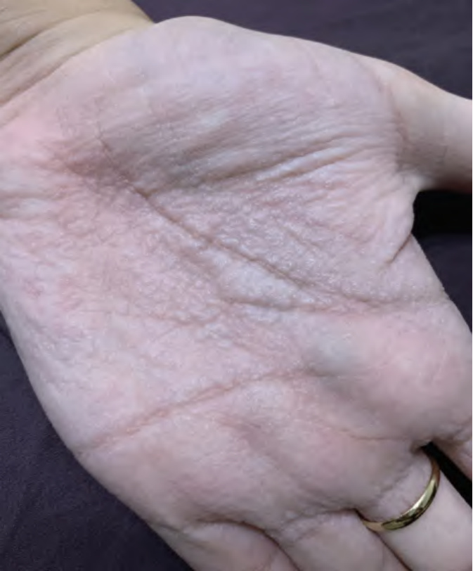
Fig. 58.19 Aquagenic wrinkling of the palms.
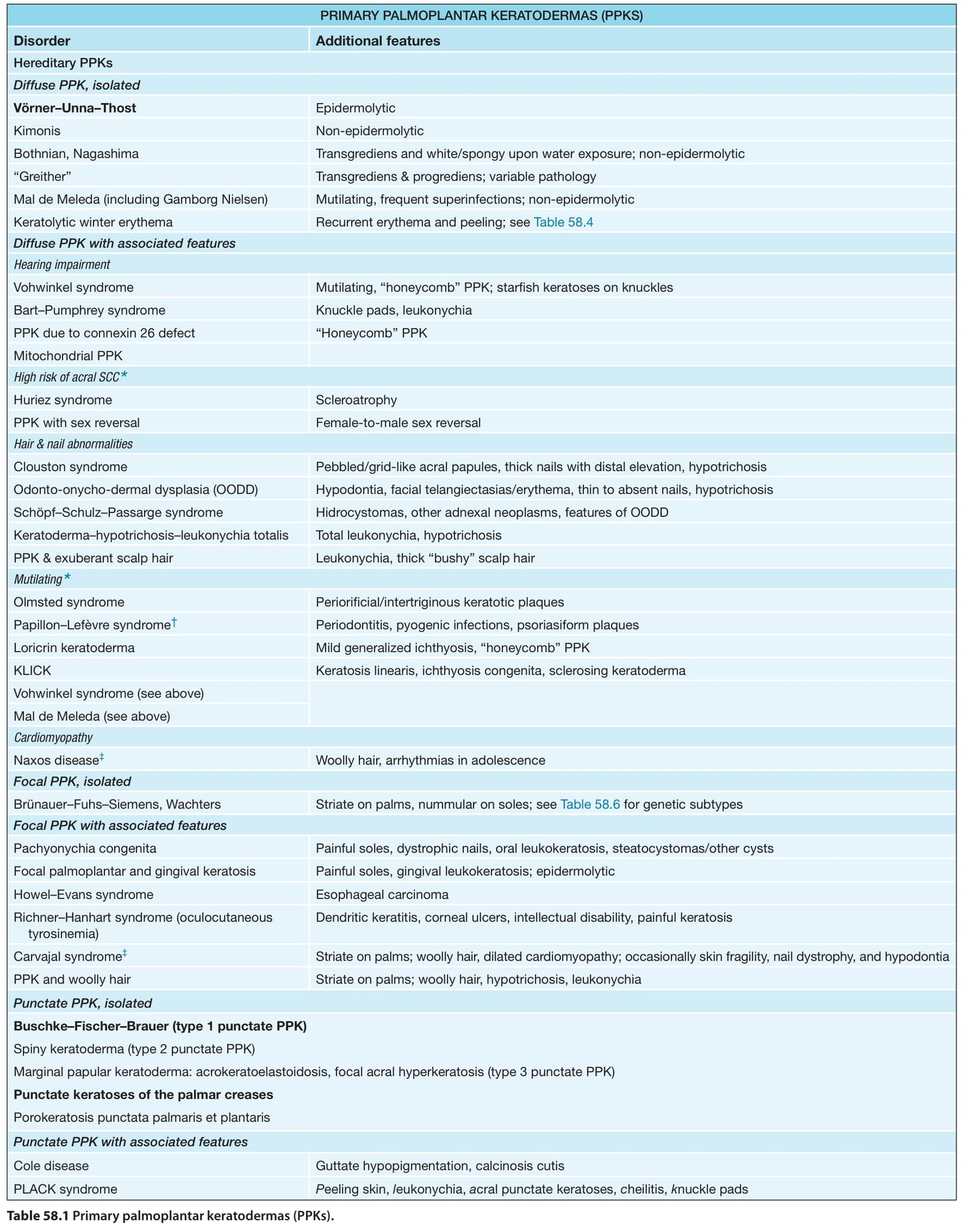
Table 58.1 Primary palmoplantar keratodermas (PPKs).
sole (Fig. 58.20). Lesions are solitary, or rarely few in number, and have a maximum diameter of 3 cm. They typically develop in women 40–85 years of age, although congenital lesions have been described. Sometimes there is a history of prior trauma or a burn at the site. The clinical differential diagnosis may include Bowen disease or porokeratosis.
Histologically, abrupt thinning of the stratum corneum over a diminished granular layer forms a sharp drop-off between normal and involved skin (see Fig. 58.20). To date, malignant transformation has not been reported. The etiology remains unknown, with postulated roles for trauma and loss of the keratinocytes’ ability to undergo palmoplantar differentiation. The latter is supported by the observation of diminished keratin 9 expression. Molecular studies have failed to detect human papillomavirus (HPV) apart from a single report of HPV type 4 DNA.
Treatments that have been reported include topical calcipotriene or 5-fluorouracil, cryotherapy, photodynamic therapy, fractional carbon dioxide laser, and excision.
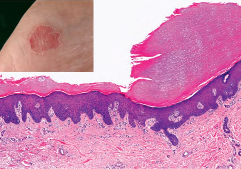
Fig. 58.20 Circumscribed palmar hypokeratosis. A sharp drop-off is seen between the lesional epidermis, which has a very thin stratum corneum, and the surrounding skin. Clinically, a well-circumscribed pink depression is seen (inset). Courtesy Luis Requena, MD.