ATAXIA–TELANGIECTASIA
Synonym: Louis–Bar syndrome
Key features
Progressive cerebellar ataxia
Oculocutaneous telangiectasias, initially of the bulbar conjunctivae
Selective deficiency of humoral and cell-mediated immunity, leading to sinopulmonary infections
Increased sensitivity to ionizing radiation
Leukemias and lymphomas
Introduction
Ataxia–telangiectasia (AT) is characterized by oculocutaneous telangiectasias, progressive cerebellar ataxia beginning in infancy, a variable immunodeficiency with a tendency to develop sinopulmonary infections, and chromosomal instability with persistent DNA damage after exposure to ionizing radiation.
Epidemiology
AT is an autosomal recessive disorder that occurs in 1 in 40 000 to 100 000 live births, with a carrier rate of up to 1% of the population.
Pathogenesis
AT results from mutations in the ataxia–telangiectasia mutated gene (ATM), which encodes a phosphatidylinositol 3-kinase-like serine/ threonine protein kinase that plays a central role in activating apoptotic and cell cycle responses to DNA damage, particularly double-stranded breaks. These DNA breaks are sensed directly by the MRN (MRE11– RAD50–nibrin [NBN]) complex, which recruits ATM and triggers its dissociation from inactive multimers to active monomers. Autophosphorylated ATM monomers subsequently activate (via phosphorylation) a variety of targets, including p53, BRCA1, FANCD2, and Artemis in addition to NBS1 and MRE11. The result is cell cycle arrest and facilitation of DNA repair, both in the setting of external insults (e.g. ionizing radiation) and in the processing of physiologic DNA breaks that occur during V(D)J recombination in lymphocytes, telomere maintenance, and meiosis. This provides an explanation for the sensitivity to ionizing radiation, immunodeficiency, premature aging, and defective spermatogenesis that are observed in patients with AT. Progressive neurologic deterioration in affected individuals likely reflects defective DNA repair in a cell population that cannot replicate. Oxidative stress related to ATM dysfunction has also been implicated in the pathogenesis of the disease.
Of note, mutations in genes encoding components of the MRN complex cause conditions with manifestations similar to AT. MRE11 mutations underlie the AT-like disorder, which features radiosensitivity and neurologic manifestations but no telangiectasias, and NBN or rarely RAD50 mutations result in Nijmegen breakage syndrome characterized by microcephaly, immunodeficiency, chromosomal instability, and cancer predisposition.
Clinical Features
The first manifestation of AT is usually ataxia, which typically appears when the affected child begins to walk. However, the diagnosis is often not recognized until the oculocutaneous telangiectasias are well developed, at a median age of 6 years. The telangiectasias first appear at 3–6 years of age on the lateral and medial bulbar conjunctivae as red, symmetric horizontal streaks (Fig. 60.1A). Telangiectasias subsequently develop on the ears, eyelids, malar prominences, antecubital and popliteal fossae, and presternal area, and less commonly on the dorsal aspects of the hands and feet as well as the hard and soft palate. The non-facial cutaneous telangiectasias are often subtle, resembling fine petechiae (Fig. 60.1B).
Progeric changes of the skin and hair are noted in almost 90% of patients. Subcutaneous fat is lost early and the facial skin tends to become atrophic and sclerotic. Gray hairs frequently appear in young children, and diffuse graying of the hair may occur by adolescence. Another common manifestation of AT is cutaneous granulomas (Fig. 60.1C), which are often associated with persistent rubella virus
infection (vaccine strain > wild type). These granulomatous plaques tend to be persistent and ulcerate, leading to significant discomfort. Children with AT often have hyper- or hypopigmented macules as well as hyper- and/or hypopigmentation along the lines of Blaschko, with chromosomal instability likely explaining their predisposition to pigmentary mosaicism. Other dermatologic findings that have been described in individuals with AT include a facial papulosquamous rash, poikiloderma, hypertrichosis favoring the arms, warts, and acanthosis nigricans.
Cerebellar ataxia usually begins during infancy and is characterized by swaying of the head and trunk. Choreoathetosis, dysarthric speech, oculomotor abnormalities, and myoclonic jerks often become prominent during childhood. Despite good muscle strength, patients are typically confined to a wheelchair by 11 years of age. The facies become dull and hypotonic, with mask-like changes developing concurrently with progressive progeric features. Of note, milder AT with onset of neurologic manifestations in adulthood has been described in individuals with ATM mutations that result in residual kinase activity.
Chronic or recurrent sinopulmonary infections occur in >80% of AT patients, with the most common cause of death being bronchiectasis with respiratory failure. The majority of affected individuals also have evidence of glucose intolerance, hyperinsulinemia, and insulin resistance. Additional manifestations of AT include growth delay (>70%), hypogonadism (especially in female patients), and intellectual disability (~30%).
Although lymphoid malignancy has been reported as the presenting sign of AT during infancy, most neoplasias occur in young adults. Patients who survive into their late teenage years have an up to 40% risk of developing a malignancy, especially leukemia (70-fold increase compared to unaffected individuals) and lymphoma (200-fold increase). Occasionally, patients develop basal cell carcinomas (BCCs) during the third decade of life.
Carriers of heterozygous ATM mutations have a two- to threefold increase in both the risk of developing breast cancer and the likelihood of death from cancer, including malignancies of the stomach, colon, and lung as well as the breast. There is greater excess mortality in individuals <50 years of age.
Laboratory Findings
Immunologic defects in patients with AT include: (1) decreased serum levels of IgE, IgA, and IgG (especially IgG and IgG) in ~80%, 70%, and 60% of patients, respectively; (2) low-molecular-weight (8 S) IgM (in 80% of patients) and increased serum levels of IgM (in a minority of patients); and (3) defects in cell-mediated immunity such as lymphopenia (in 70% of patients) and deficient in vitro responses to antigens and mitogens. Affected individuals tend to have a relative deficiency of CD4+ T cells and an excess of γ/δ T cells as well as elevated serum levels of interleukin-8 (IL-8), a chemokine that may contribute to pathogenic inflammation in AT. Most patients have an absent or abnormal thymus. Individuals with no ATM activity have more severe immunologic deficits than those with low levels of ATM activity.
Spontaneous chromosomal abnormalities such as fragments, breaks, gaps, and translocations occur 2–18 times more frequently in patients with AT than in unaffected individuals, and an increased rate of telomeric shortening is also observed. Rearrangements of chromosomes 7 and 14 are particularly common and may predict the development of lymphoma. Fibroblast DNA from both patients and carriers is extremely sensitive to ionizing radiation and to radiomimetic agents such as bleomycin.
Almost all patients with AT have elevated levels of α-fetoprotein and carcinoembryonic antigen, and measuring the former can support the diagnosis in individuals ≥2 years of age. Hepatic transaminase levels are also mildly elevated in approximately half of patients, and MRI often shows cerebellar atrophy by early childhood. Additional traditional diagnostic clues include radiosensitivity testing with the colony survival assay, analysis of radioresistant DNA synthesis (which demonstrates an abnormal S phase checkpoint), and karyotyping to identify 7;14 translocations. Sequencing the ATM gene or immunoblotting for the ATM protein may be utilized to confirm the diagnosis. Prenatal diagnosis of AT can be performed when parental mutations have been identified.
Differential Diagnosis
Children with AT may be thought to have Friedreich ataxia until the ocular telangiectasias become apparent. The conjunctival telangiectasias are occasionally misdiagnosed as conjunctivitis, but the chronicity and size of the telangiectatic vessels distinguish AT. Bloom syndrome can present with facial and occasionally conjunctival telangiectasias, café-au-lait macules, decreased immunoglobulin levels, recurrent respiratory infections, and hematologic malignancies, but patients do not have neurologic abnormalities (see Ch. 87). FILS syndrome – facial dysmorphism, immunodeficiency, livedo, and short stature – due to polymerase ε 1 (POLE1) mutations has overlapping features and can also present with telangiectasias or poikiloderma. RIDDLE syndrome is an autosomal recessive condition that features radiosensitivity, immunodeficiency, facial dysmorphism, difficulty learning, abnormal motor control, and short stature due to mutations in RNF168, which encodes a protein that mediates ubiquitin-dependent signaling at sites of DNA double-stranded breaks.
Treatment
The median lifespan in patients with AT is now >25 years. More than half of patients die from chronic sinopulmonary disease and up to a third succumb to a malignancy or complications of cancer treatment. Some of the patients who survive into adulthood show improvement in neurologic and immunologic status.
Therapy is supportive:
●antibiotics for infections; prophylactic antibiotics and IVIg replacement therapy for patients with severe immunodeficiency
●avoidance of sun exposure and use of sunscreens
●early physiotherapy for patients with bronchiectasis and consideration of systemic corticosteroid therapy for interstitial lung disease
●physical therapy to prevent contractures related to neurologic dysfunction
●screening for the development of malignancy. If malignancy occurs, chemotherapy is typically necessary; radiation and radiomimetic chemotherapeutic agents, especially bleomycin, should be avoided. AT patients have a decreased threshold for postradiation erythema, tissue necrosis, and radiation-induced cutaneous malignancy. If radiation is necessary, restriction of the fractions to 1 Gy and total radiation to <20 Gy is recommended. Allogeneic hematopoietic stem cell transplantation with a reduced intensity conditioning regimen has been utilized in AT patients with hematopoietic malignancies or severe manifestations of immunodeficiency, including destructive rubella virusassociated cutaneous granulomas. Because oxidative stress is thought to contribute to chromosomal instability, neurodegeneration, and malignancy in patients with AT, administration of antioxidants such as α-lipoic acid is often recommended; however, treatment with the latter together with nicotinamide did not improve neurologic or pulmonary function in a small controlled study. In patients with truncating ATM mutations, treatment with aminoglycosides can induce read-through of premature termination codons and restore production of functional ATM protein, providing the basis for a potential therapeutic approach.
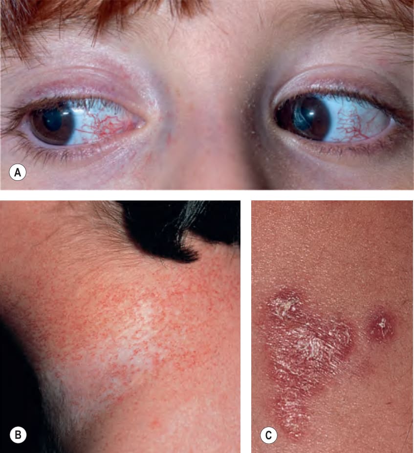
Fig. 60.1 Ataxia–telangiectasia (AT).A Linear telangiectasias on the medial and lateral bulbar conjunctivae of a girl with AT. Punctate and smaller linear telangiectasias are also present on the eyelids. B Extensive telangiectasias on the neck of a young woman with AT. C Persistent granulomatous plaques on the leg of a child. These lesions often ulcerate and are difficult to manage. A, Courtesy Edward Cowen, MD.
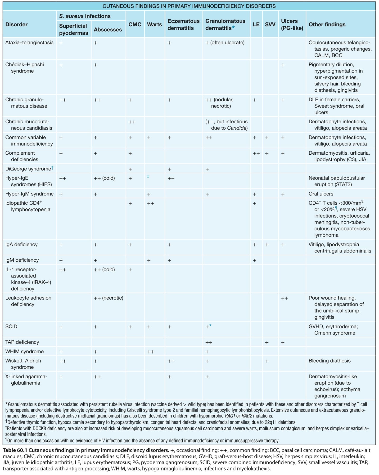
Table 60.1 Cutaneous findings in primary immunodeficiency disorders.+, occasional finding; ++, common finding; BCC, basal cell carcinoma; CALM, café-au-lait macules; CMC, chronic mucocutaneous candidiasis; DLE, discoid lupus erythematosus; GVHD, graft-versus-host disease; HSV, herpes simplex virus; IL, interleukin; JIA, juvenile idiopathic arthritis; LE, lupus erythematosus; PG, pyoderma gangrenosum; SCID, severe combined immunodeficiency; SVV, small vessel vasculitis; TAP, transporter associated with antigen processing; WHIM, warts, hypogammaglobulinemia, infections and myelokathexis.
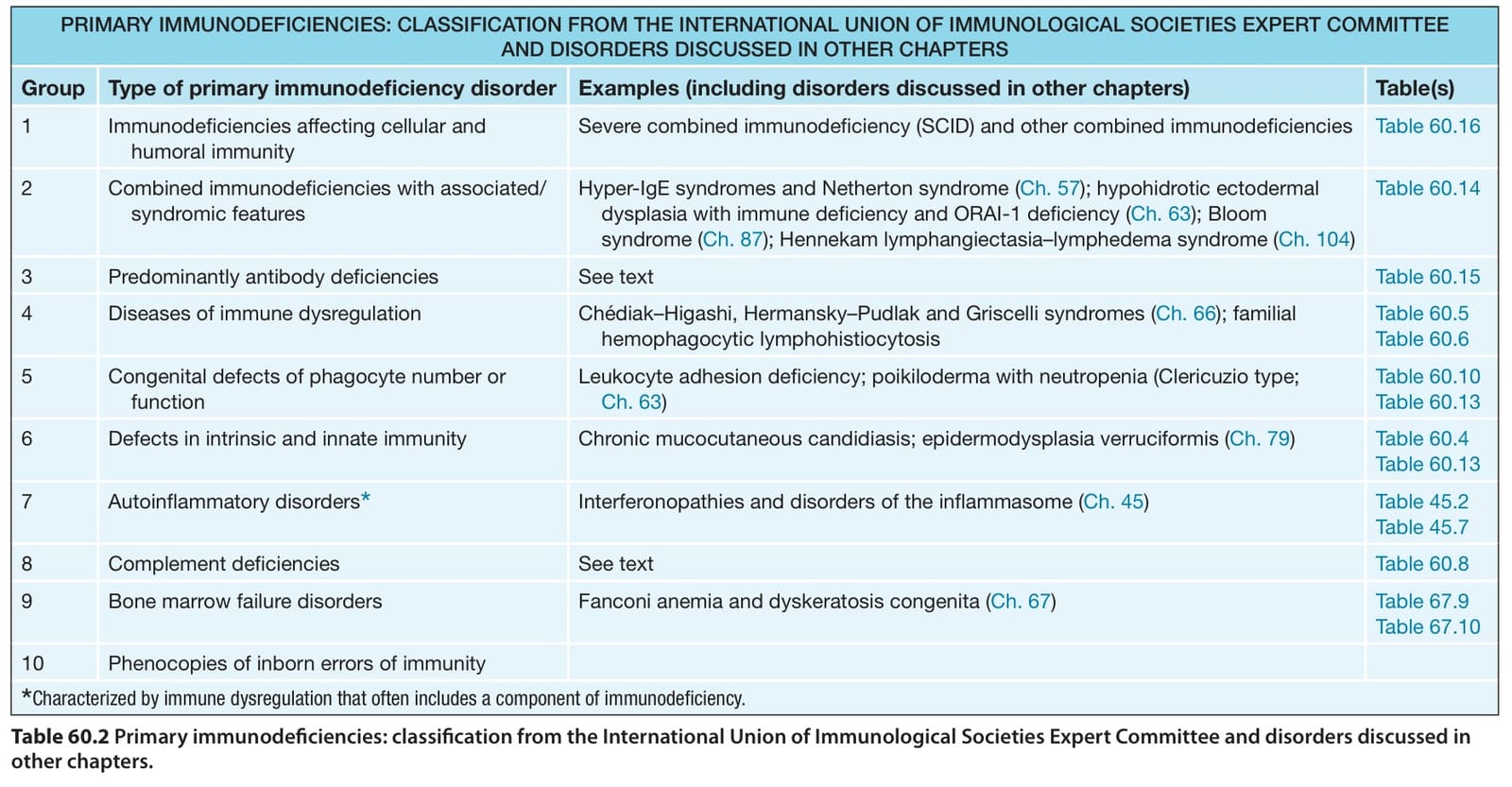
Table 60.2 Primary immunodeficiencies: classification from the International Union of Immunological Societies Expert Committee and disorders discussed in other chapters.
Table 60.3 Screening laboratory tests to assess for a primary immunodeficiency in a patient with recurrent cutaneous infections.