CHÉDIAK–HIGASHI SYNDROME
Synonyms: Béguez César–Steinbrinck syndrome Chédiak– Higashi disease Chédiak–Steinbrinck–Higashi syndrome Chédiak– Steinbrinck anomaly
Key features
Autosomal recessive disorder of vesicle trafficking that results in giant organelles, including melanosomes, leukocyte granules, and platelet dense granules
Silvery hair is accompanied by mild diffuse pigmentary dilution, often with an admixture of hyper- and hypopigmentation in sun-exposed sites; a variable degree of photophobia and nystagmus
Recurrent pyogenic infections and a mild bleeding diathesis
An “accelerated phase” with pancytopenia and organomegaly due to lymphohistiocytic infiltration, which is fatal without hematopoietic stem cell transplantation
Progressive neurologic deterioration in survivors
Epidemiology
Chédiak–Higashi syndrome (CHS) is a rare autosomal recessive disease reported most often in individuals of Northern European, Spanish, Middle Eastern, and Japanese ancestry. Patients of African descent have also been described.
Pathogenesis
CHS represents a disorder of vesicle trafficking and is caused by mutations in the lysosomal trafficking regulator gene (LYST). Most patients have frameshift or nonsense mutations that result in a truncated protein and lead to a severe phenotype with progression to a fatal lymphoproliferative “accelerated phase” during childhood, but 10%–15% of affected individuals have missense mutations that are associated with a milder phenotype and survival to adulthood.
The giant intracytoplasmic granules that characterize CHS result from dysregulated fission/fusion of lysosome-related organelles, which include melanosomes, platelet dense granules, and leukocyte cytolytic granules. The latter granules cannot effectively migrate or discharge their peroxidative and proteolytic enzymes, leading to defective target cell killing. Localization of cytotoxic T lymphocyte-associated antigen (CTLA-4) to enlarged vesicles rather than the cell surface may play a role in the development of lymphoproliferative disease. In addition, defective autophagic lysosome reformation in neurons, generalized impairment in plasma membrane repair, and abnormal antigen presentation by B cells may contribute to the pathogenesis of CHS.
Clinical Features
Patients with CHS usually present in infancy with mild diffuse pigmentary dilution of the skin, hair, and eyes. In individuals with relatively dark constitutive pigmentation, areas of bronze to slate-gray hyperpigmentation may be evident in sun-exposed sites, often admixed with guttate hypopigmented macules. Hair color is variable, but a silvery, metallic sheen is characteristic. Decreased ocular pigment can result in photophobia, nystagmus, and strabismus, but visual acuity is usually normal.
Infections primarily involve the skin and respiratory tract, and they are typically caused by Staphylococcus aureus, Streptococcus pyogenes, and Streptococcus pneumoniae. The skin infections are most often superficial pyodermas. Cutaneous ulcerations resembling pyoderma gangrenosum, gingivitis, and ulceration of the oral mucosa have also been described. Although the bleeding diathesis of CHS is generally mild, some patients experience easy bruising, petechiae, and epistaxis.
Approximately 85% of patients with CHS enter a lymphoproliferative “accelerated phase” characterized by pancytopenia and lymphohistiocytic infiltration of the liver, spleen, lymph nodes, oral mucosa, and other internal organs. This “hemophagocytic syndrome” results from uncontrolled T cell and macrophage activation, possibly associated with Epstein–Barr virus (EBV) infection, and typically leads to death by age 10 years from overwhelming infection or hemorrhage unless hematopoietic stem cell transplantation is performed.
Patients with CHS who survive the first decade of life experience progressive neurologic deterioration with abnormal gait, paresthesias, and developmental delay. Peripheral and cranial neuropathies, spinocerebellar degeneration, spastic paraplegia, parkinsonism, and dementia can manifest in adult patients who were treated with hematopoietic stem cell transplantation as children as well as in those with otherwise mild disease.
Pathology
The silvery hair of patients with CHS is characterized by clumps of melanin microscopically (Fig. 60.5), and giant melanosomes may be evident within melanocytes in skin biopsy specimens. A peripheral blood
smear demonstrating abnormally large granules in the perinuclear area of granulocytes can assist in establishing the diagnosis. Immunologic defects in CHS include neutropenia, diminished leukocyte chemotaxis, impaired antibody-dependent cell-mediated cytotoxicity, reduced cytotoxic and regulatory T cell activity, and markedly decreased natural killer (NK) cell function.
Differential Diagnosis
The features of CHS and the three types of Griscelli syndrome (GS), another autosomal recessive disorder that can present with immune dysfunction and silvery hair, are compared in Table 60.5 (see also Ch. 66). Although leukocytes generally have a normal appearance in GS, hypogammaglobulinemia can be observed. Hermansky–Pudlak syndrome (HPS) types 2, 9, and 10 (AP3B1, BLOC1S6, and AP3D1 mutations, respectively; see Table 65.1) can result in recurrent infections due to abnormal cytotoxic T cell function; additional manifestations include a bleeding diathesis, neurologic manifestations (in HPS9 & HPS10), and diffuse pigmentary dilution, but not silvery hair. Tricho-hepato-enteric syndrome, an autosomal recessive disorder caused by mutations in SKIC2 or SKIC3 which encode the SKI2 and SKI3 subunits of superkiller complex, can present with diffuse pigmentary dilution of the skin and hair, immunodeficiency, and platelet abnormalities; unlike CHS, it is also characterized by brittle hair with trichorrhexis nodosa, intractable diarrhea during infancy, primary liver disease, facial dysmorphism, and cardiac defects. Other inherited disorders characterized by lymphoproliferation due to immune dysregulation are presented in Table 60.6.
Treatment
Hematopoietic stem cell transplantation (HSCT) can reverse the immunodeficiency of CHS and may also be performed following immunochemotherapy to abrogate the lymphoproliferative accelerated phase. However, HSCT has no effect on pigmentary alteration and does not halt neurologic degeneration. Management of the CHS is otherwise largely supportive, with administration of prophylactic antibiotics to prevent recurrent infections.
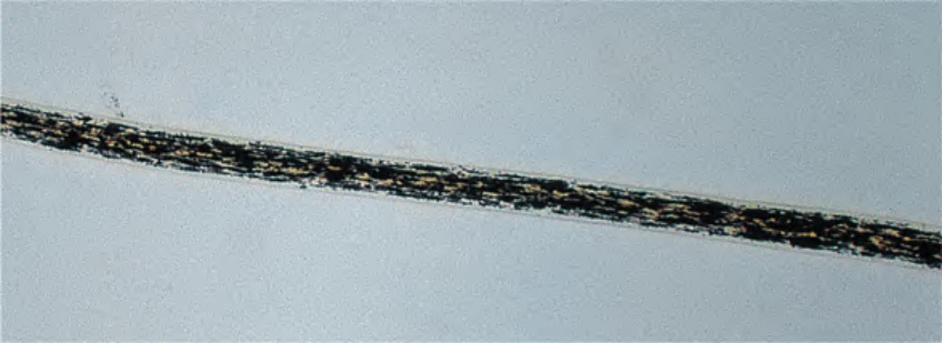
Fig. 60.5 Chédiak–Higashi syndrome. Light microscopic examination of hair shows small, regularly spaced clumps of melanin in patients with Chédiak– Higashi syndrome.
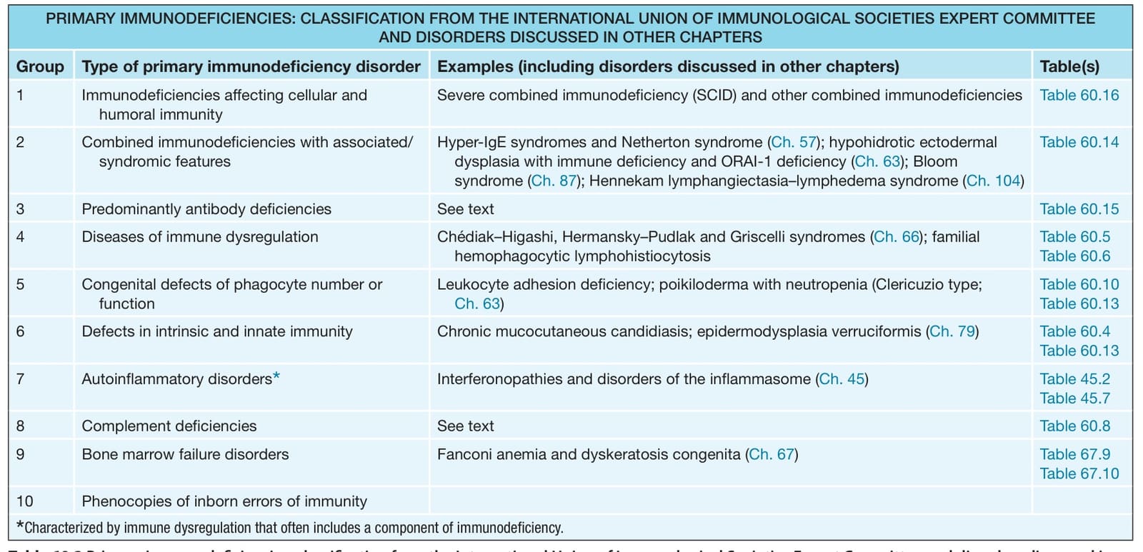
Table 60.5 Features of Chédiak–Higashi syndrome (CHS) and Griscelli syndrome (GS). The latter is caused by defective attachment of organelles to the actin cytoskeleton (see Ch. 65). Elejalde syndrome (neuroectodermal melanolysosomal disease), which presents with pigmentary features of GS plus severe neurologic dysfunction, likely represents a variant of GS1. CNS, central nervous system.
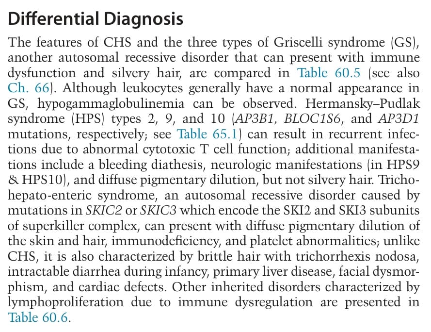
Table 60.6 Inherited disorders characterized by lymphoproliferation due to immune dysregulation. Hemophagocytic lymphohistiocytosis (HLH) results from uncontrolled T cell and macrophage activation, manifesting with fever, pancytopenia, hepatosplenomegaly (HSM), lymphadenopathy (LAN), hypertriglyceridemia, and hypofibrinogenemia. AD, autosomal dominant; AR, autosomal recessive; CTL, cytotoxic T lymphocytes; Inh, inheritance; LE, lupus erythematosus; NK, natural killer; XR, X-linked recessive.