CHRONIC GRANULOMATOUS DISEASE
Synonyms: Chronic granulomatous disorder Bridges–Good syndrome Quie syndrome
Key features
Inability to kill intracellular organisms through generation of oxidative metabolites
X-linked recessive or autosomal recessive inheritance
Recurrent pneumonias and cutaneous infections, lymph adenopathy, and hepatosplenomegaly
Patients develop granulomas as a compensatory effort to confine organisms
Introduction
Chronic granulomatous disease (CGD) is a group of disorders characterized by severe, recurrent infections due to an inability of leukocytes to kill phagocytosed bacteria and fungi by generating oxidative metabolites. The activity of the nicotinamide dinucleotide phosphate (NADPH) oxidase complex is reduced in all forms of CGD.
Epidemiology
The incidence of CGD is ~1 in 200 000 live births. Ninety percent of patients are boys. Approximately three-quarters of cases have X-linked recessive transmission, while the remainder are autosomal recessive. A small subset of male patients with X-linked CGD due to a contiguous gene deletion also have McLeod neuroacanthocytosis syndrome, with additional central nervous system, neuromuscular, cardiovascular, and hematologic (e.g. reduced Kell antigens on red blood cells) manifestations.
Pathogenesis
The defects in CGD involve subunits of the NADPH oxidase found in phagocytes. These include: (1) membrane-bound gp91phox (phagocyte oxidase) and p22phox as well as the EROS (essential for reactive oxygen species) chaperone protein required for their expression; and (2) cytoplasmic p47phox, p67phox, and p40phox (Table 60.10). The failure of microbial killing that characterizes CGD results from an inability of the NADPH oxidase system to rapidly generate toxic reactive oxygen species (ROS) by transferring electrons from NADPH to molecular O (the “respiratory burst”) after phagocytosis of a pathogen. Regardless of the gene affected in patients with CGD, having residual NADPH oxidase activity and the resultant ability to generate ROS is associated with less severe disease and longer survival.
NADPH oxidase activity results in the activation of antimicrobial proteases within the phagosome and the formation of microbicidal “neutrophil extracellular traps” (NETs). In addition to killing and degrading microorganisms, ROS produced by the NADPH oxidase system have roles in the regulation of cytokine synthesis as well as induction of neutrophil apoptosis, which prevents tissue damage at sites of inflammation. Animal models of CGD have shown that a lack of ROS leads to excess inflammation via decreased regulatory T cell activity, unrestrained γ/δ T cell activity, and augmented production of cytokines such as IL-1β, IL-8, and IL-17. NADPH oxidase also has a role in modulating MHC class II antigen presentation by B cells.
Clinical Features
The skin, perianal area, lymph nodes, and lungs are the sites most often affected by CGD. In general, the X-linked form tends to be more severe than the autosomal recessive forms, with a younger mean age at diagnosis (3 years vs 8 years). Organisms that commonly cause infections in patients with CGD are listed in Table 60.11. Most carriers of CGD do not have an increased susceptibility to infections.
The earliest manifestations of CGD are usually staphylococcal infections of the skin around the ears and nose, which may begin in the neonatal period and progress to extensive purulent dermatitis with regional lymphadenopathy during infancy. Initial presentation of CGD as ecthyma gangrenosum in the neonatal period has also been described. Cutaneous abscesses occur in 40% of patients, usually due to S. aureus but also Serratia marcescens; these infections sometimes present as large, poorly healing cutaneous ulcers. Non-infectious purulent inflammatory reactions may develop at sites of minor cutaneous trauma or regional lymph node drainage and heal slowly with scarring. Sterile cutaneous granulomas, which are often nodular and necrotic, occur less frequently than infections.
Additional cutaneous manifestations include acute or chronic cutaneous lupus erythematosus-like skin lesions, most commonly discoid (Fig. 60.7A,B); Sweet syndrome-like lesions; ulcers involving the oral mucosa (resembling aphthous stomatitis), the perioral area, and other cutaneous sites (Fig. 60.7C); and seborrheic dermatitis or folliculitis of the scalp. Female carriers of X-linked CGD occasionally present with discoid lesions, lymphocytic infiltrate of Jessner, photosensitivity, Raynaud phenomenon, severe aphthous stomatitis, and granulomatous cheilitis.
The extracutaneous organs most frequently involved in CGD are the lymph nodes, lungs, liver, spleen, and gastrointestinal tract (Table 60.12). Suppurative lymphadenitis develops in half of patients, most often affecting cervical nodes and leading to abscess and fistula
Less frequent manifestations include central nervous system vasculitis, pleuritis, pericarditis, Raynaud phenomenon, thrombocytopenia, anti-Sm or anti-RNP antibodies, circulating immune complexes and a positive RPR. ACLE, acute cutaneous lupus erythematosus; DLE, discoid lupus erythematosus; SCLE, subacute cutaneous lupus erythematosus. Adapted with permission from Torrelo A. Schachner and Hansen’s Pediatric Dermatology, 5th edn. London: Mosby, 2023 formation. Granulomas of the lungs, liver, spleen, and gastrointestinal and genitourinary tracts occur more frequently than those of the skin. These granulomas can lead to obstruction of the gastric outlet or urinary tract. Excessive inflammatory responses may also lead to wound dehiscence, pneumonitis, and hemophagocytic lymphohistiocytosis. Approximately 40%–80% of CGD patients develop gastrointestinal manifestations (e.g. chronic diarrhea, fistulas) similar to those of inflammatory bowel disease; clinical presentations can also mimic sarcoidosis, rheumatoid arthritis, and IgA nephropathy.
Pathology
Common nonspecific abnormalities in CGD include leukocytosis, anemia, elevated ESR, hypergammaglobulinemia, a decreased number of T cells, and an abnormal chest radiograph. Skin testing for delayedtype hypersensitivity is normal, as are studies of phagocytosis and chemotaxis.
Biopsy specimens from cutaneous granulomas that develop in CGD patients demonstrate histiocytic infiltrates associated with foreign body giant cells and accumulation of neutrophils with necrosis. Lupus erythematosus-like skin lesions in CGD patients and carriers may have histologic features similar to classic discoid lesions, but they sometimes lack vacuolar degeneration of basal keratinocytes; immunofluorescence examination of lesional skin is negative in most cases.
The traditional screening test for CGD was the nitroblue tetrazolium (NBT) reduction assay. NBT is yellow in its soluble, oxidized form; when reduced, the dye precipitates and becomes blue (formazan precipitate). Only
5%–10% of leukocytes from patients with CGD are able to reduce NBT during phagocytosis, compared to 80%–90% of leukocytes from unaffected individuals and ~20%–70% of leukocytes from carriers of X-linked CGD. The dihydrorhodamine (DHR) flow cytometric assay is more accurate and quantitative in measuring the neutrophil respiratory burst, and it is commercially available for verifying the diagnosis of CGD. However, the DHR assay is not sensitive for detecting p40phox-related CGD.
Immunoblot analysis may demonstrate lack of the gp91phox and p22phox proteins; however, gene sequencing must still be done to ascertain which gene is affected, since mutations resulting in the absence of one of these proteins or EROS lead to the absence of the other(s). A lack of the p47phox, p67phox, or p40phox protein by immunoblot analysis indicates the affected gene.
Differential Diagnosis
Laboratory tests allow differentiation of CGD from other immunodeficiency disorders characterized by increased susceptibility to bacterial infections. Additional inherited phagocyte disorders are listed in Table 60.13.
Treatment
The use of antibiotics and antifungals has markedly reduced the morbidity and mortality rates of CGD. Although cutaneous and nodal infections are often readily apparent, localized foci of internal infection (which may or may not be associated with fever) can be difficult to detect. Thorough periodic evaluation of the lungs, liver, and bones by routine radiographs, ultrasounds or CT, MRI, positron emission tomography (PET), and bone scans often uncovers occult foci of inflammation or infection. Cultures should be performed to identify the infectious agent, and invasive procedures may be necessary to obtain adequate tissue samples. While awaiting culture results or in situations where material from the affected site cannot be obtained, patients with evidence of infection should be treated empirically with broad-spectrum parenteral antibiotics that cover S. aureus as well as Gram-negative bacteria, with a high index of suspicion for fungal infection if despite antibiotics symptoms persist. Surgical interventions such as debridement, irrigation, and prolonged drainage are important for deeper infections.
Chronic prophylaxis with trimethoprim–sulfamethoxazole has been shown to decrease the incidence of bacterial infection in CGD patients.
The frequency and mortality of invasive fungal infections (e.g. with Aspergillus spp.) have likewise been reduced with itraconazole prophylaxis as well as treatment with other azoles (e.g. voriconazole, posaconazole, isavuconazonium sulfate). However, invasive fungal infections remain the most common cause of death in CGD. Administration of interferon-γ, which likely augments oxidant-independent antimicrobial pathways, may help to decrease the risk of serious infections. Granulocyte transfusions have been used for rapidly progressive, lifethreatening infections.
Short courses of systemic corticosteroids have been helpful for patients with obstructive granulomas of the bronchopulmonary, gastrointestinal, or genitourinary tract. Additional immunomodulatory therapies with potential benefit for inflammatory manifestations of CGD include azathioprine, hydroxychloroquine, anakinra, thalidomide, pioglitazone (increases ROS production and efferocytosis), ustekinumab, and sirolimus; tumor necrosis factor (TNF) inhibitors may improve colitis but increase the risk of infectious complications.
Hematopoietic stem cell transplantation represents a potentially curative treatment for CGD. Although younger patients without infection at the time of transplantation have the best outcomes (survival >95%), use of reduced-intensity conditioning regimens has enabled successful transplantation of higher-risk patients such as adults and individuals with recalcitrant infections or inflammatory manifestations. Children with CGD who undergo transplantation have improved growth and fewer infections, surgical interventions, and hospital admissions than those managed conservatively.
In the 1990s, gene therapy was performed in five adults with the p47phox-deficient form of CGD, and a single infusion of transduced CD34+ peripheral blood stem cells led to peak levels of corrected granulocytes in 3–6 weeks with persistence for as long as 6 months. Subsequently, two young men with X-linked recessive CGD were treated with non-myeloablative conditioning prior to the infusion of CD34+ peripheral blood stem cells transduced ex vivo with a retroviral vector expressing gp91phox; this led to sustained engraftment of functionally corrected phagocytes. However, following initial resolution of infections, the transgene was silenced due to methylation of the viral promoter in both patients, and within 3 years they developed myelodysplasia with monosomy 7 as a result of insertional activation of ecotropic viral integration site 1 (EVI1). To increase the safety and efficacy of gene therapy for CGD, current investigations are utilizing approaches such as self-inactivating lentiviral vectors, targeted integration of transgenes into a genomic “safe harbor” site, or the CRISPR-Cas9 site-specific nuclease system to encourage repair of the endogenous gene via homologous recombination (see Ch. 3). In a recent study, 6 of 9 patients with severe X-linked CGD who received ex vivo autologous CD34+ hematopoietic stem/progenitor cell-based lentiviral gene therapy following myeloablative conditioning recovered functional oxidase activity, with no new CGD-related infections or evidence of genotoxicity after a follow-up period of 1 year.
DHR flow cytometric studies may be utilized in determining the carrier status of the sisters and other female relatives of patients with X-linked CGD, which is important for genetic counseling prior to pregnancy. Prenatal diagnosis of CGD is also possible.
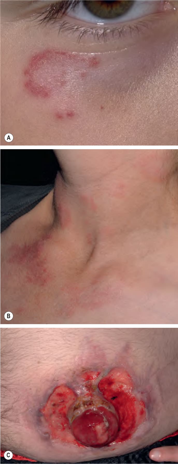
Fig. 60.7 Chronic granulomatous disease. Lupus erythematosus-like annular plaques and papules on the infraorbital cheek (A) and neck (B). Pyoderma gangrenosum-like ulcers at an ostomy stoma site (C). Courtesy Edward Cowen, MD.
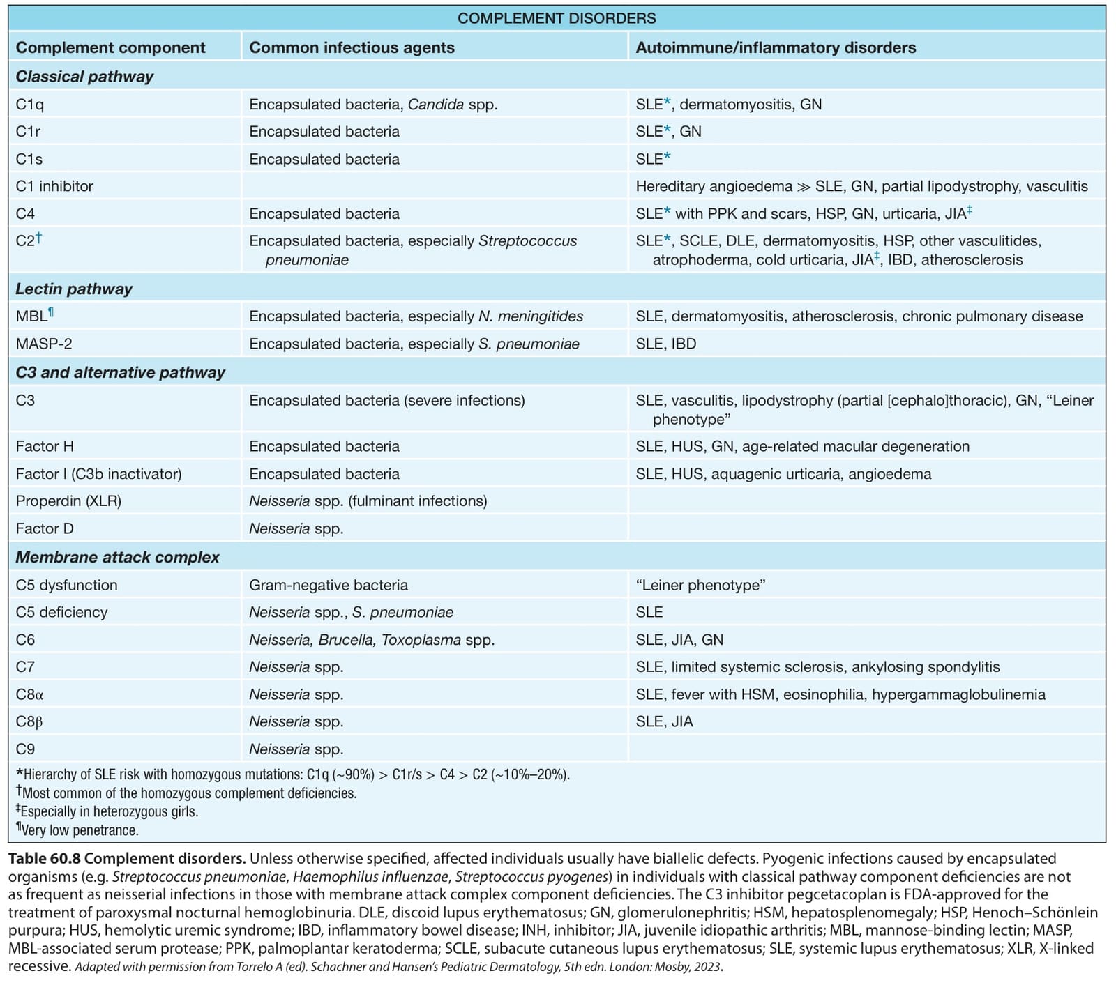
Table 60.8 Complement disorders. Unless otherwise specified, affected individuals usually have biallelic defects. Pyogenic infections caused by encapsulated organisms (e. g. Streptococcus pneumoniae, Haemophilus influenzae, Streptococcus pyogenes) in individuals with classical pathway component deficiencies are not as frequent as neisserial infections in those with membrane attack complex component deficiencies. The C3 inhibitor pegcetacoplan is FDA-approved for the treatment of paroxysmal nocturnal hemoglobinuria. DLE, discoid lupus erythematosus; GN, glomerulonephritis; HSM, hepatosplenomegaly; HSP, Henoch–Schönlein purpura; HUS, hemolytic uremic syndrome; IBD, inflammatory bowel disease; INH, inhibitor; JIA, juvenile idiopathic arthritis; MBL, mannose-binding lectin; MASP, MBL-associated serum protease; PPK, palmoplantar keratoderma; SCLE, subacute cutaneous lupus erythematosus; SLE, systemic lupus erythematosus; XLR, X-linked recessive. Adapted with permission from Torrelo A (ed). Schachner and Hansen’s Pediatric Dermatology, 5th edn. London: Mosby, 2023
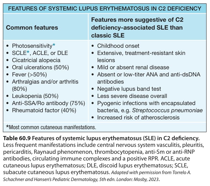
Table 60.9 Features of systemic lupus erythematosus (SLE) in C2 deficiency.
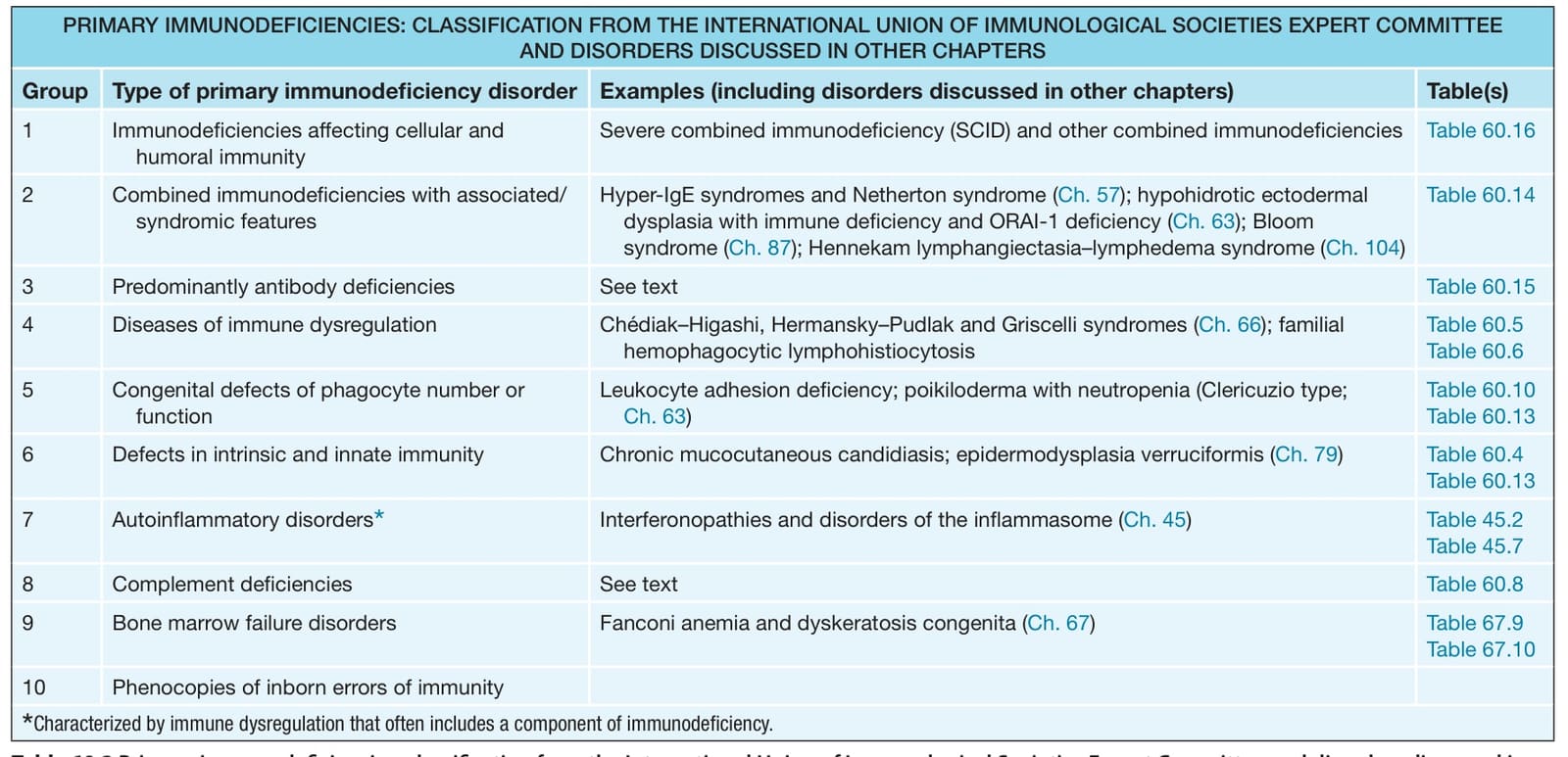
Table 60.10 Genetic defects affecting components of the phagocyte NADPH oxidase. CGD, chronic granulomatous disease; CYBC1, cytochrome b-245 chaperone 1; Eros, essential for reactive oxygen species; NCF, neutrophil cytosolic factor; RhoGDI, Rho GDP-dissociation inhibitor.
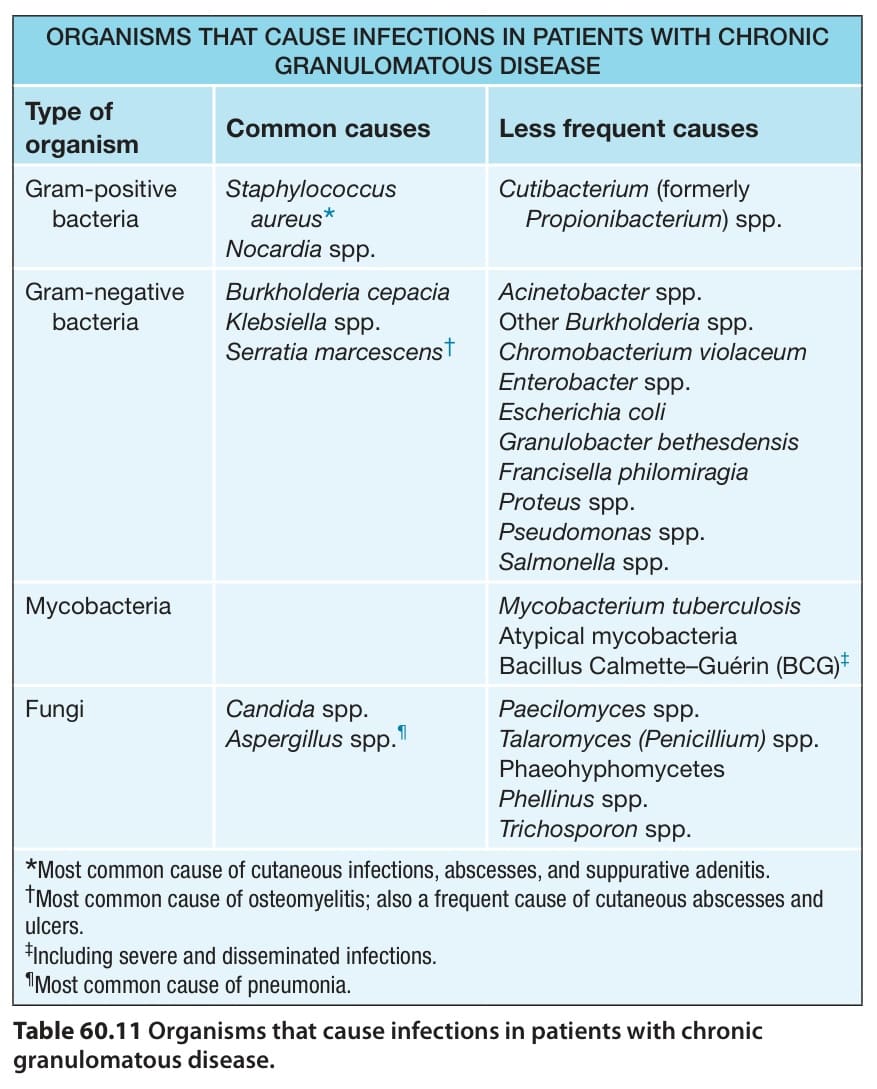
Table 60.11 Organisms that cause infections in patients with chronic granulomatous disease.
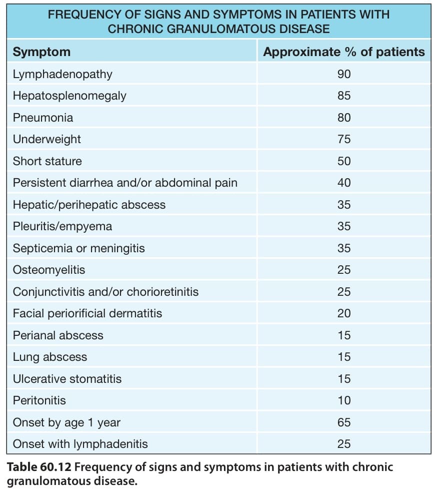
Table 60.12 Frequency of signs and symptoms in patients with chronic granulomatous disease.
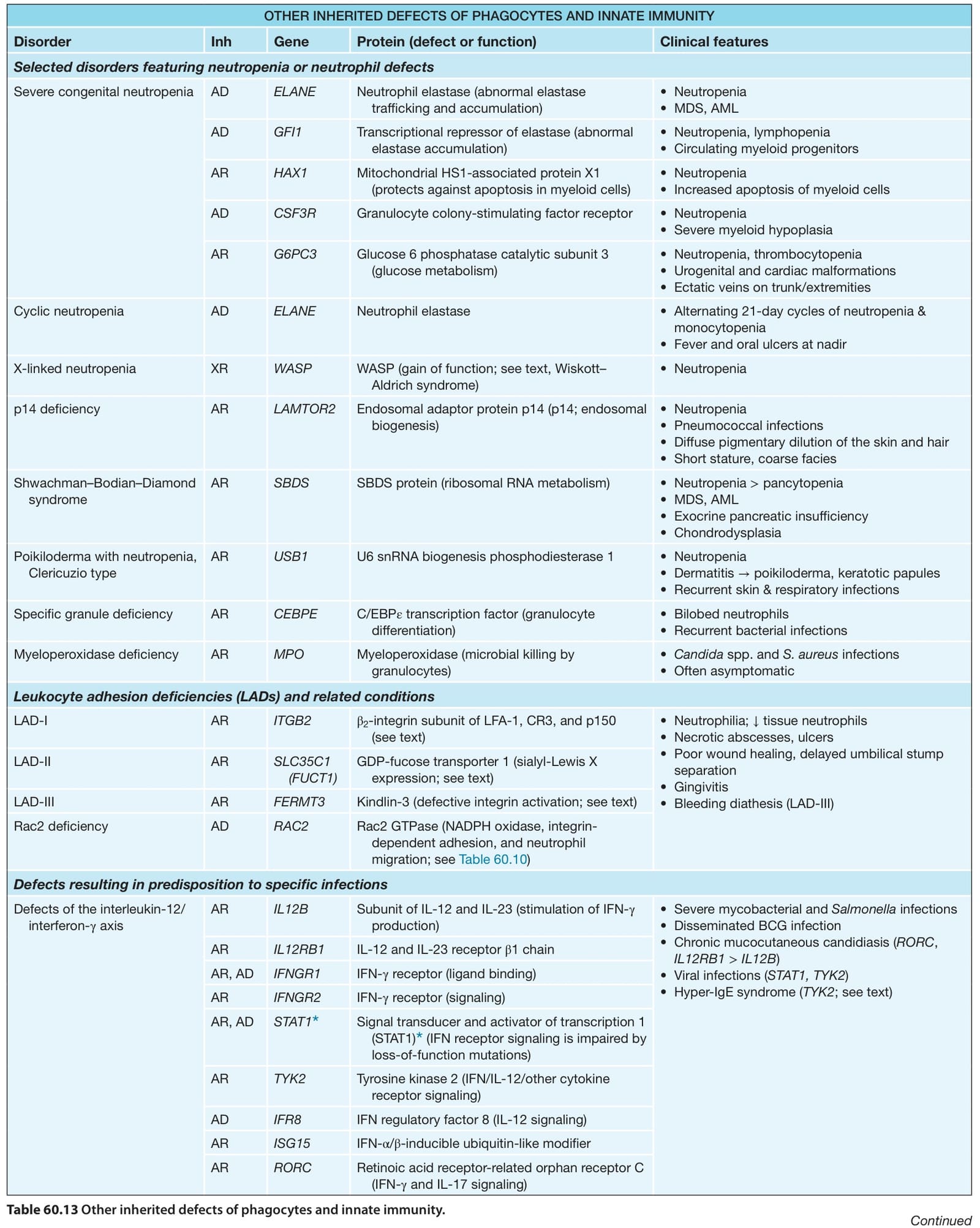
Table 60.13 Other inherited defects of phagocytes and innate immunity.