HYPERIMMUNOGLOBULIN E SYNDROMES
Synonyms: Hyper-IgE syndromes Job syndrome Buckley syndrome
Key features
Characterized by recurrent skin and sinopulmonary tract infections, early-onset eczematous dermatitis, and extremely elevated IgE levels
The classic form, an autosomal dominant disorder due to STAT3 deficiency, features skin infections caused primarily by S. aureus (in particular “cold abscesses”) and, especially during infancy, C. albicans, in addition to a neonatal papulopustular eruption, progressively coarse facies, osteopenia with fractures, scoliosis, and dental abnormalities
Autosomal recessive forms have a broader spectrum of infectious complications
Introduction and History
Hyperimmunoglobulin E syndromes (HIESs) are characterized by the triad of recurrent cutaneous and sinopulmonary infections, dermatitis beginning in infancy or early childhood, and extremely elevated IgE levels. Job syndrome is a subgroup of autosomal dominant (AD) HIES that was originally described in female patients with fair skin, red hair, and hyperextensible joints as well as the typical features of HIES. In 2007, classic AD HIES was found to result from STAT3 deficiency (see Pathogenesis). Since then, multiple additional forms of HIES caused by defects in proteins with roles in STAT3 signaling or other pathways of immune regulation have been identified.
Epidemiology
As noted above, classic HIES is inherited in an AD fashion with variable expression. Additional AD and autosomal recessive (AR) forms of HIES with variable clinical features have also been described.
Pathogenesis
Heterozygous mutations in the gene encoding STAT3 (signal transducer and activator of transcription 3) cause classic AD HIES, commonly referred to as STAT3-HIES. Different combinations of receptorassociated Janus kinases (JAKs) and STAT proteins transduce cytokine signals; upon phosphorylation by JAKs, the latter dimerize, translocate to the nucleus, and activate target genes (see Fig. 128.11). Susceptibility to bacterial and candidal infections in HIES reflects the roles of STAT3-dependent cytokines (e.g. IL-6, IL-21, IL-23) in the differentiation of IL-17-producing CD4+ T cells that help to defend against these organisms (see Fig. 4.10). A lack of stimulation of β-defensin production by IL-22, which is produced by Th17 cells and signals via STAT3, may contribute to the development of infections at epithelial surfaces (e.g. skin, lungs) in STAT3-HIES.
STAT3 is critical to the signaling pathways for both IL-6, which promotes acute phase responses and acts as a pyrogen, and IL-10, an anti-inflammatory cytokine. STAT3 dysfunction provides an explanation for the “cold” abscesses and destructive inflammation (e.g. in the lung) that characterize HIES. Defective generation of IL-10-induced tolerogenic dendritic cells and FOXP3+-induced regulatory T cells may contribute to other inflammatory features of HIES, such as the atopic-like dermatitis and high IgE levels. STAT3 also downregulates differentiation of osteoclasts, and osteoporosis represents a feature of STAT3 deficiency in mice as well as HIES. Lastly, leukocytes from STAT3-deficient individuals produce higher levels of interferon-γ and tumor necrosis factor (TNF) upon stimulation with IL-12 and lipopolysaccharide/interferon-γ, respectively, than do leukocytes from unaffected controls.
Other variants of HIES can also result from disruption of STAT3 signaling, including deficiencies in the following proteins: the IL-6 receptor (AR) and IL-6 signal transducer (gp130; AD or AR); phosphoglucomutase 3 (PGM3; AR), which promotes gp130 expression; ZNF341 (AR), which regulates STAT3 expression; and ERBIN (AD), expression of which is stimulated by STAT3. In general, these disorders have clinical findings that overlap with those of STAT3-HIES, with additional manifestations such as autoimmunity and neurocognitive abnormalities in PGM3 deficiency and craniosynostosis in AR gp130 deficiency.
DOCK8 (dedicator of cytokinesis 8) deficiency is an AR condition with features of HIES (see below). The DOCK8 protein interacts with the Wiskott–Aldrich syndrome protein (WASP) in a complex that links T cell receptors to the actin cytoskeleton. DOCK8 deficiency results in an increase in Th2 cells and production of the pruritogen IL-31, but a decrease in Th1 and Th17 cells.
CADINS (CARD11-associated atopy with dominant interference of NF-κB signaling) results from heterozygous dominant negative mutations in CARD11, which encodes a scaffolding protein involved in lymphocyte receptor signaling. Patients present with the characteristic HIES triad together with additional atopic manifestations and B cell lymphopenia. The features of HIES have also been identified in a subset of patients with homozygous mutations in the gene encoding tyrosine kinase 2 (TYK2), a member of the JAK family. Affected individuals have defects in signaling pathways for IL-12 and interferon-α/β, which lead to susceptibility to mycobacterial and viral infections (see Table 60.13), as well as IL-6 and IL-10.
Clinical Features
Patients with STAT3-HIES typically present during the first month of life with a non-infectious, folliculocentric papulopustular eruption involving the face, scalp, neck, axillae, and diaper area. Chronic candidiasis of the oral mucosa, periungual areas, and nails occurs in ~80% of HIES patients and may represent the initial infectious manifestation during infancy.
Cutaneous staphylococcal infections include impetiginized plaques and retroauricular fissures, folliculitis, furunculosis, abscesses, cellulitis, lymphangitis, and paronychia leading to nail dystrophy. The cutaneous abscesses are often large, and they most commonly involve the neck, scalp (Fig. 60.8A), periorbital area, axillae, and groin. Such lesions are referred to as “cold abscesses” because they are not as red or tender as those that develop in healthy individuals. Patients are often afebrile or have only a low-grade fever. Although the abscesses tend to be staphylococcal, many patients have recurrent skin infections with other organisms, including Str. pyogenes as well as C. albicans.
The eczematous rash of HIES shares many clinical features with atopic dermatitis, including severe pruritus, lichenification, and staphylococcal superinfection. It is almost always present in young children with STAT3-HIES but frequently clears by adolescence. Unlike individuals with atopic dermatitis, patients with STAT3-HIES do not commonly exhibit allergic rhinitis, asthma, or other cutaneous signs of atopy.
Although some STAT3-HIES patients have only cutaneous manifestations, most also develop recurrent bronchitis and pneumonias. S. aureus and Haemophilus influenzae usually cause the pulmonary infections, which may result in empyema, bronchiectasis, and pneumatocele formation. The pneumatoceles tend to persist and become the site of further infections with bacterial or fungal organisms, particularly Aspergillus
spp. Massive hemoptysis occasionally ensues. Pneumocystis jirovecii pneumonia can also occur in infants and children with STAT3-HIES. Other common sites of infection include the ears, oral mucosa, sinuses, and eyes. Visceral infections other than pneumonia are unusual.
STAT3-HIES patients develop progressive facial coarsening with thickened doughy skin, large follicular ostia, pitted scarring, a broad nasal bridge, a wide fleshy nasal tip, deep-set eyes, a prominent forehead, and irregularly proportioned cheeks and jaw (Fig. 60.8B). Osteopenia is frequently present and is associated with an increased risk of fractures of the long bones, ribs, and pelvis. More than half of adolescents and adults with STAT3-HIES have had at least three fractures, often due to unrecognized or minor trauma. Scoliosis occurs in 75% of patients ≥16 years of age, and joint hyperextensibility affects ~70% of patients. Approximately half of individuals with STAT3-HIES have a high-arched palate, and retention of primary teeth due to failure of root resorption and lack of eruption of secondary teeth are characteristic dental manifestations. Several brain abnormalities have been described, including Chiari malformations, lacunar infarctions, and focal hyperintensities in white matter on T2-weighted MRI sequences. Development of coronary artery aneurysms during adulthood has also been reported. HIES is associated with an increased risk of non-Hodgkin lymphoma of B cell origin.
DOCK8 deficiency shares some features with STAT3-HIES, including highly elevated serum IgE levels, peripheral eosinophilia, chronic eczematous dermatitis (Fig. 60.9A), recurrent staphylococcal skin infections (including cold abscesses), respiratory tract infections, and mucocutaneous candidiasis. However, skeletal and dental abnormalities, facial coarsening, and pneumatoceles are not characteristic features. Patients with DOCK8 deficiency are predisposed to the development of warts, molluscum contagiosum, severe herpes simplex and varicella–zoster viral infections, and opportunistic infections (Fig. 60.9B,C). Additional manifestations include asthma, food allergies resulting in anaphylaxis, autoimmunity, cerebral vasculitis, mucocutaneous squamous cell carcinomas, and lymphomas. Up to half of patients die by 20 years of age from infection, malignancy, or CNS disease.
Laboratory Findings and Pathology
HIES patients have markedly elevated serum levels of polyclonal IgE, usually peaking at >2000 IU/ml and sometimes declining during adulthood. Patients develop particularly high levels of anti-staphylococcal and anti-candidal IgE antibodies and often have immediate whealand-flare reactions to a variety of foods and environmental allergens. Many affected individuals also have eosinophilia of the peripheral blood and sputum. Cell-mediated immunity is often abnormal, as manifested by anergy to skin testing and impaired in vitro lymphoproliferative responses to specific antigens. Serum levels of IgG, IgA and IgM are usually normal in STAT3-HIES, as are lymphocyte subsets. In contrast, DOCK8 deficiency represents a combined immunodeficiency that is characterized by lymphopenia with a paucity of CD4+ T cells > CD8+ T cells > B cells, low IgM levels, and variable IgG levels as well as elevated IgE levels and peripheral eosinophilia.
The infantile papulopustular eruption of STAT3-HIES is characterized histologically by eosinophilic folliculitis, eosinophilic spongiosis, and a superficial and deep perivascular infiltrate with abundant eosinophils.
Differential Diagnosis
Current diagnostic criteria for STAT3-HIES include an IgE level >1000 IU/ml and a weighted score of five clinical features (Table 60.14); these characteristics plus a lack of Th17 cells or the detection of a heterozygous STAT3 mutation allow for a probable or definitive diagnosis, respectively.
HIES must be differentiated from a number of other disorders that can feature elevated IgE levels and a cutaneous eruption, including atopic dermatitis, Wiskott–Aldrich syndrome, Netherton syndrome, Omenn syndrome, DiGeorge syndrome, IPEX syndrome, ARPC1B (actin-related protein 2/3 complex subunit 1B) deficiency, Loeys– Dietz syndrome, and GVHD. Atopic dermatitis and Wiskott–Aldrich syndrome are most easily confused with HIES because they present with eczematous dermatitis and staphylococcal infections. However, the development of cold abscesses, recurrent pneumonia, coarse facial features, and osteopenia help to differentiate STAT3-HIES from these disorders, and platelet abnormalities distinguish Wiskott–Aldrich syndrome as well as ARPC1B syndrome. In a series of pediatric patients who had a serum IgE level of >2000 IU/ml, 69% (48/70) had atopic dermatitis and only 8% (6/70) had HIES; no correlation was observed between the IgE level and diagnosis of HIES. Elevated IgE levels, susceptibility to pyogenic infections, and eczematous dermatitis have
A Eczematous dermatitis with prurigo nodularis lesions. B Extensive molluscum contagiosum. C Numerous coalescing facial warts. A, B, Courtesy Edward Cowen, MD.
also been described in patients with prolidase deficiency, an autosomal recessive condition due to PEPD mutations that also features chronic leg ulcers, facial dysmorphism, and intellectual disability. Patients with CGD and myeloperoxidase deficiency develop bacterial and candidal abscesses, but they do not typically have markedly elevated levels of IgE.
In addition to DOCK8 deficiency and the other six conditions noted in Table 60.1, primary immunodeficiencies that result in increased susceptibility to warts include epidermodysplasia verruciformis (see Ch. 79), Netherton syndrome (see Ch. 57), and activated phosphoinositide- 3-kinase δ syndrome (see Table 60.15) as well as deficiencies of serine/ threonine kinase 4 (STK4), GATA2 (see Table 60.13), TNF-like weak inducer of apoptosis (TWEAK; see Table 60.15), coronin 1 A (see Table 60.16), RhoH GTPase, minichromosome maintenance complex component 4 (MCM4; also herpesvirus infections), and capping protein regulator and myosin 1 linker 2 (CARMIL2; also severe molluscum contagiosum). Of note, CD4+ lymphocytopenia can be a prominent feature of DOCK8 deficiency and other wart-prone immunodeficiency syndromes.
Treatment
Treatment of the infectious complications of HIES includes incision and drainage of abscesses as well as therapeutic and prophylactic administration of antibiotics. Interferon-γ has been shown to help control infections, and IVIg may improve the dermatitis, prevent infections, and lower IgE levels. Treatment with dupilumab and omalizumab has been reported to improve the eczematous dermatitis of HIES.
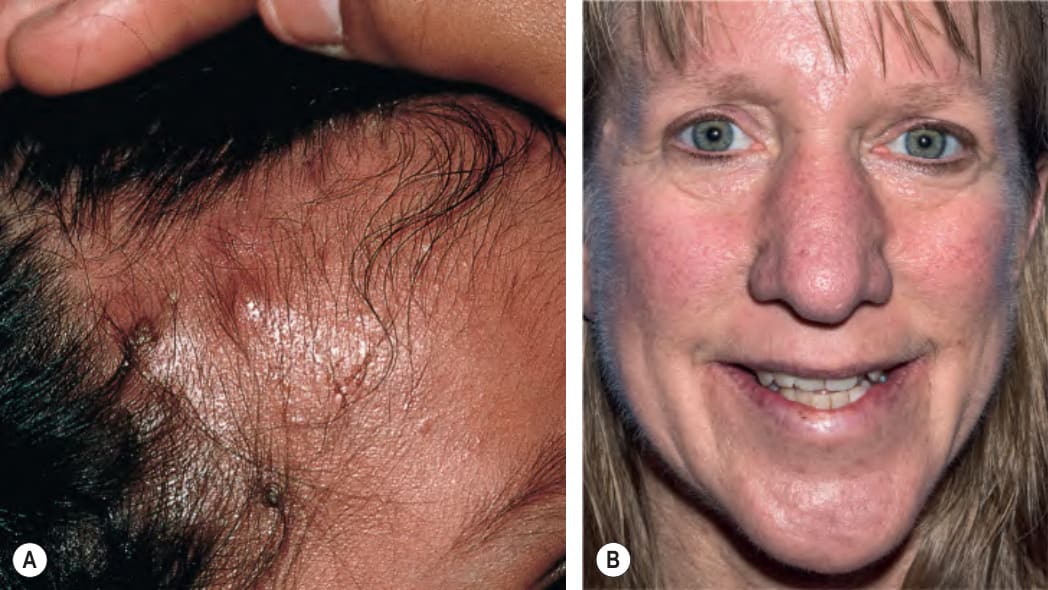
Fig. 60.8 STAT3-hyperimmunoglobulin E syndrome.A Several erythematous, slightly purulent “cold” abscesses are apparent on the forehead and scalp of this infant. B Coarse facial features that developed progressively over time in an affected woman. Note the prominent follicular ostia, doughy skin, and broad nasal bridge. B, Courtesy Edward Cowen, MD.
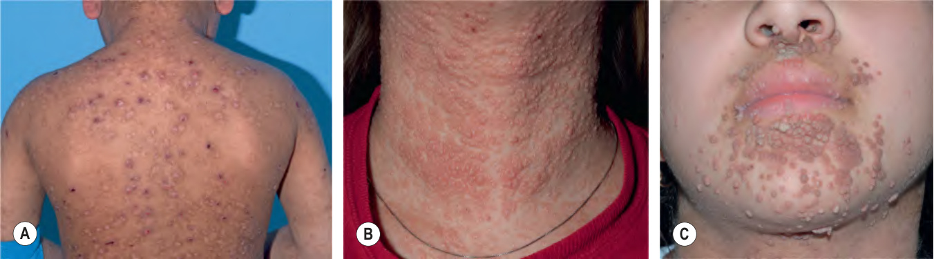
Fig. 60.9 DOCK8 deficiency.
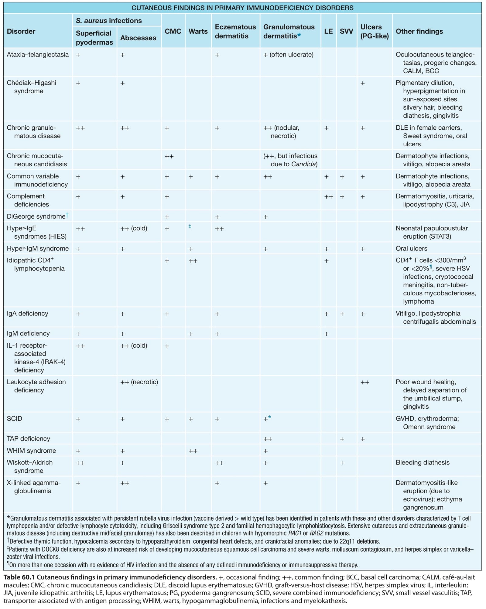
Table 60.1 Cutaneous findings in primary immunodeficiency disorders.+, occasional finding; ++, common finding; BCC, basal cell carcinoma; CALM, café-au-lait macules; CMC, chronic mucocutaneous candidiasis; DLE, discoid lupus erythematosus; GVHD, graft-versus-host disease; HSV, herpes simplex virus; IL, interleukin; JIA, juvenile idiopathic arthritis; LE, lupus erythematosus; PG, pyoderma gangrenosum; SCID, severe combined immunodeficiency; SVV, small vessel vasculitis; TAP, transporter associated with antigen processing; WHIM, warts, hypogammaglobulinemia, infections and myelokathexis.
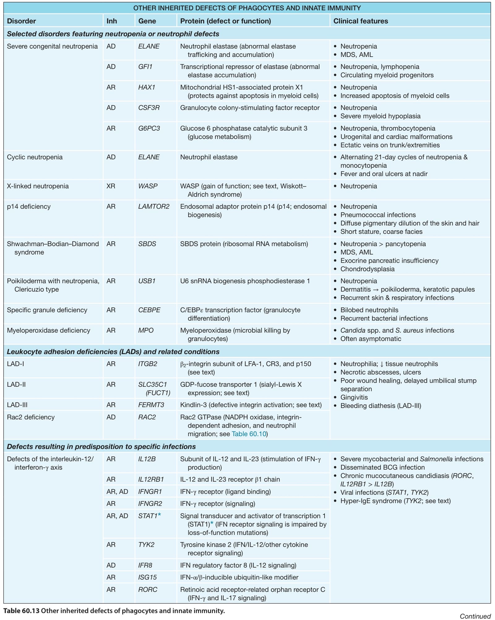
Table 60.13 Other inherited defects of phagocytes and innate immunity.
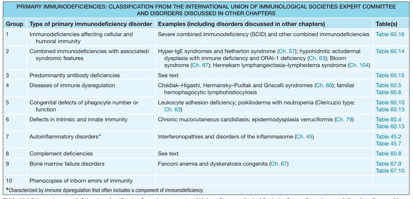
Table 60.14 Diagnostic guidelines for autosomal dominant hyperimmunoglobulin E syndrome (AD-HIES) due to STAT3 mutations.
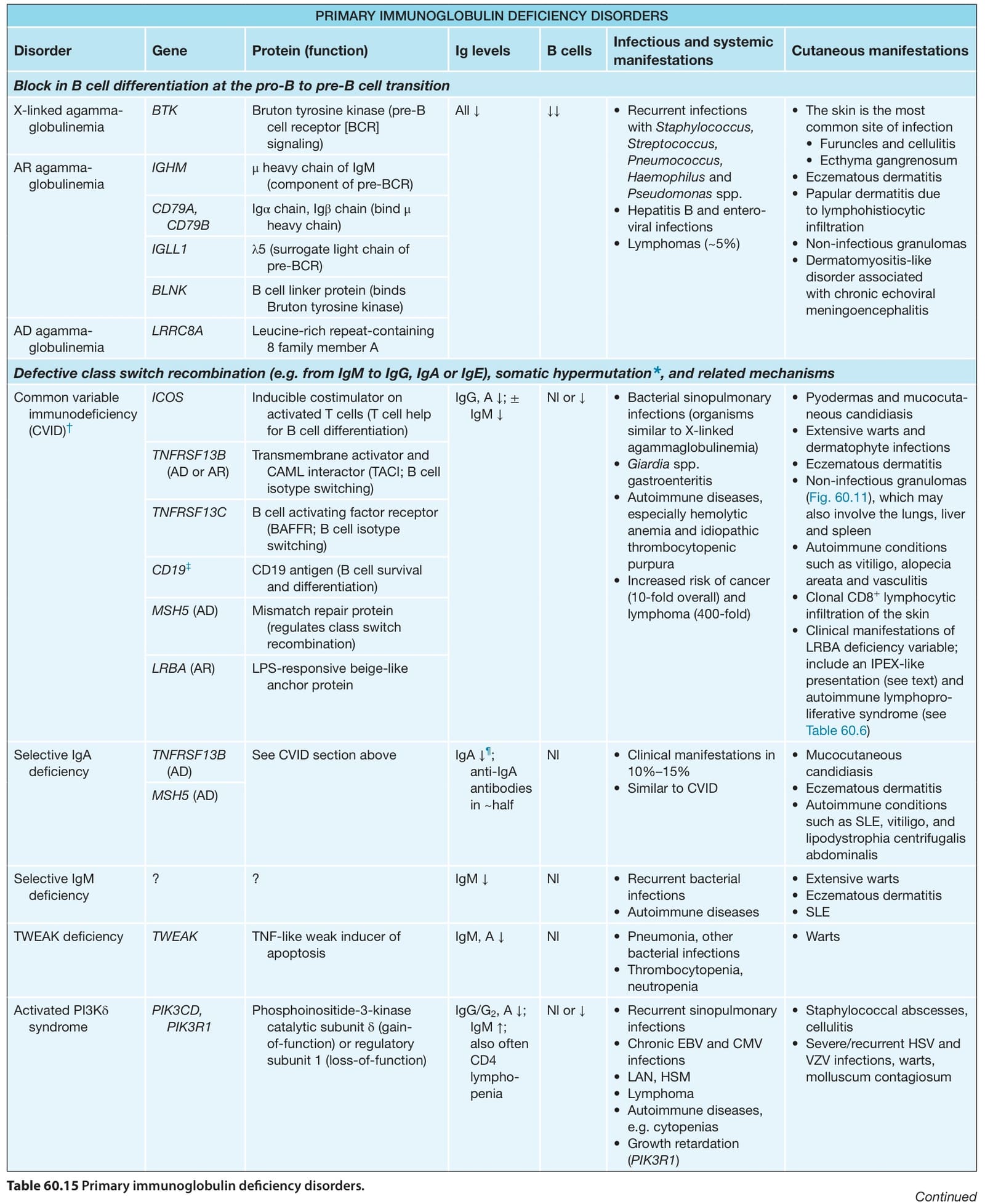
Table 60.15 Primary immunoglobulin deficiency disorders.
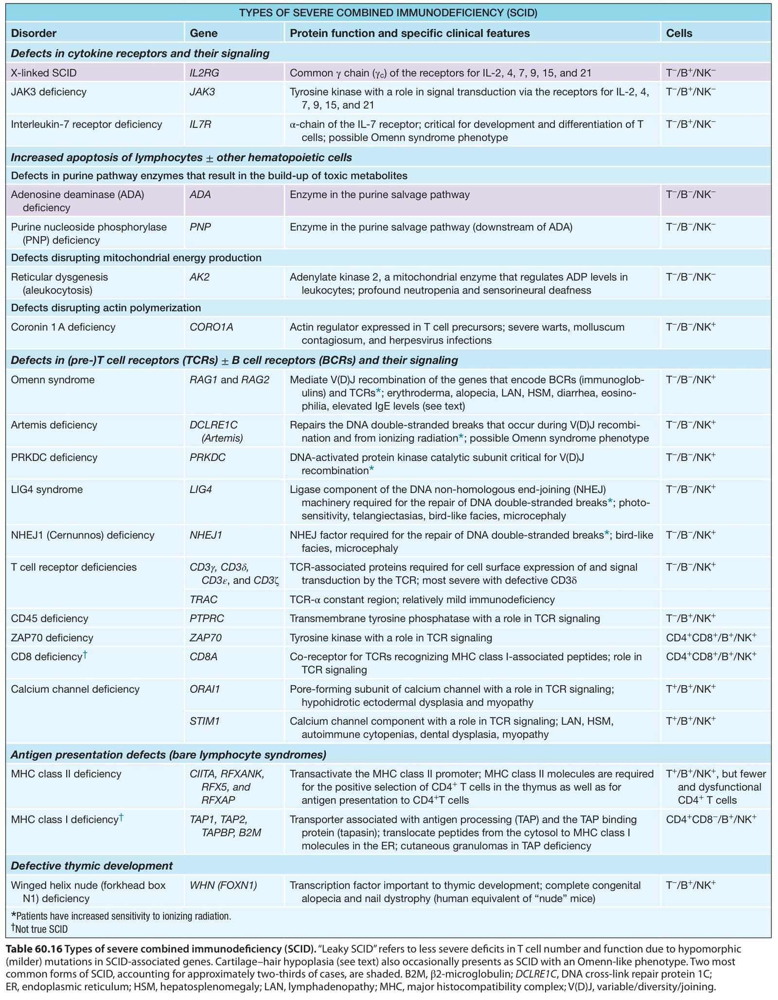
Table 60.16 Types of severe combined immunodeficiency (SCID). “Leaky SCID” refers to less severe deficits in T cell number and function due to hypomorphic (milder) mutations in SCID-associated genes. Cartilage–hair hypoplasia (see text) also occasionally presents as SCID with an Omenn-like phenotype. Two most common forms of SCID, accounting for approximately two-thirds of cases, are shaded. B2M, β2-microglobulin; DCLRE1C, DNA cross-link repair protein 1C; ER, endoplasmic reticulum; HSM, hepatosplenomegaly; LAN, lymphadenopathy; MHC, major histocompatibility complex; V(D)J, variable/diversity/joining.