IMMUNOGLOBULIN DEFICIENCIES
Synonyms: X-linked agammaglobulinemia Bruton agamma globulinemia Bruton–Gitlin syndrome Congenital hypogamma globulinemia X-linked hypogammaglobulinemia
Key features
The inheritance pattern is X-linked recessive in ~90% of patients and autosomal recessive in ~10%
Recurrent bacterial infections begin in the first few years of life
Heterogeneous group of disorders with defects in both humoral and cell-mediated immunity
May manifest during childhood or adulthood, with clinical onset at an average age of 30 years
Variable severity of infectious and autoimmune complications
Most common immunoglobulin deficiency, but usually asymptomatic, with clinical manifestations in only 10%–15% of affected individuals
Bacterial sinopulmonary infections and autoimmune disorders may occur
Usually has an X-linked recessive inheritance pattern, but autosomal recessive forms also exist
Recurrent sinopulmonary and gastrointestinal infections with pyogenic bacteria and opportunistic organisms
Oral ulcerations and verrucae may develop
The most common immunoglobulin deficiency is selective IgA deficiency, overall found in ~1 in 500 persons with an equal sex distribution. Panhypogammaglobulinemia has a prevalence of 1 in 25 000, with X-linked agammaglobulinemia (affecting primarily boys) and common variable immunodeficiency (CVID; no sexual predilection) representing the most frequent forms. CVID has its clinical onset at a mean age of 30 years whereas X-linked agammaglobulinemia often manifests during the first few years of life, after levels of maternal antibodies wane at ~6 months of age. Approximately 15% of patients with CVID have family members with selective IgA deficiency. The pathogenesis and clinical features of immunoglobulin deficiencies are presented in Fig. 60.10 and Table 60.15.
The treatment of hypogammaglobulinemia is antibody replacement by IVIg or subcutaneous immunoglobulins and antibiotic therapy for infections. However, IVIg and other blood products containing IgA should be avoided in individuals with profound IgA deficiency, who may develop anti-IgA antibodies; fatal anaphylactic reactions have been reported. Recalcitrant non-infectious cutaneous and extracutaneous granulomas in patients with CVID may respond to treatment with TNF inhibitors.
Female carriers of X-linked agammaglobulinemia may be detected via examination of B cell X-inactivation patterns, which show skewing towards cells with inactivation of the mutated X chromosome. DNA-based prenatal diagnosis is possible when the genetic defect in affected family members has been identified.
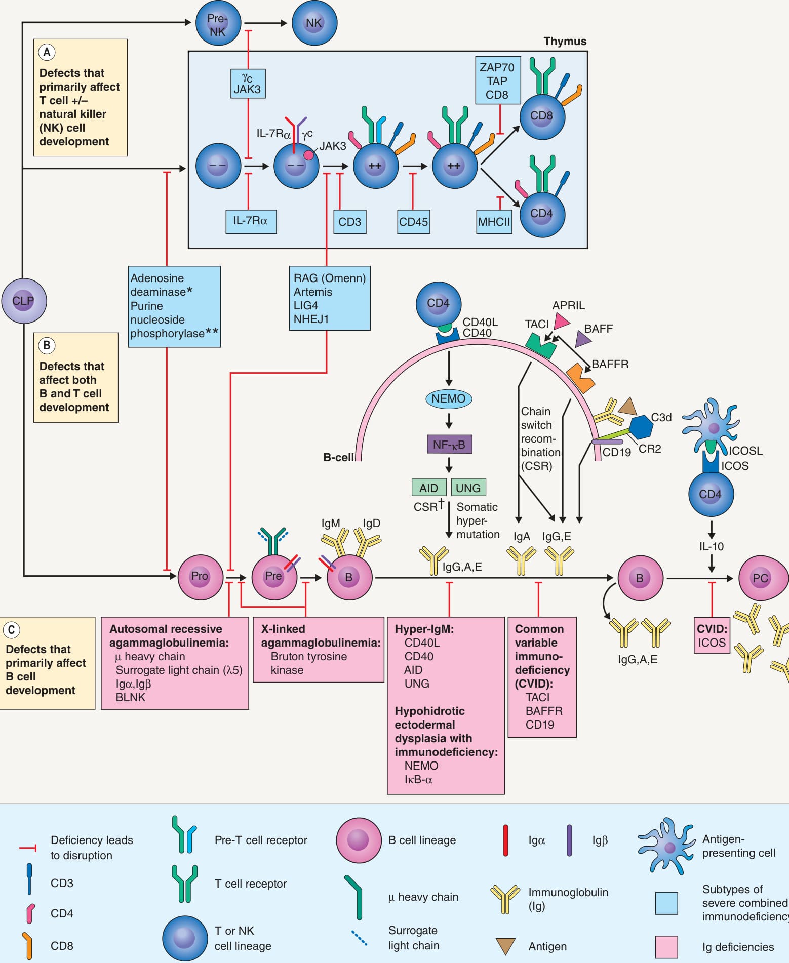
**Fig. 60.10 Molecular defects resulting in immunoglobulin deficiencies and severe combined immunodeficiency (SCID). *Also important for B cell maturation in germinal centers. Affects T cells more than B cells. †Switch from IgM to IgG, IgA, or IgE. AID, activation-induced cytidine deaminase; APRIL, a proliferation-inducing ligand; BAFF(R), B cell activating factor (receptor); BLNK, B cell linker protein (binds Bruton tyrosine kinase); CD40L, CD40 ligand; CLP, common lymphoid precursor; CR2, complement receptor 2; γc, common γ chain; ICOS(L), inducible costimulator on activated T cells (ligand); IκB-α, inhibitor of κB-α; IL, interleukin; IL-7Rα, IL-7 receptor α-chain; MHC, major histocompatibility complex; NEMO, NF-κB essential modulator; NHEJ1, non-homologous end-joining 1 (Cernunnos); NK, natural killer cell; PC, plasma cell; TACI, transmembrane activator and CAML interactor; TAP, transporter associated with antigen processing; UNG, uracil-DNA glycosylase; _ _, double-negative thymocyte; ++, double-positive thymocyte.
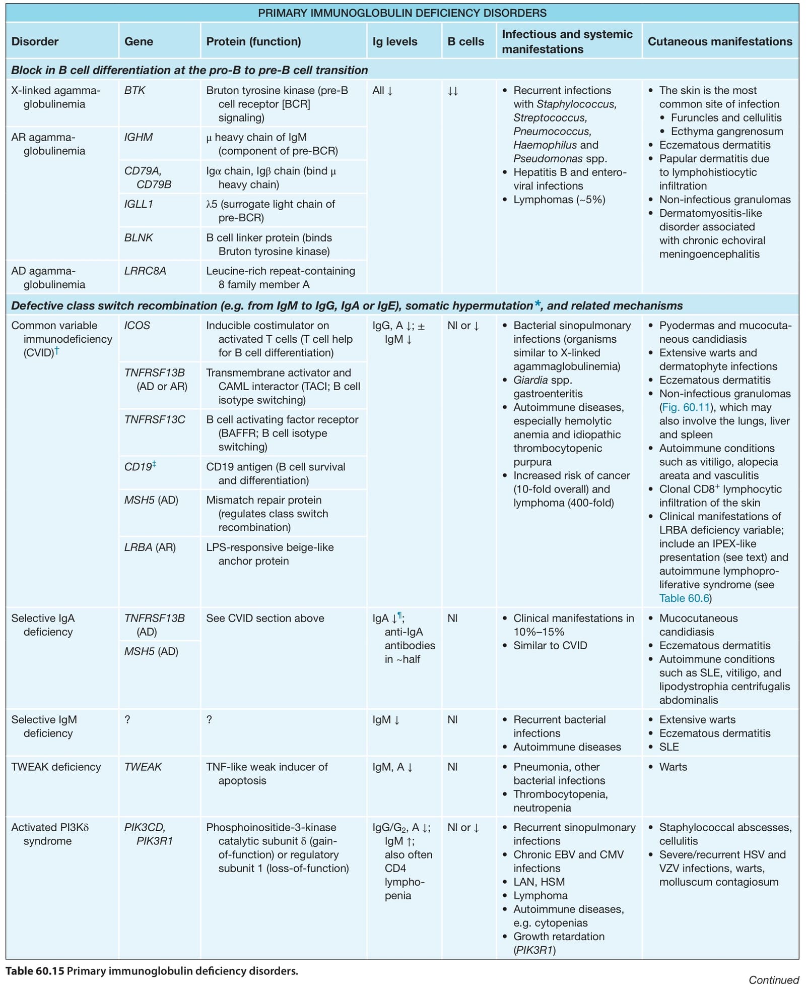
Table 60.15 Primary immunoglobulin deficiency disorders.