LEUKOCYTE ADHESION DEFICIENCY
Key features
Group of autosomal recessive disorders characterized by decreased ability of leukocytes to adhere to the vascular endothelium and migrate to sites of tissue injury and infection
Gingivitis and periodontitis
Necrotic ulcerations clinically resembling pyoderma gangrenosum
Poor wound healing; delayed separation of the umbilical stump
Life-threatening bacterial and fungal infections
Introduction
Leukocyte adhesion deficiency (LAD) comprises three autosomal recessive disorders that affect the ability of neutrophils, monocytes, and lymphocytes to adhere to the vascular endothelium and migrate to sites of infection and tissue injury.
Pathogenesis
Leukocyte adhesion requires a group of cell surface integrins that share the 95-kDa β-subunit known as CD18. This β-subunit can be linked to different α-chains to form distinct cell surface glycoproteins including: (1) lymphocyte function antigen 1 (LFA-1 – CD18 paired with CD11a); (2) complement receptor type 3 (CR3/iC3b receptor/Mac-1 – CD18 paired with CD11b); and (3) p150,95 (complement receptor type 4 – CD18 paired with CD11c). The primary ligand for these glycoproteins is intercellular adhesion molecule-1 (ICAM-1), which participates in the initiation and evolution of localized inflammation in the skin and other tissues (see Fig. 102.10). LAD-I is caused by mutations in ITGB2 which encodes CD18, and it manifests with dysfunction of all three glycoproteins. This leads to profound impairment of leukocyte firm adhesion to the vascular endothelium and mobilization into extravascular sites of inflammation, as well as defective neutrophil and monocyte chemotaxis and phagocytosis.
LAD-II is due to mutations in SLC35C1 (solute carrier family 35 member C1), which encodes the GDP-fucose transporter-1 (FUCT1) that is required for formation of sialyl-Lewis X, the fucosylated ligand for selectins on the surface of leukocytes. This causes a defect in the initial steps of tethering and rolling of leukocytes on the endothelial cell surface via interaction of sialyl-Lewis X on leukocytes with E- or
P-selectin on endothelial cells. These contacts are required for their targeting to sites of infection and inflammation.
In order to undergo firm adhesion and subsequently extravasate from the bloodstream, circulating leukocytes must activate integrins in situ to rapidly increase their affinity and avidity for endothelial ligands. Patients with LAD-III have impaired integrin activation in hematopoietic cells, which leads to impaired leukocyte β- and platelet β-integrins in addition to leukocyte β-integrins as in LAD-I. It is caused by loss-of-function mutations in the fermitin family member 3 gene (FERMT3) that encodes kindlin-3, an effector of integrin activation in hematopoietic cells.
Clinical Features
The most common manifestation of LAD is gingivitis with periodontitis, which often leads to tooth loss and alveolar bone resorption and is thought to be related to excessive production of interleukin-17. Minor injuries to the skin may rapidly expand to form large, chronic ulcerations that can resemble “burnt out” pyoderma gangrenosum (Fig. 60.13). Poor wound healing also results in paper-thin, atrophic scars. A history of delayed separation of the umbilical stump represents a clue to the diagnosis.
Patients with LAD develop recurrent otitis media, pneumonia, and cutaneous infections caused by pyogenic bacteria. The latter
often present as cellulitis and necrotic abscesses with relatively little production of purulent material, typically located in the perianal area or on the face. Life-threatening bacterial, fungal, or (less frequently) viral infections may also occur. More than three-quarters of patients with severe LAD-I (<1% of normal CD18 levels) die within the first 5 years of life, whereas approximately half of patients with moderate disease (1%–10% of normal CD18 levels) live to age 30 years.
Additional clinical manifestations of LAD-II include intellectual disability, microcephaly, hypotonia, short stature, and facial dysmorphism. Patients with LAD-III often present with a bleeding tendency as well as variable osteopetrosis and hepatosplenomegaly. Dermal hematopoiesis with a “blueberry muffin” appearance secondary to myelodysplastic syndrome has been described in infants and children with LAD-III.
Pathology
Individuals with LAD have marked peripheral blood neutrophilia (5–20 times normal levels). Although cutaneous ulcerations may resemble pyoderma gangrenosum clinically, histologic examination reveals a relative paucity of tissue neutrophils. Flow cytometric analysis reveals markedly decreased leukocyte CD18 expression in individuals with LAD-I. Patients with LAD-II have the Bombay red blood cell type due to a lack of the H blood group antigen, and those with LAD-III have abnormal platelet aggregation due to defective activation of β-integrin and less often have anemia.
Differential Diagnosis
A mutation in the gene encoding the Rac2 GTPase, which has a role in phagocyte NADPH oxidase activation as well as integrin-dependent adhesion and neutrophil migration, can lead to clinical features similar to those of LAD, including delayed separation of the umbilical stump, poor wound healing, neutrophilia, and recurrent perirectal abscesses with an absence of pus (see Tables 60.10 & 60.13).
Treatment
Soft tissue infections in LAD patients require prolonged courses of antimicrobial therapy and, in some cases, surgical debridement. Meticulous dental hygiene is important in reducing the severity of the periodontitis. Granulocyte transfusions have potential benefit in LAD-I and -III, and oral administration of fucose may improve immune function in patients with LAD-II. In LAD-I, healing of chronic oral and cutaneous ulcers following administration of IVIg or ustekinumab has been reported. Hematopoietic stem cell transplantation represents the only definitive therapy for LAD, but it is limited by infectious complications and GVHD.
Two patients with severe LAD-I were treated without prior conditioning with an infusion of autologous hematopoietic stem cells that had been corrected ex vivo with a retroviral vector encoding CD18, but circulating CD18+ cells persisted for <2 months. However, canine LAD-I has been successfully treated with non-myeloablative conditioning followed by infusion of autologous hematopoietic stem cells transduced ex vivo by a CD18-expressing foamy virus vector, which has less potential for genotoxicity than a retroviral vector; this led to complete reversal of the LAD phenotype that was sustained for more than 4 years. Preclinical studies have also investigated lentiviral-mediated gene therapy for LAD-I, with correction of the phenotype in a mouse model.
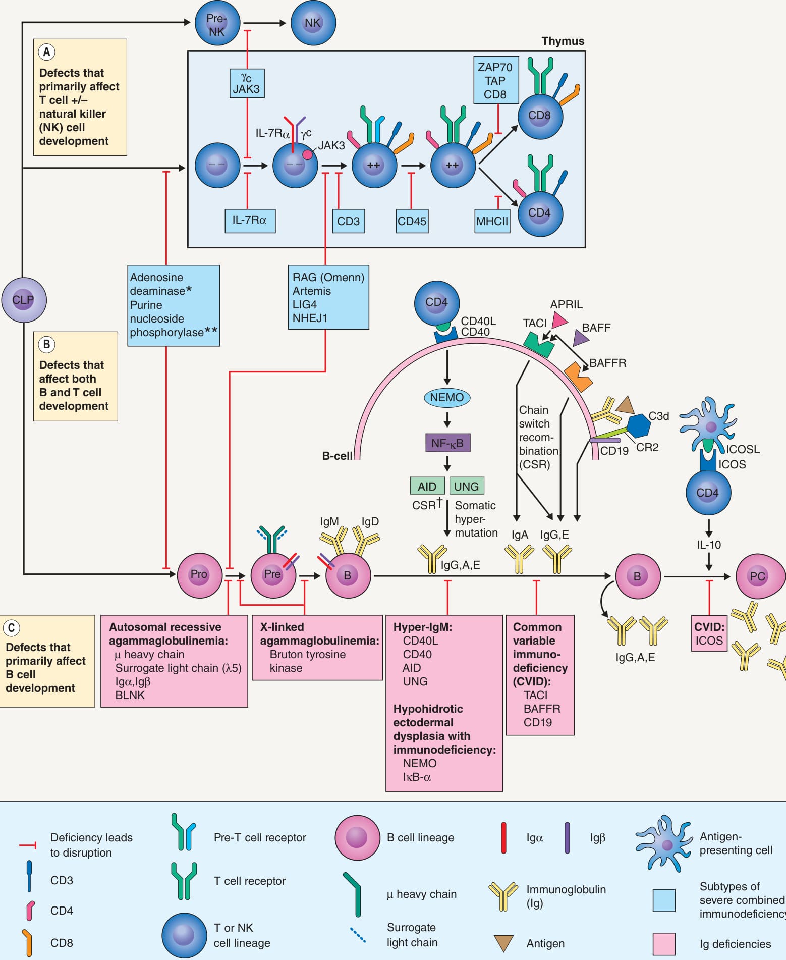
**Fig. 60.10 Molecular defects resulting in immunoglobulin deficiencies and severe combined immunodeficiency (SCID). *Also important for B cell maturation in germinal centers. Affects T cells more than B cells. †Switch from IgM to IgG, IgA, or IgE. AID, activation-induced cytidine deaminase; APRIL, a proliferation-inducing ligand; BAFF(R), B cell activating factor (receptor); BLNK, B cell linker protein (binds Bruton tyrosine kinase); CD40L, CD40 ligand; CLP, common lymphoid precursor; CR2, complement receptor 2; γc, common γ chain; ICOS(L), inducible costimulator on activated T cells (ligand); IκB-α, inhibitor of κB-α; IL, interleukin; IL-7Rα, IL-7 receptor α-chain; MHC, major histocompatibility complex; NEMO, NF-κB essential modulator; NHEJ1, non-homologous end-joining 1 (Cernunnos); NK, natural killer cell; PC, plasma cell; TACI, transmembrane activator and CAML interactor; TAP, transporter associated with antigen processing; UNG, uracil-DNA glycosylase; _ _, double-negative thymocyte; ++, double-positive thymocyte.
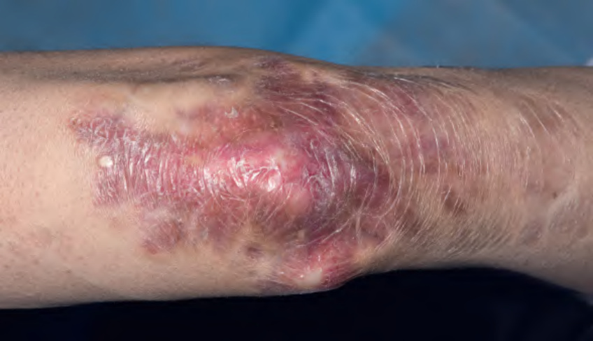
Fig. 60.11 Persistent granulomatous plaques in a patient with common variable immunodeficiency (CVID).Courtesy Edward Cowen, MD.
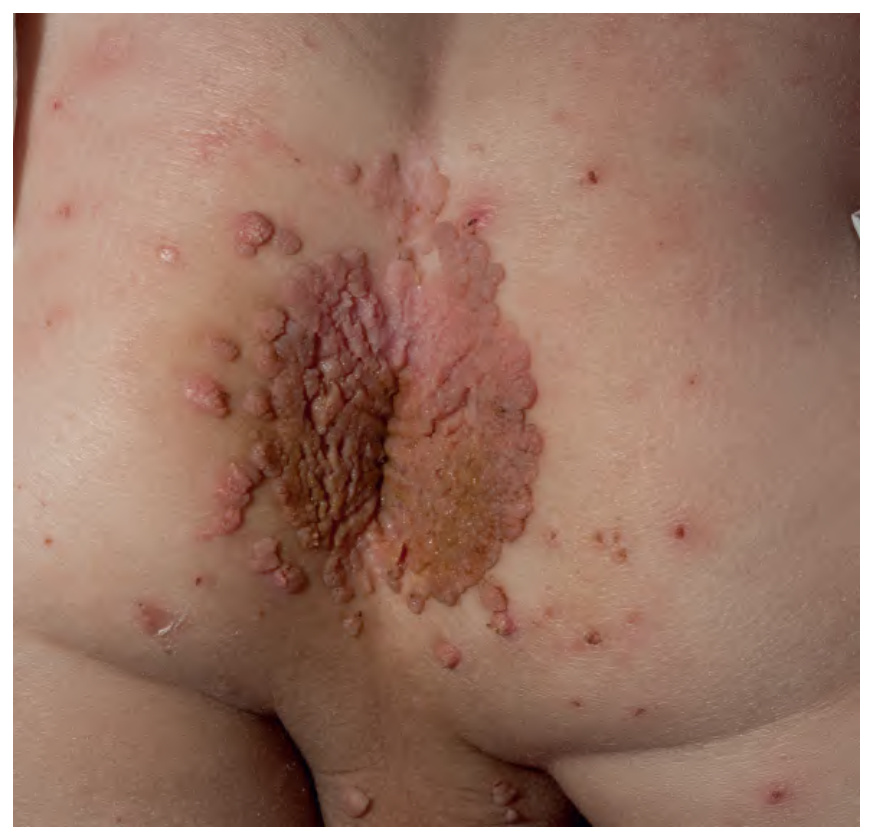
Fig. 60.12 Extensive, recalcitrant perianal warts in a child with WHIM syndrome.Courtesy Edward Cowen, MD.
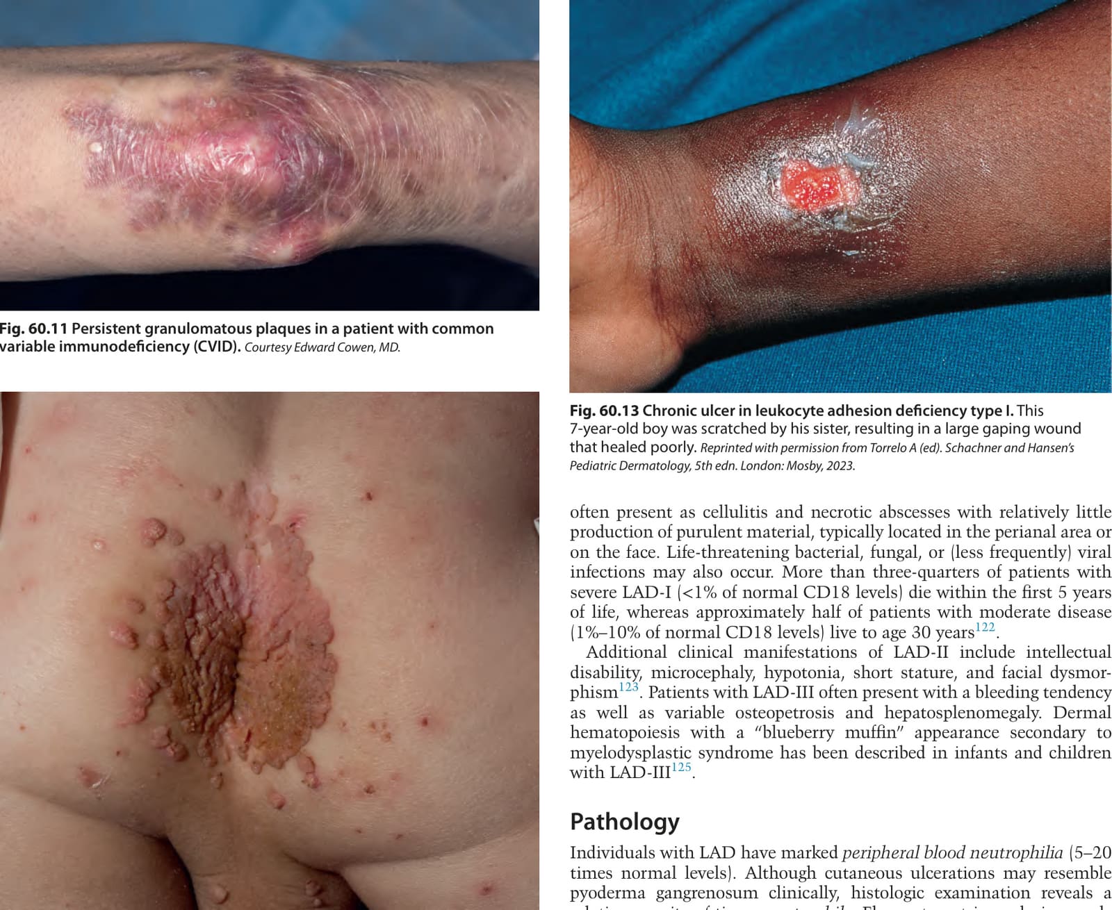
Fig. 60.13 Chronic ulcer in leukocyte adhesion deficiency type I. This 7-year-old boy was scratched by his sister, resulting in a large gaping wound that healed poorly. Reprinted with permission from Torrelo A (ed). Schachner and Hansen’s Pediatric Dermatology, 5th edn. London: Mosby, 2023.
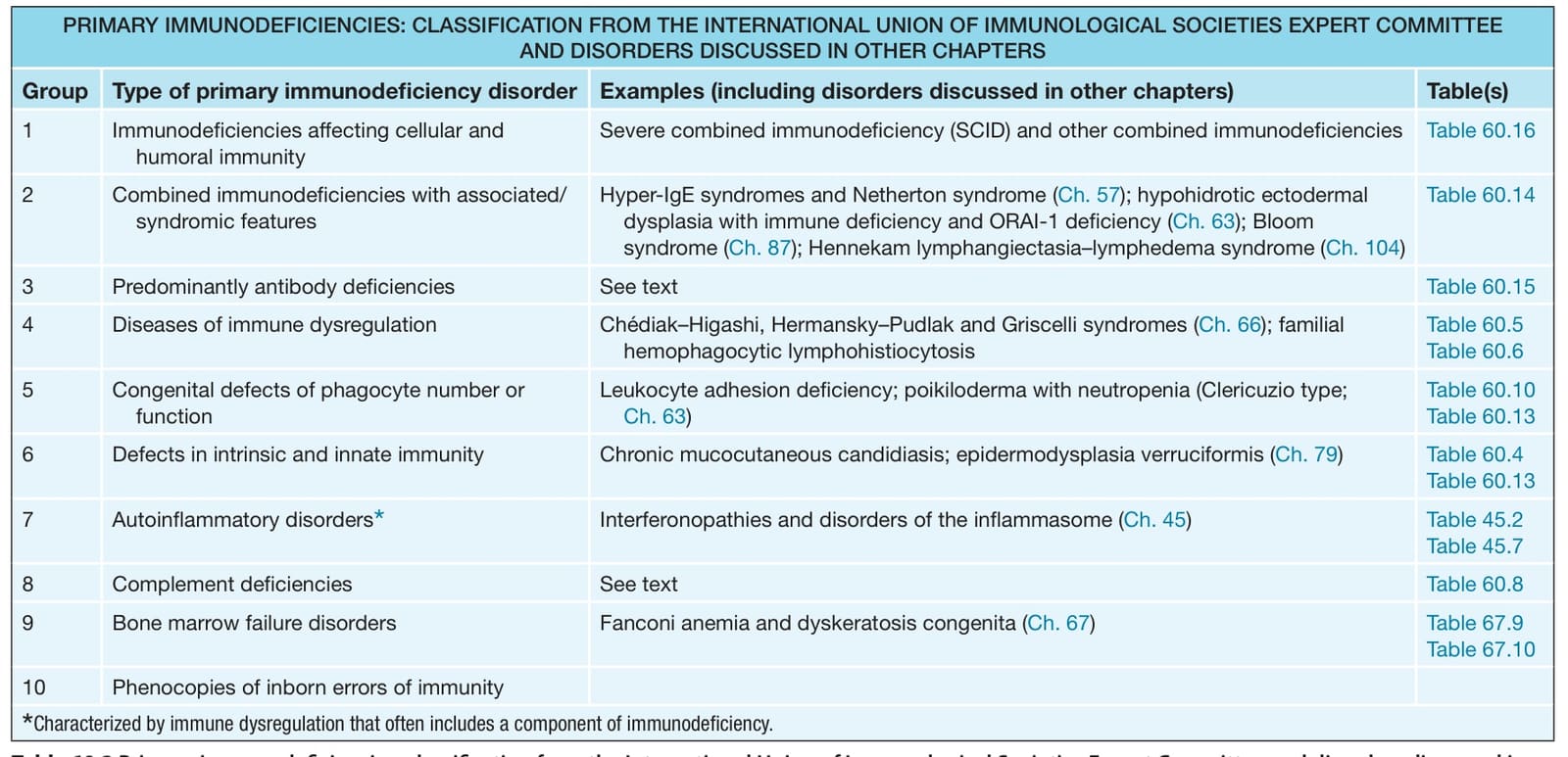
Table 60.10 Genetic defects affecting components of the phagocyte NADPH oxidase. CGD, chronic granulomatous disease; CYBC1, cytochrome b-245 chaperone 1; Eros, essential for reactive oxygen species; NCF, neutrophil cytosolic factor; RhoGDI, Rho GDP-dissociation inhibitor.
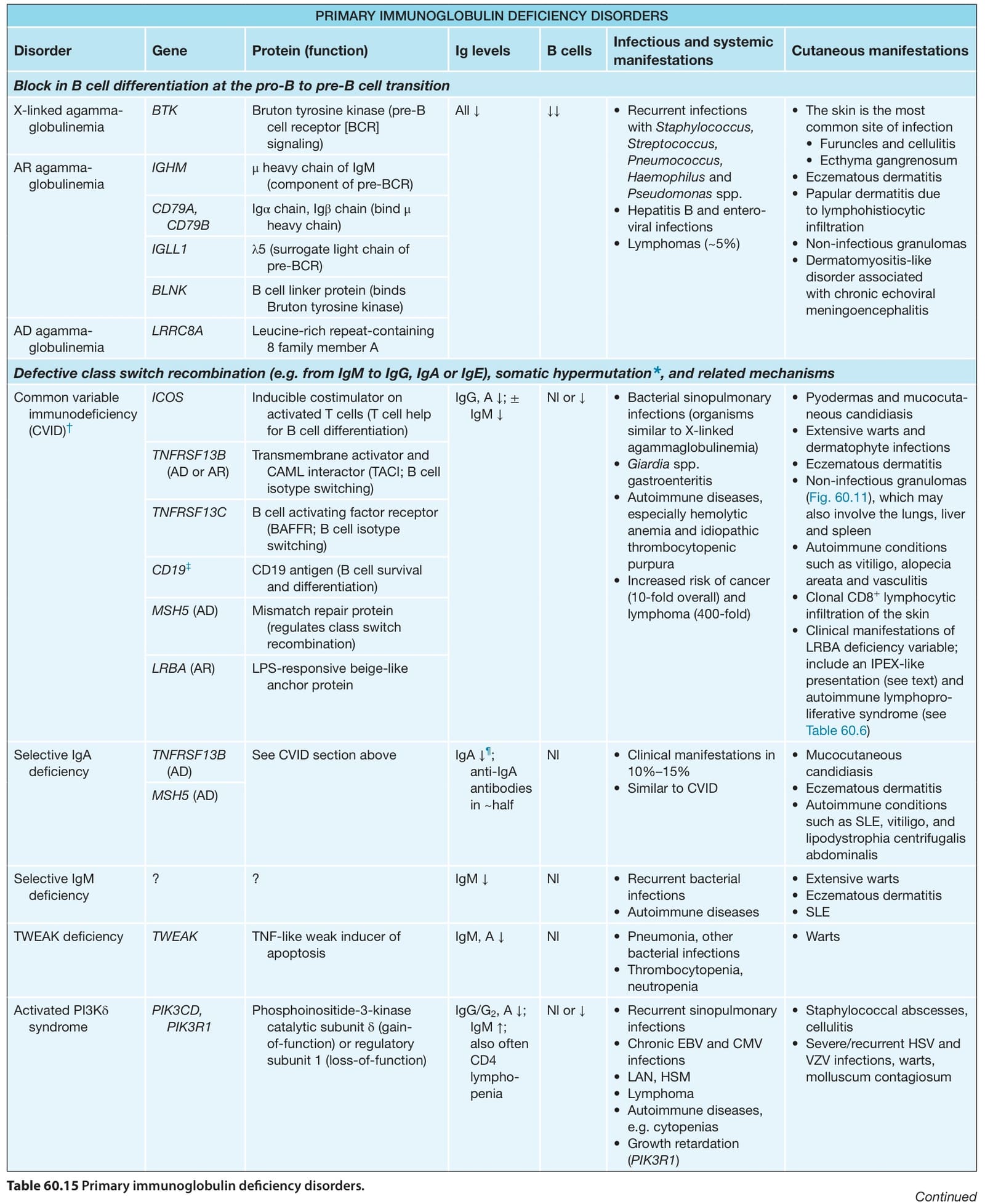
Table 60.15 Primary immunoglobulin deficiency disorders.