WISKOTT–ALDRICH SYNDROME
Synonyms: Wiskott–Aldrich–Huntley syndrome Eczema–thrombocytopenia–immunodeficiency syndrome
Key features
X-linked recessive immunodeficiency disorder characterized by the classic triad of atopic-like dermatitis, a bleeding tendency due to microthrombocytopenia, and recurrent sinopulmonary infections
Onset of bacterial infections is early in life, with later development of viral and P. jirovecii infections
Patients are also at risk of autoimmune diseases and lymphomas
Introduction
Wiskott–Aldrich syndrome (WAS) is characterized by recurrent pyogenic infections, bleeding due to thrombocytopenia and platelet dysfunction, and recalcitrant eczematous dermatitis. The full triad develops in a minority of patients, with platelet abnormalities representing the most constant feature.
Epidemiology
Most patients with this X-linked recessive immunodeficiency are boys, but girls are occasionally affected in the setting of non-random X-chromosome inactivation or homozygosity for a mild mutation. WAS has an incidence of ~1 in 250 000 live male births in European populations; it is less common in Black individuals and Asians.
Pathogenesis
WAS is caused by loss-of-function mutations in WASP, which is constitutively expressed in hematopoietic cell lineages. The WAS protein (WASP) controls the assembly of actin filaments, which are required for microvesicle and pro-platelet formation as well as T cell activation and polarization toward antigen-presenting cells. Loss-of-function WASP mutations also cause isolated X-linked recessive thrombocytopenia, while gain-of-function WASP mutations can lead to X-linked congenital neutropenia.
Abnormal T cell function is central to the immunologic defect in WAS, with resultant diminution of both cell-mediated and humoral responses. Although the number of T cells in WAS patients has been observed to decrease with age, affected infants also have reduced numbers of T cells (especially naive T cells) and B cells. This suggests that impaired lymphocyte maturation and output from the thymus are central to the immunodeficiency of WAS. WASP function is required for the activation of regulatory T cells, helping to explain the autoimmune manifestations of WAS. Defects in B cell migration, differentiation, and organization into germinal centers have roles in the impaired humoral immunity of WAS, with reduced expression of the CD21 and CD35 complement receptors involved in antigen capture and presentation also potentially contributing to autoimmunity. Although NK cells are not decreased in number, their cytolytic activity is abnormal in WAS. Lastly, small, structurally abnormal platelets with a reduced half-life (due in part to splenic destruction) account for the thrombocytopenia of WAS.
Clinical Features
Because thrombocytopenia and platelet dysfunction are present from birth, the first clinical signs of WAS are usually petechiae and ecchymoses of the skin and oral mucosa. Spontaneous bleeding from the oral cavity, epistaxis, hematemesis, melena, and hematuria are commonly observed.
The dermatitis of WAS typically develops during the first few months of life, and it fulfills the diagnostic criteria for atopic dermatitis (see Table 12.1). The face, scalp, and flexural areas are most severely affected, although patients often have widespread involvement with progressive lichenification. Exfoliative dermatitis occasionally develops. Excoriated areas frequently have serosanguineous crusts and petechiae (Fig. 60.15). Secondary bacterial infections are common, as are eczema herpeticum and molluscum contagiosum.
Recurrent bacterial infections begin during the first 3 months of life, coincident with diminishing levels of placentally transmitted maternal antibodies. These infections include otitis externa and media, pneumonia, sinusitis, conjunctivitis, furunculosis, meningitis, and septicemia. Encapsulated bacteria such as Str. pneumoniae, H. influenza, and Neisseria meningitidis are the predominant organisms. With advancing age, there is increased susceptibility to infections with viruses (e.g. herpes simplex) and P. jirovecii.
Most children with WAS develop at least one autoimmune disease, most commonly cutaneous small vessel vasculitis (often associated with painful edema), autoimmune cytopenias, arthritis, inflammatory bowel disease, and cerebral vasculitis. IgE-mediated conditions such as urticaria, food allergies, and asthma also occur with increased frequency. Lymphomas develop at a mean age of 10 years in ~15% of WAS patients who do not undergo successful HSCT, especially those with a history of autoimmune disease. Non-Hodgkin lymphomas predominate, most often diffuse large B cell types with extranodal and brain involvement as in AIDS-related lymphomas.
The median survival for WAS patients who do not receive a HSCT is ~15 years, with infection (50%), bleeding (25%), and malignancy (25%) representing the most common causes of death.
Laboratory Findings and Pathology
The most frequent laboratory abnormalities in WAS are persistent thrombocytopenia (platelet counts <70 000/mm) and low mean platelet volume (<5.0 fl). WAS patients may also have lymphopenia and eosinophilia. Serum IgM and IgG levels are typically low, while IgA, IgE, and IgD levels tend to be elevated. Antibody responses to polysaccharide antigens are markedly diminished. Delayed-type hypersensitivity skin test reactions are usually absent, and responses to mitogens are often depressed, especially in older patients. Monocyte and neutrophil chemotaxis may also be defective.
Assessment of WASP expression in peripheral blood mononuclear cells by flow cytometry-based assays or immunoblot analysis can be performed in patients suspected to have WAS prior to mutational analysis. Patients with a complete lack of WASP expression in circulating cells tend to have a more severe phenotype.
Differential Diagnosis
Although several other immunodeficiency syndromes feature eczematous dermatitis together with susceptibility to infection (see HIES
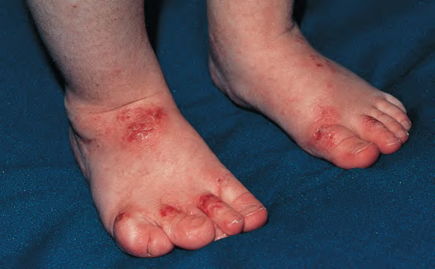
Fig. 60.15 Severe atopic dermatitis in an infant with Wiskott–Aldrich syndrome. This young boy was successfully treated with bone marrow transplantation, and his dermatitis virtually cleared owing to the T cell engraftment. Reprinted with permission from Torrelo A (ed). Schachner and Hansen’s Pediatric Dermatology, 5th edn. London: Mosby, 2023.
Differential Diagnosis), WAS can usually be differentiated by the bleeding tendency and laboratory evidence of microthrombocytopenia. Of note, deficiency of the actin-related protein 2/3 complex subunit 1b (ARPC1B), which interacts with WASP, presents with overlapping features including eczematous dermatitis, thrombocytopenia (with variably sized platelets), recurrent infections, autoimmune manifestations, vasculitis, and colitis.
An approach to the differential diagnosis of infantile erythroderma is presented in Fig. 10.11.
HSCT is the treatment of choice for patients with WAS. Full engraftment results in normal platelet numbers and function, restoration of immunologic status, and clearance of the dermatitis. Affected children who receive a HSCT have an overall survival rate of >90%, with lower rates in those transplanted at >5 years of age with an unrelated donor. Gene therapy using lentivirally transduced, WASP-reconstituted, autologous CD34+ cells can lead to sustained resolution of the dermatitis and susceptibility to infection as well as improvement in the bleeding diathesis and autoimmunity, without the high risk of leukemic transformation that was observed with utilization of a retroviral vector.
Prophylactic use of antibiotics can decrease the risk of fatal infections in WAS patients. IVIg therapy also helps to prevent infections and may potentially improve the dermatitis. Topical corticosteroids remain a mainstay of treatment for the latter. Although splenectomy may reduce bleeding complications in patients with recurrent severe hemorrhage, it further increases the risk of infections with encapsulated organisms. Platelet transfusions may be given prior to surgery and in the setting of life-threatening hemorrhage. Systemic corticosteroids, rituximab, and other immunosuppressive agents may be utilized for autoimmune disorders.
Genetic counseling is important for the sisters and other female relatives of WAS patients. Female carriers may be detected by genetic analysis or detection of selective inactivation of the abnormal X chromosome in lymphocytes and platelets. Prenatal diagnosis can be performed by mutational analysis.