Incontinentia Pigmenti
Synonym:▪ Bloch–Sulzberger syndrome
Key features
An X-linked dominant disorder with skin lesions that follow the lines of Blaschko
Four cutaneous stages: (1) inflammatory/vesicular; (2) verrucous; (3)
hyperpigmented; and (4) hypopigmented/atrophic
Other ectodermal abnormalities may be present, e. g. alopecia, nail dystrophy, pegged or missing teeth
May also have ocular (~20–40%) and neurologic (~30%)
involvement
Caused by mutations in IKBKG (NEMO)
Introduction
Incontinentia pigmenti (IP) is a multisystem disorder with an estimated birth prevalence of 1 : 140 000 and a female : male ratio of ~20 : 1. Patients may present to neonatologists, neurologists, ophthalmologists, or dentists as well as dermatologists. However, the diagnosis typically rests on recognition of the cutaneous findings17b. The name refers to the pathologic finding of pigmentary incontinence, i.e. dermal melanophages, in the third stage of the disease. The linear skin lesions reflect functional mosaicism secondary to X inactivation (lyonization).
History
Although Garrod probably first described IP in 1906, Bloch’s 1926 report of a “previously unreported pigment disorder” and that 2 years later by Sulzberger are remembered eponymously. In 2000, the International Incontinentia Pigmenti Consortium established that IP is caused by mutations in IKBKG, which encodes inhibitor of nuclear factor κB kinase regulatory subunit γ, also known as NEMO (NF-κB essential modulator).
Genetics
IP is an X-linked dominant disorder, usually antenatally lethal in boys, that is caused by mutations in IKBKG located at Xq28. Approximately 80% of patients have a common large deletion of exons 4–10 that tends to arise during paternal meiosis (male gametogenesis). Occasional occurrences of clinically typical IP in boys are attributed to Klinefelter syndrome (~10% of male patients) or to genomic mosaicism due to a half-chromatid or postzygotic NEMO mutation (Table 62.4). In the latter patients, the
distribution of the skin lesions may be relatively limited. A large genetic biobank has been established to facilitate IP research19a.
Pathogenesis
The NEMO protein is a subunit of a kinase that activates NF-κB, a transcription factor that protects against TNF-α-induced apoptosis (Fig. 62.4). Defective linear ubiquitination has been described in association with a truncated NEMO protein19b. The idea of IP as a pro-apoptotic state explains male lethality, destruction of epidermal cells, and progressive replacement of cells expressing the mutant X chromosome by those expressing the normal allele. In one report, a liveborn boy whose mother had classic IP died hours after birth due to lethal hematopoietic and immunologic disturbances. Milder hypomorphic NEMO mutations are responsible for hypohidrotic ectodermal dysplasia with immune deficiency (HED-ID) in boys, whose mothers often have relatively subtle features of IP (see Table 62.4 and Ch. 63).
Clinical features
IP typically presents with linear erythema and vesicles (stage 1) during the first few weeks to months of life in an otherwise healthy baby girl (see Fig. 34.13). Vesiculobullous lesions are most common on the limbs and scalp, frequent on the trunk, and rare on the face (Fig. 62.5A). They resolve within days to weeks and are often replaced by verrucous linear plaques (stage 2) that favor the extremities (Fig. 62.5B) and usually disappear later in infancy. Streaks and swirls of reticulate grayish-brown hyperpigmentation (stage 3) develop next and have a predilection for the trunk and intertriginous sites, with scalloped edges that are thought to reflect the growth of normal keratinocytes into affected skin (Fig. 62.5B, C). Gray-brown dots in a linear array represent a characteristic dermoscopic finding of stage 3 IP. The inflammatory sequence sometimes recurs within pigmented areas during later infancy or childhood together with intercurrent febrile illnesses. In addition, acral keratotic nodules (including subungual lesions) occasionally arise after puberty (Fig. 62.5D). The hyperpigmented streaks tend to fade by adolescence, although a few areas of slate-gray pigmentation may persist lifelong. From puberty onward, linear hypopigmented bands lacking hair and sweat glands (stage 4) appear on the posterior aspects of the limbs (especially the calves; Fig. 62.5E) and may be the only stigmata of the disease during adulthood. Individual stages of IP may be absent or overlap. Extracutaneous manifestations of this condition are listed in Table 62.5.
Pathology
The early inflammatory phase of IP demonstrates eosinophilic spongiosis and scattered dyskeratotic keratinocytes. The epidermis of verrucous lesions is acanthotic with hyperkeratosis and foci of
dyskeratosis. In stage 3, there is pigmentary incontinence as well as variable vacuolization of basal keratinocytes, whereas stage 4 is characterized by a thinned epidermis and dermis devoid of adnexa.
Differential diagnosis
Neonatal IP can usually be distinguished from infectious conditions (e.g. herpes zoster, varicella, herpes simplex viral infections) by the wellbeing of the child and characteristic patterning of the skin lesions. Peripheral eosinophilia and leukocytosis are common in neonates with IP, and histologic examination can confirm the diagnosis. In Blaschko-linear hyper- or hypopigmentation due to pigmentary mosaicism, there is no preceding inflammatory phase, the hyperpigmentation is epidermal, and adnexa are typically normal.
Treatment
Baseline and longitudinal ophthalmologic (especially during infancy) and neurologic evaluations represent the most important aspects of IP management early in life17b. Prompt laser therapy can reduce the likelihood of visual sequelae in patients with retinal vasculopathy23a. MRI and electroencephalogram are indicated for infants with neurologic symptoms, and European consensus guidelines also recommend a brain MRI at ~2 years of age in children without neurologic manifestations17b. Initial dental assessment is recommended at 2-3 years of age, with further intervention by specialists as indicated. The mother should be examined for subtle atrophic streaks, usually most visible on the calves, and genetic testing can assist with family planning. Rare cases of transmission from a mosaic father to a daughter have been reported23b.
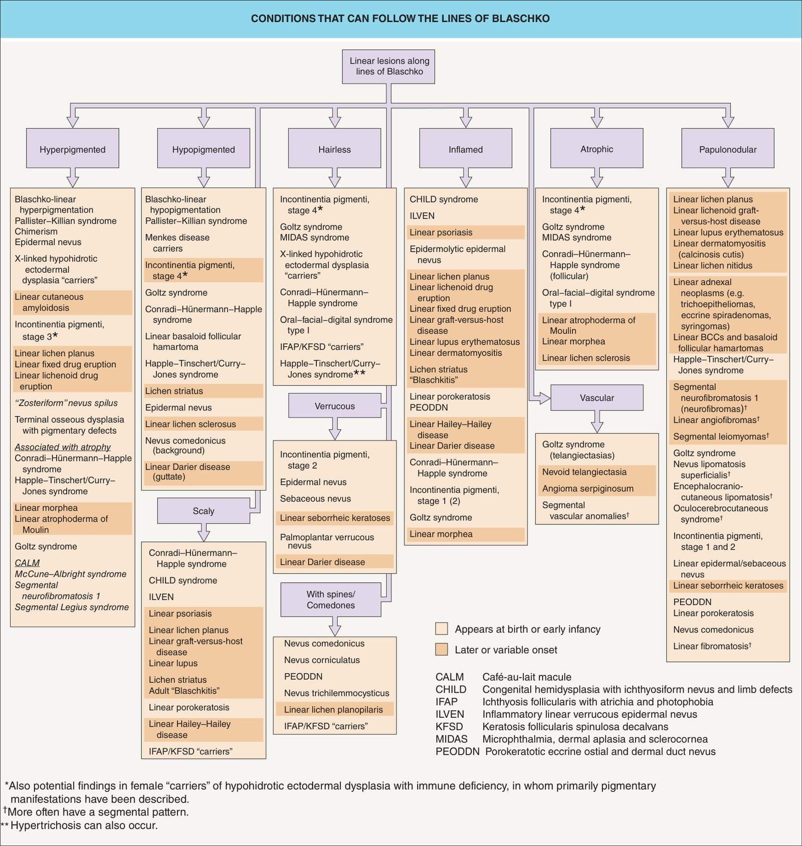
Fig. 62.3 Conditions that can follow the lines of Blaschko. Most of these conditions occur in narrow bands as shown in Fig. 62.2A-E. However, those in italics typically have the segmental pattern with quadrilateral shapes shown in Fig. 62.2G, which includes broad bands along the lines of Blaschko.
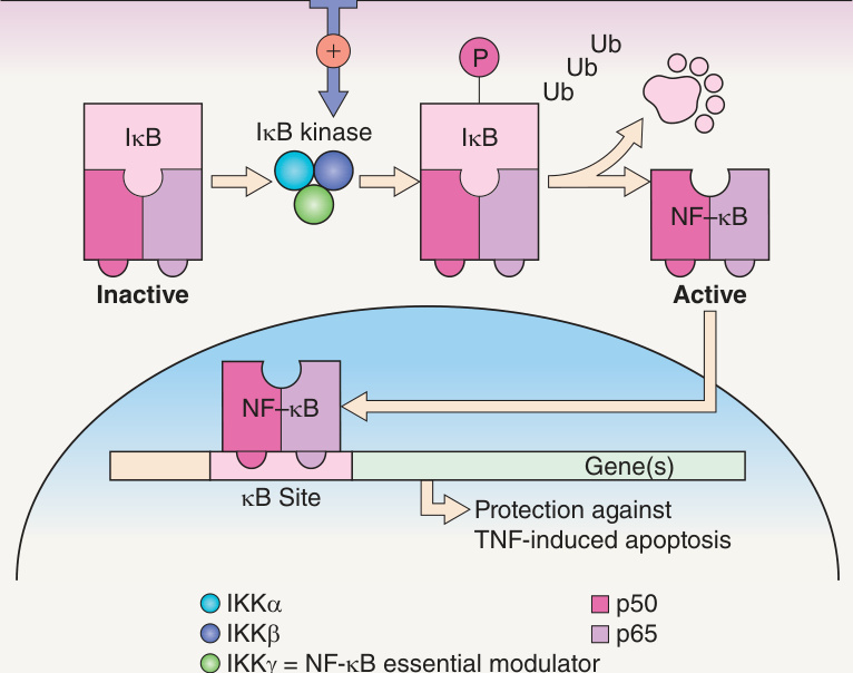
Fig. 62.4 Function of NEMO. In incontinentia pigmenti, lack of NEMO (NF-κB essential modulator; also known as IKBKG [inhibitor of nuclear factor κB kinase regulatory subunit γ]) results in failure to activate NF-κB, which normally protects against tumor necrosis factor (TNF)-induced apoptosis.
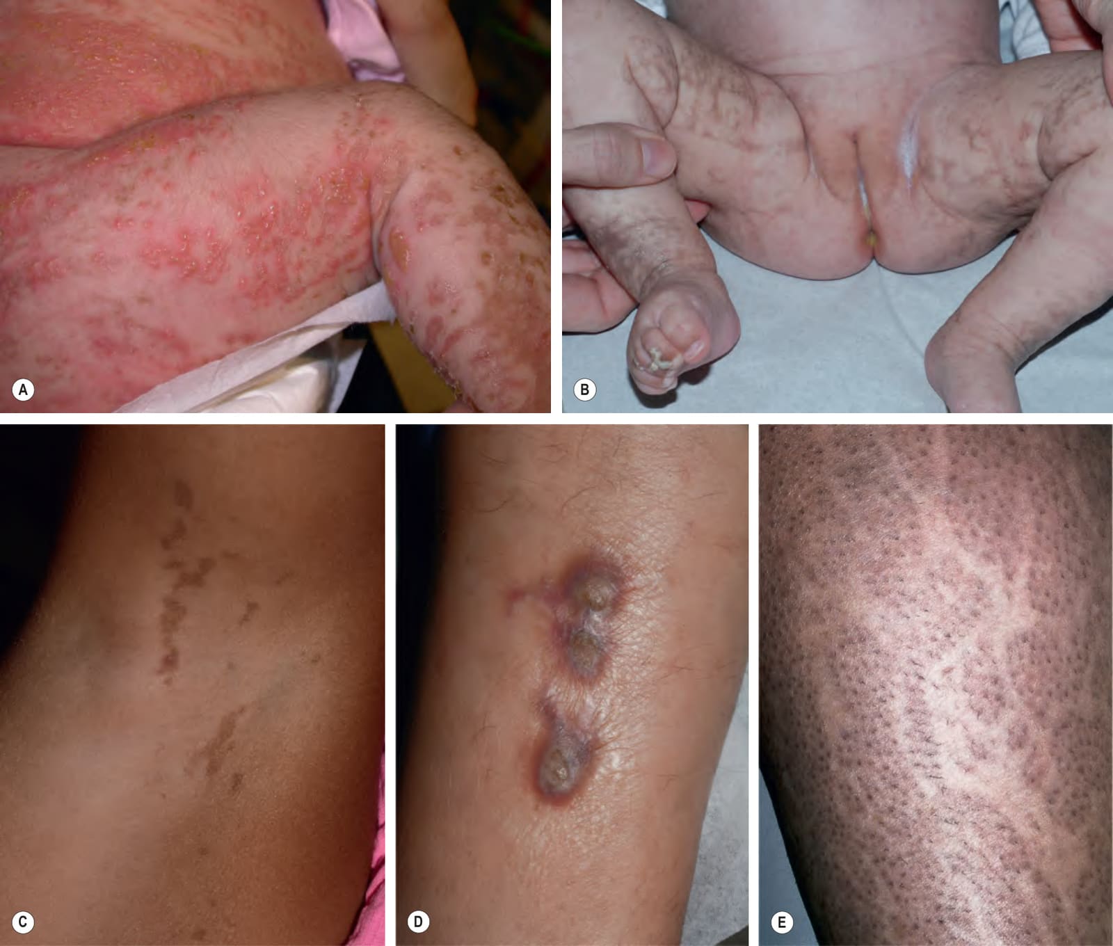
Fig. 62.5 Incontinentia pigmenti.A Stage 1 with streaks of erythema and vesicles in a neonate. B Stages 2 (verrucous) and 3 (hyperpigmented). Note the keratotic lesions on the toes (B). C Residual stage 3 involvement in the axilla of a young girl. The scalloped edges of the hyperpigmented streaks are thought to reflect growth of normal keratinocytes into areas of apoptosis. D Hyperkeratotic nodules on the leg of a teenage girl. E Stage 4 (atrophic and hypopigmented). Note absence of hairs within the streaks on the calf. A, Courtesy Robert A. Silverman, MD; B, E, Courtesy Celia Moss, MBBS, MA, DM.
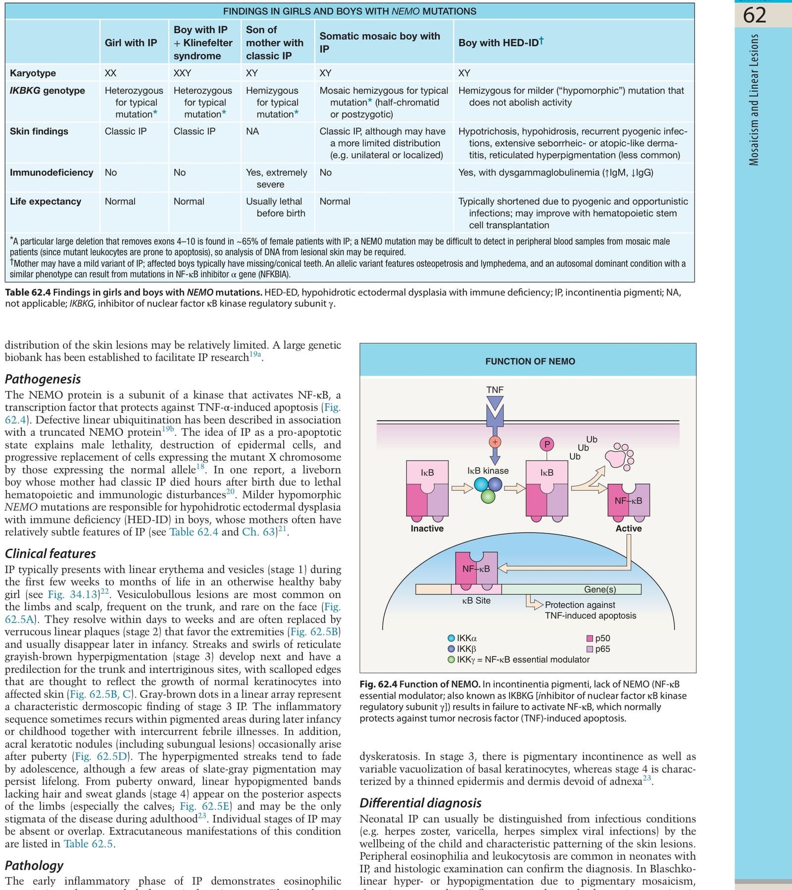
Table 62.4 Findings in girls and boys with NEMO mutations. HED-ED, hypohidrotic ectodermal dysplasia with immune deficiency; IP, incontinentia pigmenti; NA, not applicable; IKBKG, inhibitor of nuclear factor κB kinase regulatory subunit γ.
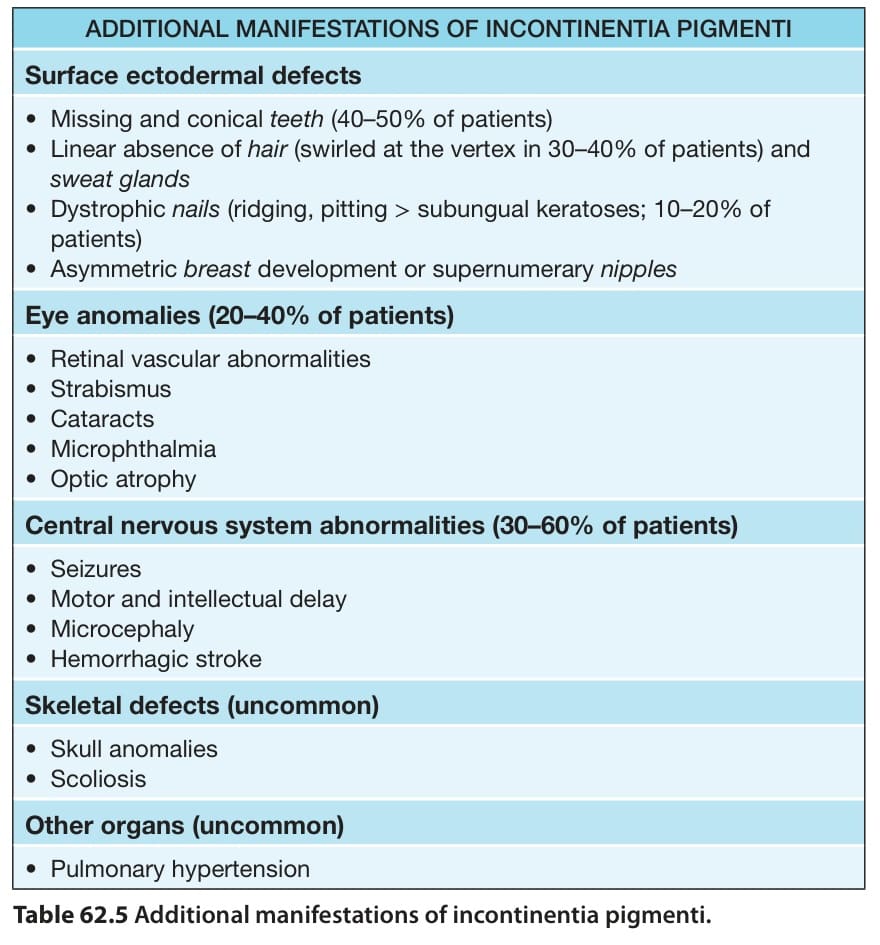
Table 62.5 Additional manifestations of incontinentia pigmenti.