Goltz Syndrome (Focal Dermal Hypoplasia)
Synonym: ▪ Goltz–Gorlin syndrome
Key features
X-linked dominant condition in which skin lesions follow the lines of Blaschko and consist of dermal atrophy in association with variable fat “herniation”, telangiectasias, and pigmentary changes
“Raspberry-like” papillomas favor the perioral and anogenital regions
Nail dystrophy, ocular defects, dental anomalies, and limb malfor- mations such as ectrodactyly (split hand/foot) are common
Caused by mutations in PORCN
Introduction
This uncommon genetic disorder was first described by Goltz in 1962 and ectodermal and mesodermal structures – primarily the skin, eyes, teeth, and bones – are affected in a mosaic pattern.
Genetics
Goltz syndrome is an X-linked dominant condition caused by mutations in PORCN, with the mosaic distribution of skin lesions in female patients reflecting lyonization. Although Goltz syndrome is antenatally lethal in boys with non-mosaic hemizygous mutations (as in IP; see Table 62.4), male patients with genomic (e.g. due to a postzygotic mutation) or functional (in the setting of Klinefelter syndrome) mosaicism account for ~10% of affected individuals.
Approximately 95% of all Goltz syndrome cases are sporadic, which likely reflects both a decreased likelihood of reproduction by severely affected women and the lethality of PORCN mutations with widespread expression. Some authors have postulated that either preferential inactivation of the mutant X chromosome or a postzygotic mutation is required for survival of affected female fetuses. Rare familial cases often show anticipation, with the offspring being more severely affected than the parent and having a higher proportion of cells expressing the mutant X chromosome. The possibility that an apparently normal mother is a carrier must therefore be considered.
Pathogenesis
PORCN belongs to the evolutionarily conserved porcupine gene family and is expressed in the skin, craniofacial and long bones, tooth buds, and eyes – structures affected in Goltz syndrome. PORCN is an endoplasmic reticulum protein with multiple transmembrane domains. It functions as an O-acyltransferase involved in palmitoylation and secretion of Wnt, a morphogen important in ectomesodermal tissue development. Wnt signaling stimulates fibroblast proliferation, inhibits adipogenesis, and induces osteogenesis, explaining the dermal hypoplasia, fat “herniations”, and osteopathia striata (streaks of decreased bone density) observed in Goltz syndrome. Wnt also has key roles in early limb patterning and tooth formation.
Although the primary cutaneous abnormality in Goltz syndrome is in the dermis, the distribution of skin lesions along the lines of Blaschko reflects embryonic migration pathways of epidermal cells. Ectoderm-specific deletion of PORCN in mice has shown that expression of this gene in the epidermis and resultant Wnt signaling regulate development of the underlying dermis and fat. Of note, patients with Goltz syndrome have a thin epidermis and abnormal adnexal structures. This is not surprising considering that Wnt signaling, including β-catenin-dependent pathways, is known to have critical roles in epidermal regeneration and adnexal morphogenesis/ maintenance (see Fig. 55.6).
In 3 of 24 patients in one series, a microdeletion affected both PORCN and the adjacent EBP gene responsible for Conradi–Hünermann–Happle syndrome (see below). Curiously, however, these patients showed no features of the latter disorder. This implies that the PORCN-related hypoplastic skin phenotype predominates over the ichthyosis and follicular changes of Conradi–Hünermann–Happle syndrome.
Clinical features
The phenotype of Goltz syndrome is highly variable, depending on the proportion and distribution of cells expressing the mutant X chromosome. Streaks of vermiculate dermal atrophy and/or telangiectasias are often present from birth (Fig. 62.6A–C). Later, hypo- and hyperpigmentation as well as “herniations” of fat develop. Although “raspberry-like” papillomas with a verrucous surface and fibrovascular core may appear in any location, they favor the anogenital region as well as the lips (Fig. 62.6D), larynx, and acral sites. Limb malformations affect >80% of patients and include ectrodactyly (split hand/foot malformation), asymmetric syndactyly, oligodactyly, and long bone reduction defects. Radiographs of the midportion of the lower extremities characteristically show osteopathia striata. Eye abnormalities, often unilateral, include iritic and chorioretinal colobomas, microphthalmia, anophthalmia, and cataracts. Dystrophic nails (e.g. longitudinal fissures, hypoplasia), sparse hair, abnormal teeth (e.g. vertical grooving, hypodontia), and dysmorphic facies (e.g. notched nasal alae, pointed chin, large malformed ears) may be observed. Other reported features include aplasia cutis congenita, ventral body wall defects, cleft lip/palate, obstructive sleep apnea, gastrointestinal dysfunction, impaired hearing, intellectual impairment, and anomalies of the CNS, spine, urinary tract, or uterus.
Pathology
Affected skin shows a marked reduction in the thickness of the dermis, with fewer adnexal structures. As a consequence of the decreased dermal thickness, adipose tissue may be found just below the epidermis. An increase in papillary dermal blood vessels may also be seen.
Differential diagnosis
The distinctive cutaneous and extracutaneous findings of Goltz syndrome, especially dermal atrophy/fat “herniations” and osteopathia striata, distinguish it from other X-linked dominant genodermatoses. Examples of such conditions include MIDAS (which does not involve the distal extremities), Conradi–Hünermann–Happle, and oral–facial– digital type 1 syndromes. Although an inflammatory component is occasionally observed, the skin lesions of Goltz syndrome are relatively static compared to the evolving lesions of IP.
Treatment
Treatment is supportive, with appropriate subspecialist referral based on the associated abnormalities. The telangiectasias may be improved by pulsed dye laser treatment. Troublesome exophytic papillomas can be treated with curettage or photodynamic therapy.
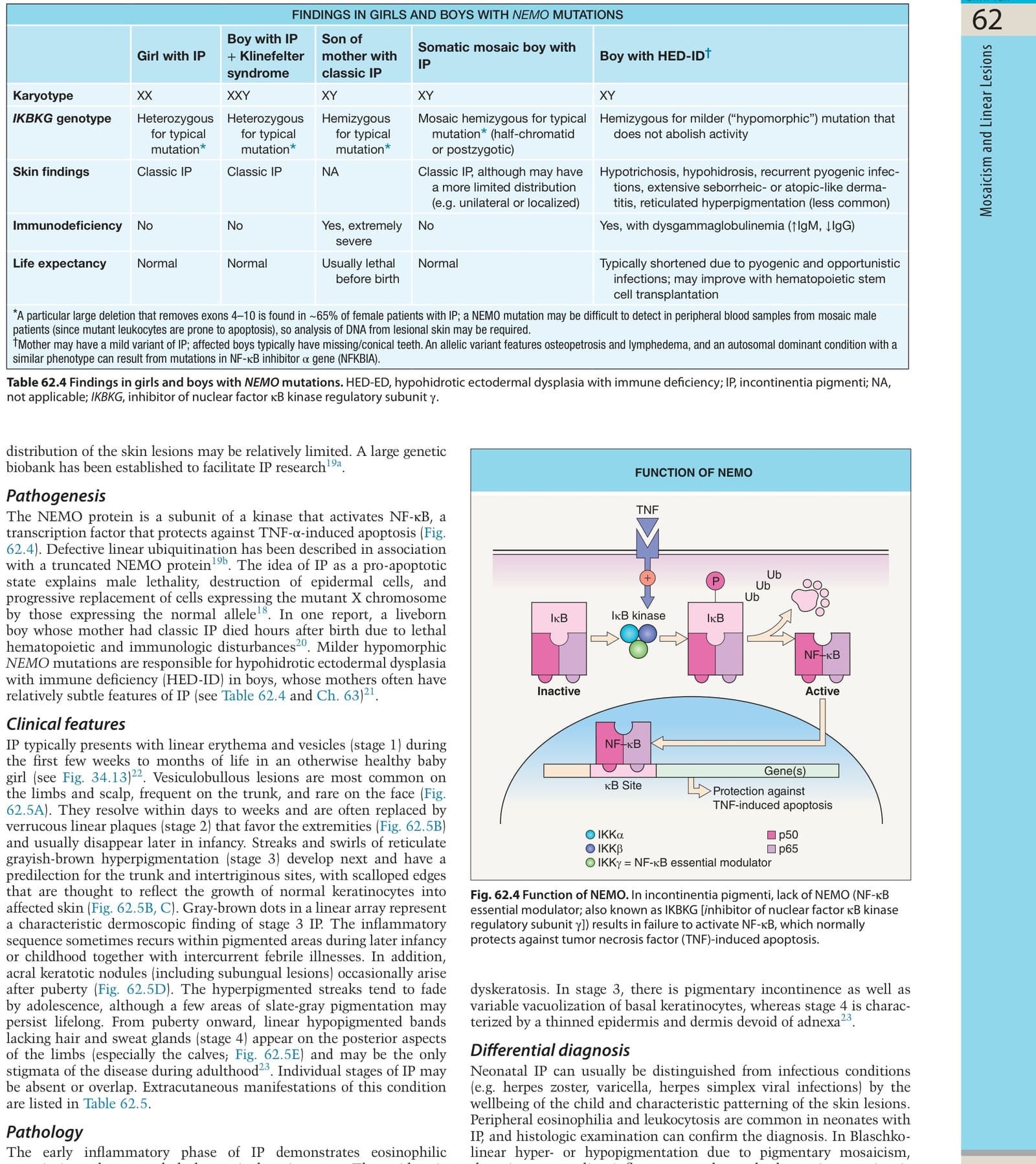
Table 62.4 Findings in girls and boys with NEMO mutations. HED-ED, hypohidrotic ectodermal dysplasia with immune deficiency; IP, incontinentia pigmenti; NA, not applicable; IKBKG, inhibitor of nuclear factor κB kinase regulatory subunit γ.
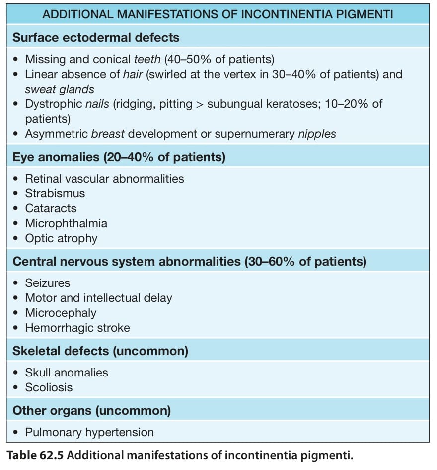
Table 62.5 Additional manifestations of incontinentia pigmenti.