Conradi–Hünermann–Happle Syndrome (X-Linked Dominant Chondrodysplasia Punctata)
This rare X-linked dominant disorder presents in female neonates with ichthyosiform erythroderma characterized by feathery, adherent scale distributed along the lines of Blaschko (see Ch. 57). Characteristic extracutaneous findings include asymmetric shortening of the proximal limbs with epiphyseal stippling (chondrodysplasia punctata) and ocular anomalies such as cataracts and microphthalmia. Patients often have swirled cicatricial alopecia and coarse, lusterless hair. After infancy, the skin changes evolve into follicular atrophoderma. Conradi– Hünermann–Happle syndrome is caused by mutations in the emopamil binding protein (EBP) gene, which lies on the X chromosome adjacent to PORCN and encodes an isomerase essential for cholesterol biosynthesis. The cardinal biochemical abnormality is increased plasma 8(9)-cholestenol and 8-dehydrocholesterol.
CHILD Syndrome (Congenital Hemidysplasia With Ichthyosiform Erythroderma [or Nevus] and Limb Defects)
CHILD syndrome is a rare X-linked dominant disorder that presents with unilateral skeletal hypoplasia ranging from amelia to mild distal
defects, chondrodysplasia punctata, and occasionally defects in internal organs (see Ch. 57). Erythematous, thickened skin with yellowish scaling may have a striking unilateral distribution with sharp midline demarcation, follow the lines of Blaschko, and/or favor skin folds. With time, lipid accumulates and lesions develop features of a verruciform xanthoma. CHILD syndrome results from mutations in NSDHL located on Xq28, which encodes a 3β-hydroxysteroid dehydrogenase enzyme involved in cholesterol biosynthesis. Pathogenesis-based topical therapy with lovastatin or simvastatin plus cholesterol is highly beneficial.
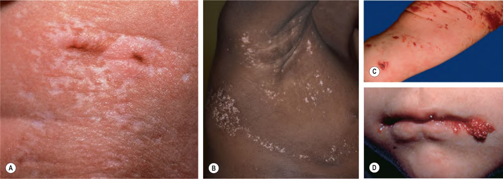
Fig. 62.6 Goltz syndrome (focal dermal hypoplasia).A, B Hypopigmented streaks of vermiculate atrophy. Note the fat herniation (A). C Streaks of telangiectasias plus erosions on the leg. D Several erythematous, raspberry-like papules on the lips. The most prominent lesion is at the angle of the mouth. Similar lesions can occur in the anogenital region and may be confused with warts. D, Courtesy Celia Moss, MBBS, MA, DM.