PIGMENTARY MOSAICISM
Both hypo- and hyperpigmentation can occur in a linear or segmental pattern as a reflection of mosaicism. Traditionally, such presentations have been split into the disorders described below, but there is considerable overlap between these heterogeneous conditions, and distinctions can become blurred.
Linear and Block-Like Hypo- and Hyperpigmentation
Synonyms/variants: Linear nevoid hypo- and hyperpigmentation Blaschkoid dyspigmentation Pigmentary mosaicism Hypomelanosis of Ito (becoming historical) Linear and whorled nevoid hypermelanosis Segmental pigmentation disorder Incontinentia pigmenti achromians (historical)
Key features
Descriptive terms rather than specific diagnoses, with a variety of genetic etiologies
Hypo- or hyperpigmentation following the lines of Blaschko or in a block-like pattern can be evident at birth or appear later
Variable extracutaneous involvement, which can include CNS, musculoskeletal, and ocular abnormalities
Introduction
Hypo- or hyperpigmentation following the lines of Blaschko reflects epidermal mosaicism and has a wide variety of genetic etiologies. Block-like hypo- or hyperpigmentation, which has been referred to as “segmental pigmentation disorder”, is also presumed to result from mosaicism. Associated extracutaneous findings are found in a minority of affected individuals with either distribution pattern.
History
The first published description of hypopigmentation along Blaschko’s lines was by Ito in 1951, resulting in the eponym “hypomelanosis of Ito.” This term is becoming historical and should be reserved for patients with extracutaneous manifestations due to the presence of the mosaic abnormality in tissues other than the skin.
Epidemiology
Linear and block-like pigmentary alterations affect both sexes and are typically easier to see in darkly pigmented skin. The areas of patterned hypo- or hyperpigmentation may be apparent at birth or become evident during childhood, especially in those with lightly pigmented skin.
Genetics and pathogenesis
Blaschko-linear hypo- or hyperpigmentation can have a variety of genetic etiologies, which range from mosaic chromosomal alterations (up to 50% of cases; e.g. trisomy 18 or 20) or point mutations (e.g. MTOR) to germline X-linked conditions (e.g. Börjeson-Forssman- Lehmann syndrome; see above) and rarely chimerism (see below). The pattern of narrow bands along Blaschko’s lines is thought to reflect mosaicism at the level of keratinocytes, with the pigmentary changes resulting from interactions between altered keratinocytes and neighboring melanocytes9a,102. This is supported by the identification of postzygotic mutations in genes such as TP63 and KITLG, which are primarily expressed by keratinocytes, in patients with Blaschko-linear hyperpigmentation9b.
Clinical features
Streaks and swirls of hypo- or hyperpigmentation along the lines of Blaschko can occur anywhere on the cutaneous surface, with unilateral or bilateral involvement. Superimposed lentigines, hypertrichosis, and alopecia are occasionally observed. Only a small proportion (<15–30%) of children with Blaschko-linear hypo- or hyperpigmentation who present to a pediatric dermatologist at a tertiary referral center have extracutaneous abnormalities, which generally became apparent during infancy. The extracutaneous abnormalities are highly variable, depending on the genetic etiology and tissues affected; they most often involve the CNS, eyes, and musculoskeletal system. In a recent series, neurodevelopmental abnormalities were significantly more common in children with Blaschko-linear hypopigmentation affecting ≥4 vs <3 of 5 body areas101a. Block-like hypo- or hyperpigmentation favors the trunk, but any site can be affected. Midline demarcation is more often present ventrally than dorsally, and the lateral borders tend to be less distinct. Segmental hyperpigmentation occasionally coexists with a light red-pink capillary malformation (“phakomatosis melanorosea”). Associated extracutaneous findings are uncommon, affecting <2–5% of patients.
Some patients have linear streaks of both hypo- and hyperpigmentation or a combination of linear and block-like hypopigmentation. When involvement is extensive, it may be difficult to determine whether the lighter or darker skin represents the background color.
Associated syndromes
The most common known single-gene cause of Blaschko-linear hypopigmentation is mosaic activating MTOR mutations. In a recent French series of 71 patients with “hypomelanosis of Ito”, 15 (21%) had an underlying mosaic MTOR mutation103a. Additional findings in affected individuals can include macrocephaly, (hemi)megalencephaly, intellectual disability, seizures, facial dysmorphism, ocular anomalies, and asymmetric overgrowth103b. RHOA-related neuroectodermal syndrome results from mosaic mutations that lead to inactivation of a RAS-related Rho GTPase. Clinical features include Blaschko-linear hypopigmentation (Fig. 62.17), scarring alopecia, leukoencephalopathy (usually asymptomatic), facial dysmorphism, and hemihypoplasia as well as digital, dental, and ocular anomalies103c. X-linked syndromes that present with Blaschko-linear hypo- or hyperpigmentation are discussed above.
Pallister-Killian syndrome due to mosaic tetrasomy 12p, which usually results from gonadal mosaicism in a parent, is a distinct disorder that presents with hypopigmentation following the lines of Blaschko or in an irregular pattern. Frontotemporal alopecia and hypohidrosis represent additional cutaneous manifestations. Extracutaneous features include facial dysmorphism, intellectual disability, and broad first digits in addition to cardiac, renal, genitourinary, and gastrointestinal abnormalities.
Phylloid hypomelanosis in a pattern of leaf-like and floral shapes represents a distinct entity due to mosaic copy number variation involving chromosome 13. Additional features may include telangiectatic macules, thick eyebrows, long eyelashes, intellectual disability, agenesis of the corpus callosum, conductive hearing loss, colobomas, and digital anomalies. A few patients with phylloid hypermelanosis have also been reported, with variable extracutaneous manifestations and underlying mosaic chromosomal aberrations (e.g. involving 5p).
Pathology
Hypopigmented skin may appear normal or have a reduced number of melanocytes, while hyperpigmented skin shows increased epidermal pigmentation.
Differential diagnosis
Fig. 62.3 shows the differential diagnosis of hypo- and hyperpigmented lesions following the lines of Blaschko. Diagnostic considerations for block-like hyperpigmentation may include McCune–Albright syndrome, which tends to have darker, sharply demarcated lesions that follow the broad lines of Blaschko, as well as a large CALM, Becker nevus, and early nevus spilus. Block-like hypopigmentation is usually distinguishable from the amelanotic lesions of segmental vitiligo.
Evaluation and management
A thorough history and physical examination should be performed to assess for extracutaneous involvement, with further evaluation directed by abnormal findings and/or recognition of distinct syndromes (see above). Growth and development should be followed in conjunction with the child’s pediatrician.
In patients with extracutaneous abnormalities, identification of the underlying genetic alteration may provide better understanding of the overall diagnosis, help direct further evaluation, and potentially impact future family planning. For example, mosaic MTOR mutations involving the gonads have been implicated in transmission of Smith-Kingsmore syndrome, which presents with megalencephaly, seizures, and intellectual disability due to heterozygous germline MTOR mutations. In addition, recognizing the genetic etiology can provide the basis for targeted therapies such as sirolimus or other mTOR inhibitors.
Although mosaic mutations are sometimes present in the peripheral blood, a sample of affected tissue (e.g. a skin biopsy specimen) is more likely to contain mutant cells. If no specific single-gene disorder is suspected, genetic testing should start with an assessment for chromosomal mosaicism, which can be performed via microarray-based tests, e.g. single nucleotide polymorphism (SNP) array or array comparative genomic hybridization (CGH). If initial evaluation of DNA from a blood sample is not diagnostic, the test can be repeated on DNA extracted from a 4 mm punch skin biopsy of affected skin. The latter is generally of higher yield than the fibroblast cultures routinely performed by cytogenetics laboratories, as the mutation may not be present in that cell type. Keratinocyte cultures may be of utility when available, and whole exome or whole genome sequencing represents an additional testing option.
Nevus Depigmentosus
A nevus depigmentosus is another hypopigmented skin lesion that is thought to reflect cutaneous mosaicism. However, it is limited in extent and generally lacks extracutaneous associations. The most common presentation is a localized patch that breaks apart into smaller macules at its periphery, resembling a splash of paint. Although classically evident at birth, a nevus depigmentosus sometimes becomes apparent later in childhood, especially in lightly pigmented individuals.
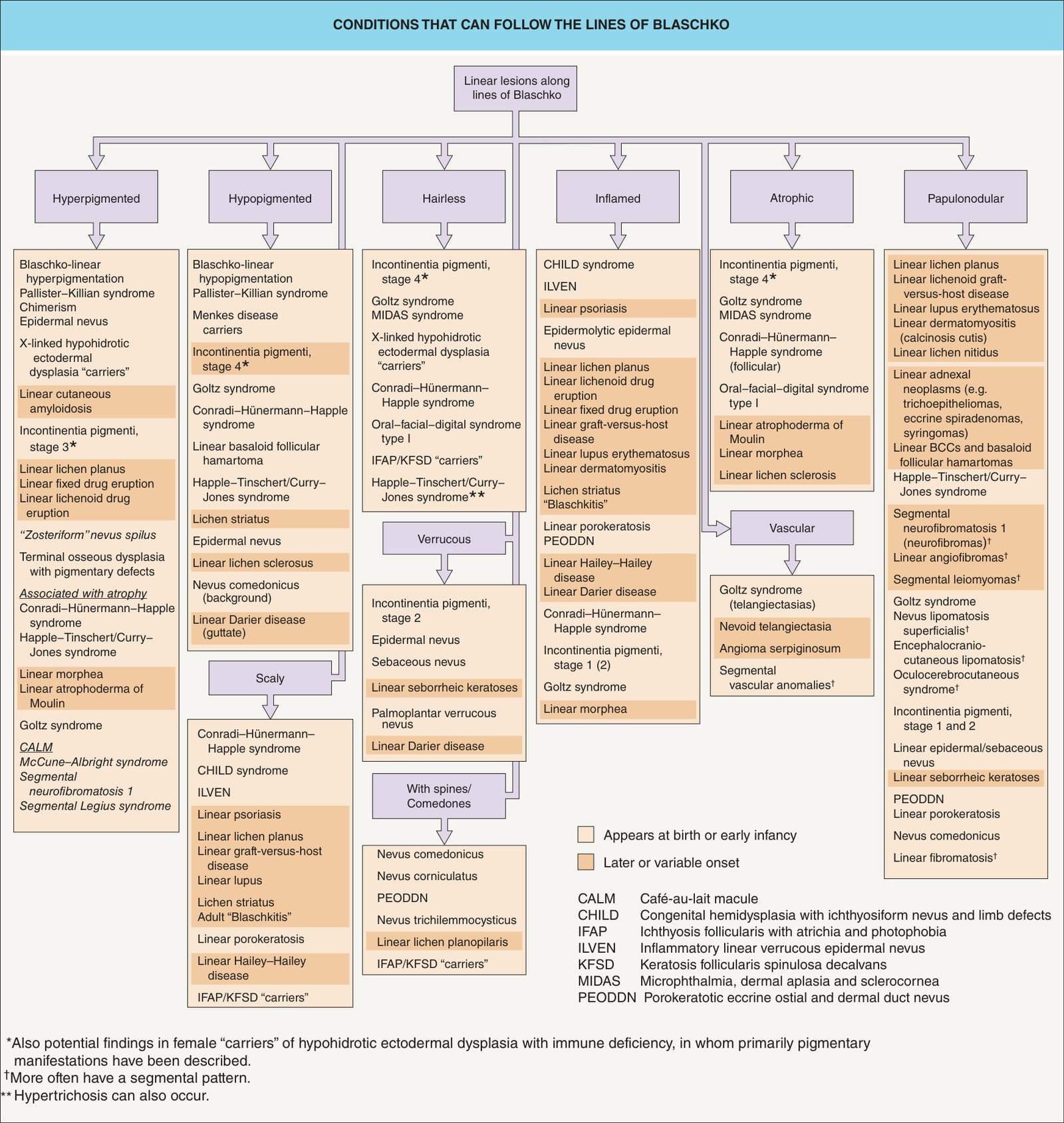
Fig. 62.3 Conditions that can follow the lines of Blaschko. Most of these conditions occur in narrow bands as shown in Fig. 62.2A-E. However, those in italics typically have the segmental pattern with quadrilateral shapes shown in Fig. 62.2G, which includes broad bands along the lines of Blaschko.
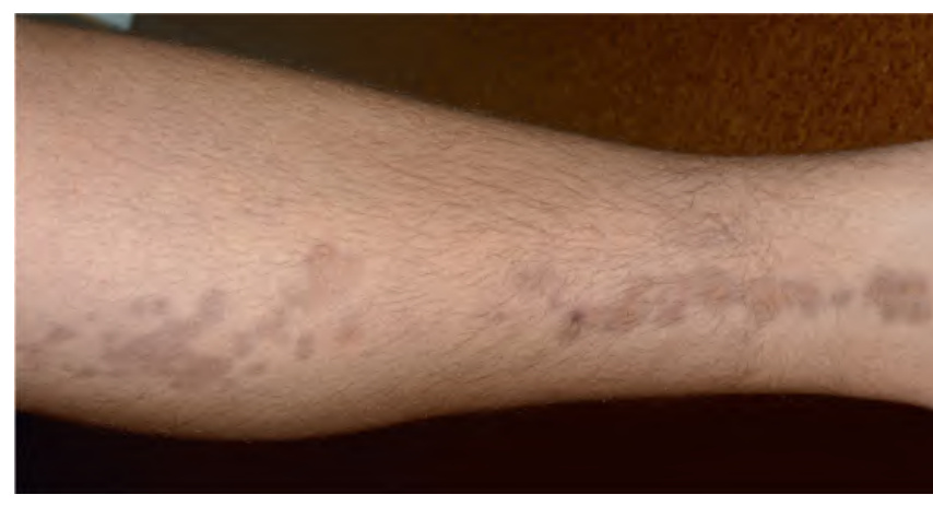
Fig. 62.16 Linear atrophoderma of Moulin. Hyperpigmented depressions with “cliff-drop” borders following the lines of Blaschko. The morphology of the lesions resembles that of atrophoderma of Pasini and Pierini.
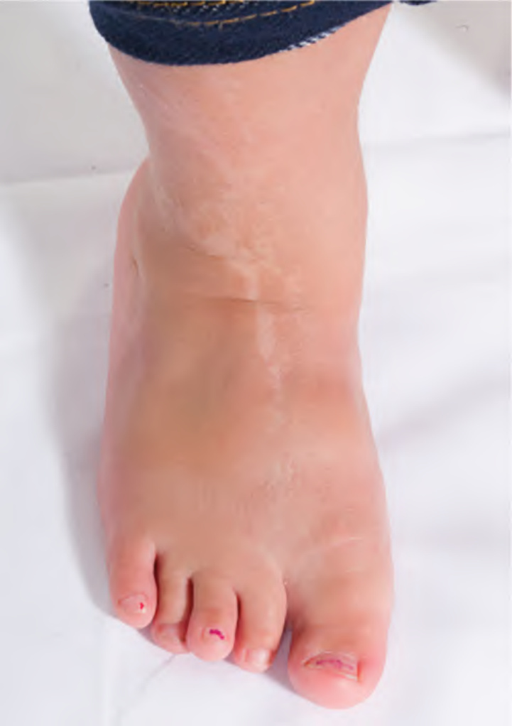
Fig. 62.17 Blaschko-linear hypopigmentation on the foot and leg of a child with a mosaic RHOA mutation.Courtesy Veronica Kinsler, MD, PhD.