GARDNER SYNDROME (VARIANT OF FAMILIAL ADENOMATOUS POLYPOSIS)
Synonym: Familial polyposis of the colon
Key features
Autosomal dominant disorder that is a variant of familial adenomatous polyposis (FAP) with prominent extraintestinal manifestations
Features the intestinal polyps and adenocarcinoma that charac- terize FAP together with epidermoid cysts, fibromas, desmoid tumors, and osteomas
Retinal pigment epithelial hamartomas represent an early sign
Caused by mutations in the APC (adenomatous polyposis coli) gene
Introduction
The main features of Gardner syndrome are premalignant intestinal polyposis, epidermoid cysts, osteomas, and desmoid or fibrous tumors of the skin and other sites. It is considered to represent a phenotypic variant of the familial adenomatous polyposis syndrome (FAP) with prominent extraintestinal involvement.
History
In 1951, Gardner described a large family in which colonic polyps and carcinoma were associated with osteomas and “surface tumors”. This FAP phenotype with prominent extraintestinal manifestations continues to bear Gardner’s name.
Epidemiology
The prevalence of FAP is estimated to be 1 in 7000 to 1 in 30 000 live births. It is inherited as an autosomal dominant trait with high penetrance of the gastrointestinal manifestations but variable expressivity of extraintestinal findings, affecting men and women equally.
Pathogenesis
FAP (including Gardner syndrome) is caused by heterozygous germline mutations in the adenomatous polyposis coli (APC) tumor suppressor gene. APC encodes a protein that downregulates the Wnt/β-catenin signaling pathway, which has important functions in cellular proliferation, differentiation, and adhesion (see Fig. 55.6). Extraintestinal manifestations (especially desmoid tumors) are more likely in patients with mutations in the distal portion of APC.
Clinical Features
Cutaneous and soft tissue manifestations
Skin lesions appear during childhood and adolescence, frequently preceding the onset of polyposis. Cutaneous epidermoid cysts affect ~50% of patients with FAP and are often multiple. The cysts typically begin to appear before puberty and favor the face, scalp, and extremities. Multiple familial pilomatricomas have also been reported as a presentation of Gardner syndrome.
Gardner-associated fibromas may occur in the skin, subcutaneous tissues, mesentery, or retroperitoneum. The fibromas typically arise during the first two decades of life, favor the back (especially the paraspinal region), and can serve as a desmoid tumor precursor. Lipomas, leiomyomas, trichoepitheliomas, and neurofibromas are less frequently observed.
Desmoid tumors (also referred to as desmoid fibromatosis) are non-encapsulated, non-metastasizing, locally aggressive tumors that are usually rare but affect 10%–30% of patients with FAP. These lesions arise from musculoaponeurotic structures and can occur spontaneously or at surgical sites. In patients with APC, desmoid tumors most often develop within the abdominal wall or intra-abdominally following colectomy, with the latter location causing greater morbidity via obstruction of the small bowel and/or ureters. Extra-abdominal lesions affecting the shoulder girdle, chest wall, or inguinal region can also occur. Although cytologically benign, desmoid tumors represent a potential cause of mortality in FAP, e.g. due to compression of vital organs.
Other extraintestinal manifestations
Retinal pigment epithelial hamartomas (RPEH) are evident in ~75%–80% of patients with FAP. These lesions may be present at birth, serving as an early and specific sign of the disorder (Fig. 63.5). Although often referred to as congenital hypertrophy of the retinal pigment epithelium (CHRPE), FAP-associated pigmented retinal lesions have irregular borders and a haphazard bilateral distribution that differentiate them from sporadic multifocal CHRPE.
Osteomas occur in ~65%–80% of patients with FAP and favor the frontal bone, mandible, and maxilla, although they can also develop in other bones of the skull and long bones. Seen in children as young as 8 years of age, osteomas are painless and can grow large enough to be clinically obvious or be detectable only by radiographic studies. Other skeletal lesions include exostoses, endostoses, and cortical thickening of the long bones. Dental anomalies are apparent in ~30% of patients with FAP and include supernumerary teeth, odontomas, unerupted teeth, and congenitally missing teeth.
Patients with FAP are at increased risk for the development of other tumors, including papillary carcinoma of the thyroid (particularly in women), hepatoblastoma (usually affecting children <3 years of age), pancreatic and biliary tract carcinomas, nasal angiofibromas, adrenal adenomas (or occasionally carcinomas), and sarcomas (e.g. fibrosarcoma, osteosarcoma). The association of brain tumors (most often medulloblastomas) with adenomatous polyposis in patients with heterozygous APC mutations has historically been referred to as Turcot syndrome type 2. The latter is distinct from the constitutional mismatch repair deficiency syndrome (previously referred to as Turcot syndrome type 1) that presents during childhood with brain tumors (primarily glioblastomas), colonic polyposis, hematologic malignancies, and multiple café- au-lait macules due to biallelic mutations in mismatch repair genes (see below and Table 61.3).
Gastrointestinal manifestations
In adults, the diagnosis of FAP is commonly made when the patient presents with gastrointestinal bleeding secondary to adenomatous polyps. Other findings may include anemia, abdominal pain, diarrhea or constipation, and weight loss. Premalignant adenomatous polyps most commonly occur in the colorectum but are also found in the small intestine and stomach in over half of patients. Polyps are typically <1 cm in diameter and are rarely seen before the age of 10 years. By the age of 20 years, 50% of patients with FAP have demonstrable polyps. Without surgical intervention, colorectal carcinoma is inevitable; it usually develops before age 40 years and arises by early adolescence in ~5% of affected individuals. Carcinomas of the small intestine (especially the duodenum) can also occur.
Pathology
Histologically, intestinal polyps show adenomatous hyperplasia of the mucosa. Cutaneous cysts in patients with FAP usually demonstrate the same microscopic changes as ordinary epidermoid cysts, with a lining of keratinizing epithelium that includes a granular layer. Pilomatricoma-like changes, with characteristic basophilic and shadow cell populations, have also been noted.
Differential Diagnosis
Although epidermoid cysts represent a common sporadic finding, multiple lesions with onset during childhood should prompt a thorough family history and evaluation for other findings of Gardner syndrome (e.g. RPEH, osteomas). Of note, mutations in the MUTYH base excision repair gene can cause an autosomal recessive form of colorectal adenomatous polyposis, with osteomas, RPEH, desmoid tumors, pilomatricomas, and sebaceous neoplasms (see below) having been described in affected individuals.
Management
The diagnosis of FAP is established by identifying a pathogenic variant in APC in an individual with multiple colorectal adenomatous polyps (≥100 for an adult pre-colectomy and without a relative with confirmed FAP). Genetic counseling is imperative, and family members should be evaluated for intestinal and extraintestinal signs of the disorder.
Because of the inevitable development of colorectal carcinoma, prophylactic colectomy is recommended when polyp formation becomes evident, and it is usually performed between 15 and 25 years of age. Initially, children with FAP should be monitored with sigmoidoscopy or colonoscopy every 1–2 years beginning at 10–12 years of age. Once polyps are detected, patients should undergo annual colonoscopy until a colectomy is done. Esophagogastroduodenoscopy is recommended every 6 months to 4 years (depending on polyp burden) beginning at 20–25 years of age, with careful inspection of the ampulla of Vater. Annual physical examination should include evaluation for neurologic deficits and other extraintestinal manifestations, including palpation of the abdomen (for desmoid tumors) and thyroid starting in the late teenage years. If there is a family history of desmoid tumors, abdominal CT or MRI beginning 1–3 years post colectomy and then every 5–10 years should be considered. Because of the increased risk of hepatoblastoma, some experts recommend abdominal ultrasound and measurement of serum α-fetoprotein every 3–6 months during the first 5 years of life.
Symptomatic epidermoid cysts can be surgically excised. Desmoid tumors are also treated by surgical excision, although there tends to be a high recurrence rate. Preoperative MRI of the abdomen can demonstrate the origin of the tumor and the extent of involvement. If wide surgical excision fails, radiotherapy, hormonal therapy (e.g. tamoxifen, raloxifene), chemotherapy, or a combination thereof can be tried. The osteomas of FAP may or may not be amenable to surgical intervention. Medications that affect prostaglandin metabolism, such as nonsteroidal anti-inflammatory drugs (NSAIDs), may reduce the risk of colorectal cancer and desmoids.
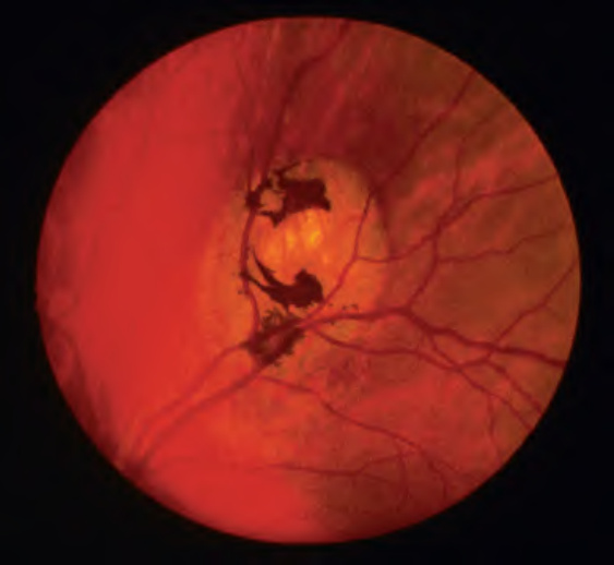
Fig. 63.5 Retinal pigment epithelial hamartomas in Gardner syndrome.Courtesy L. Tychsen, MD.