MUIR–TORRE SYNDROME (VARIANT OF LYNCH SYNDROME)
Synonym: Cutaneous sebaceous neoplasms and keratoacanthomas, with gastrointestinal and other carcinomas
Key features
An autosomal dominant disorder associated most commonly with adenocarcinoma of the colon, followed by genitourinary carcinomas
The cutaneous manifestations are sebaceous neoplasms and keratoacanthomas
Caused by mutations in DNA mismatch repair genes (MSH2>>
MLH1>MSH6, PMS2)
Introduction
Muir–Torre syndrome (MTS) is an uncommon autosomal dominant genodermatosis that is characterized by sebaceous neoplasms (adenoma, sebaceoma, or carcinoma; see Ch. 111), and internal malignancies. It is considered to represent a variant of Lynch syndrome, which is also known as hereditary non-polyposis colorectal cancer (HNPCC) syndrome.
History
Muir in 1967 and Torre in 1968 independently reported the syndrome that bears their names. Their two patients had malignancies in several organs plus either keratoacanthoma-like lesions or multiple tumors of sebaceous origin.
Epidemiology
The male : female ratio in MTS is 3 : 2, and affected individuals present with cutaneous manifestations at a mean age of 55 years. The prevalence of Lynch syndrome in the general population is estimated to be ~1 in 350. In a study of Lynch syndrome patients, MTS was observed in 28% (14/50) of families and 9% (14/152) of individuals with a confirmed germline mismatch repair gene mutation.
Pathogenesis
MTS is caused by heterozygous germline mutations in one of several DNA mismatch repair (MMR) genes, including MSH2 (90% of MTS cases), MLH1, and rarely MSH6 or PMS2. MMR proteins are responsible for detecting and repairing DNA replication errors. In patients with MTS, somatic inactivation of the other allele of the affected gene (loss of heterozygosity) results in microsatellite instability, i.e. increased variability in the lengths of repetitive “microsatellite” sequences in the genome, as well as mutations that lead to tumor formation.
Clinical Features
Sebaceous tumors can present prior to (~30%), concurrently with (~10%), or after (~60%) the diagnosis of a visceral malignancy. The skin tumors sometimes appear decades before or after a visceral malignancy develops. In general, sebaceous tumors favor the head and neck region (Fig. 63.6), and sporadic sebaceous carcinomas tend to affect the
Courtesy Dan Ring, MD.
periocular area. However, in patients with MTS, these neoplasms can occur in other locations (trunk > extremities). Sebaceous adenomas are the most common tumor type, and they present as slow-growing, yellow to pink papules or nodules, numbering from one to hundreds. Keratoacanthomas, which are keratotic skin-colored to erythematous papules or nodules that arise quickly, are seen in ~25% of patients with MTS and usually exhibit sebaceous differentiation. Fordyce granules (“free” sebaceous glands) on the vermilion lip and oral mucosa (especially the vestibule) are more common and prominent in patients with MTS than in the general population.
The most frequently observed malignancies in Lynch syndrome are colorectal (~50%–60% of patients with MSH2 or MLH1 mutations) and genitourinary (e.g. endometrium [15%–50% of women], ovary [10%–35% of women], urinary tract; highest risk with MSH2 mutations) carcinomas. Colorectal carcinoma in Lynch syndrome most often involves the proximal colon and develops at a median age of 45 years, about one decade before it typically occurs in the general population. Lynch syndrome patients are also at increased risk of neoplasms of the small intestine, stomach, pancreas, hepatobiliary system, CNS, and perhaps breast; hematologic malignancies and sarcomas have also been reported.
Diagnosis
The diagnosis of MTS was traditionally based on the presence of at least one sebaceous tumor (or keratoacanthoma with sebaceous differentiation) and a visceral malignancy without another known predisposing factor. In the absence of a sebaceous tumor, the diagnosis can be made in a patient with multiple keratoacanthomas, multiple visceral malignancies, and a family history of MTS. Findings that suggest a higher or lower likelihood of MTS in patients with sebaceous neoplasms are summarized in Table 63.5. Testing for a mismatch repair gene mutation is helpful in confirming the diagnosis and facilitating identification and surveillance of affected family members.
Pathology
Skin tumors arising as part of MTS fall into two general categories: sebaceous neoplasms and keratoacanthomas. Sebaceous adenomas (with a predominance of mature sebocytes), sebaceomas (vertically oriented with a predominance of basaloid seboblastic cells), so-called sebaceous epitheliomas (inconsistently defined term used for various lesions with seboblastic differentiation), and sebaceous carcinomas may occur (see Ch. 111). A type of keratoacanthoma with sebaceous differentiation that may be specific to this syndrome has been called a sebo(kerato)acanthoma; it has the architecture of a keratoacanthoma but contains well-differentiated sebaceous lobules (Fig. 63.7). In addition, cystic sebaceous tumors, which are well-circumscribed, deep sebaceous lesions with a cystic growth pattern, occur in some MTS patients and are a strong clue for the presence of the syndrome.
Immunohistochemical (IHC) staining for MSH2, MSH6, MLH1, and PMS2 can be a valuable screening tool for MTS-associated tumors, which frequently show a lack of expression of one or more of these mismatch repair proteins (see Table 63.5). However, abnormal IHC staining is not diagnostic of MTS, as it is sometimes seen in sporadic sebaceous
Well-circumscribed basaloid tumor with abundant secreted material, surrounded by a pseudocapsule of compressed fibrous tissue. Note the sebaceous differentiation (inset). Courtesy Luis Requena, MD.
neoplasms, especially in immunocompromised patients; conversely, IHC staining may rarely be normal in lesions from patients with a mismatch repair gene mutation (see Ch. 111). PCR-based assays can also be used to demonstrate microsatellite instability (MSI) in tissue from MTS-associated neoplasms. Confirmatory testing for a germline
mismatch repair gene mutation should be considered in patients with abnormal lesional IHC staining for these proteins, evidence of MSI, or a strong clinical suspicion of MTS.
Differential Diagnosis
Sebaceous hyperplasia is a common finding in the general population, and sebaceous adenomas and carcinomas can occur in patients without MTS. Sebaceous adenomas and carcinomas as well as colorectal carcinomas have also been described in patients with autosomal recessive colorectal adenomatous polyposis caused by mutations in the MUTYH base excision repair gene. This disorder accounts for some of the patients with sebaceous neoplasms and visceral malignancy who meet the traditional criteria for MTS, but whose tumors do not exhibit microsatellite instability. Some authors have referred to patients without evidence of microsatellite instability due to mismatch repair dysfunction as having MTS type 2. Additional manifestations of MUTYH-associated polyposis include other malignancies (e.g. small bowel, ovarian, bladder) and findings that overlap with Gardner syndrome, such as osteomas, RPEH, desmoid tumors, and pilomatricomas.
Multiple keratoacanthomas may occur as an isolated finding (i.e. without visceral malignancy), especially in patients with the Ferguson– Smith (multiple self-healing) or Grzybowski (generalized eruptive) types of keratoacanthomas. However, these patients should be evaluated to exclude the possibility of MTS.
Treatment
Patients with an internal malignancy require appropriate oncologic referral and management. Recommendations for surveillance of high-risk individuals are outlined in Table 63.6. Long-term aspirin therapy may decrease the risk of colorectal carcinoma in patients with Lynch syndrome.
Treatment options for benign sebaceous tumors include curettage, excision, and cryosurgery. Sebaceous carcinomas have metastatic potential and require wide local excision or Mohs micrographic surgery, with the latter recommended for facial lesions. Oral retinoids with or without interferon-α may be useful in the prevention of new sebaceous neoplasms.
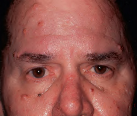
Fig. 63.6 Muir–Torre syndrome with multiple sebaceous neoplasms.
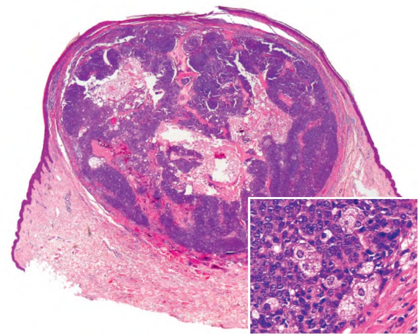
Fig. 63.7 Seboacanthoma in a patient with Muir–Torre syndrome.
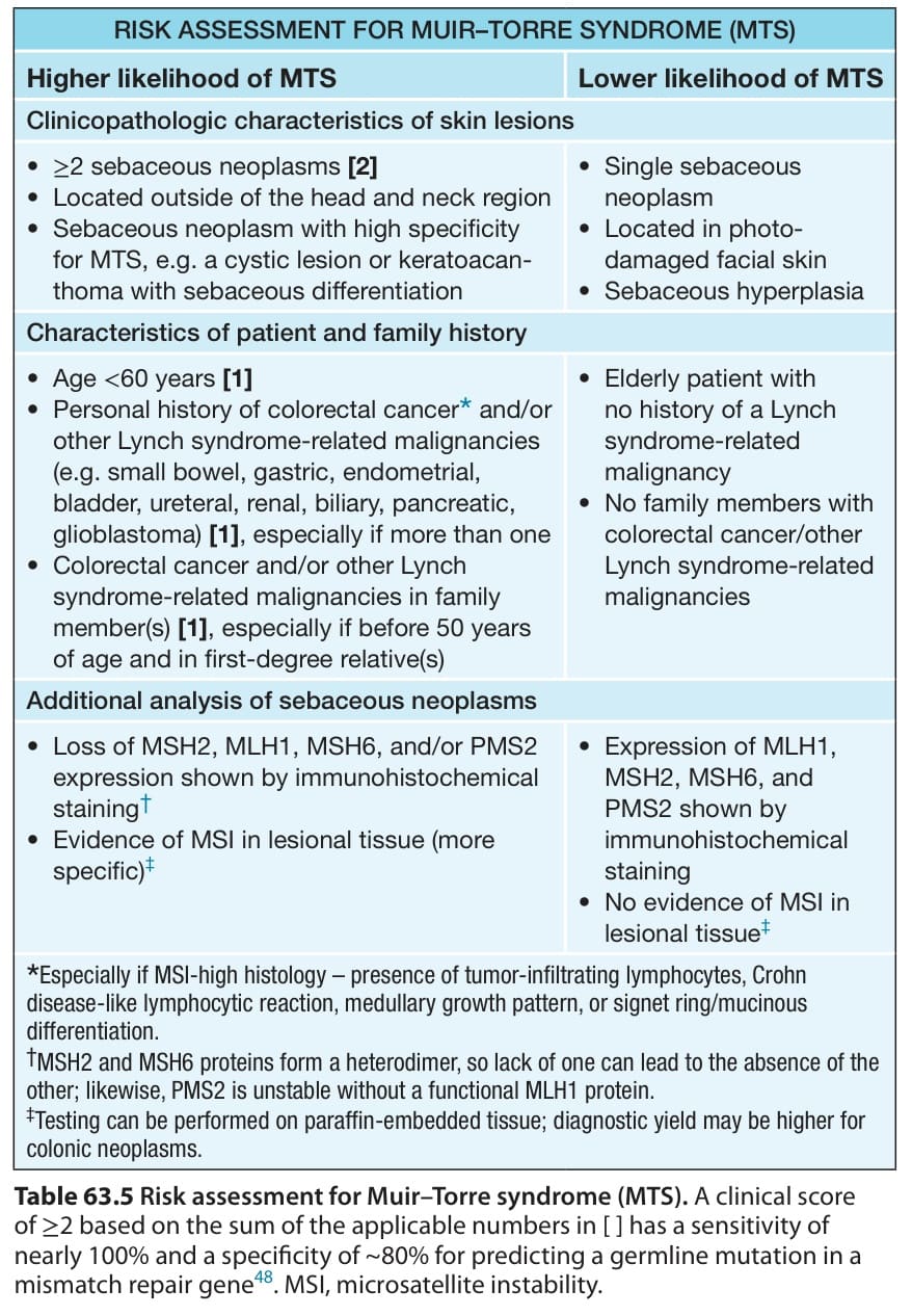
Table 63.5 Risk assessment for Muir–Torre syndrome (MTS). A clinical score of ≥2 based on the sum of the applicable numbers in [ ] has a sensitivity of nearly 100% and a specificity of ~80% for predicting a germline mutation in a mismatch repair gene. MSI, microsatellite instability.
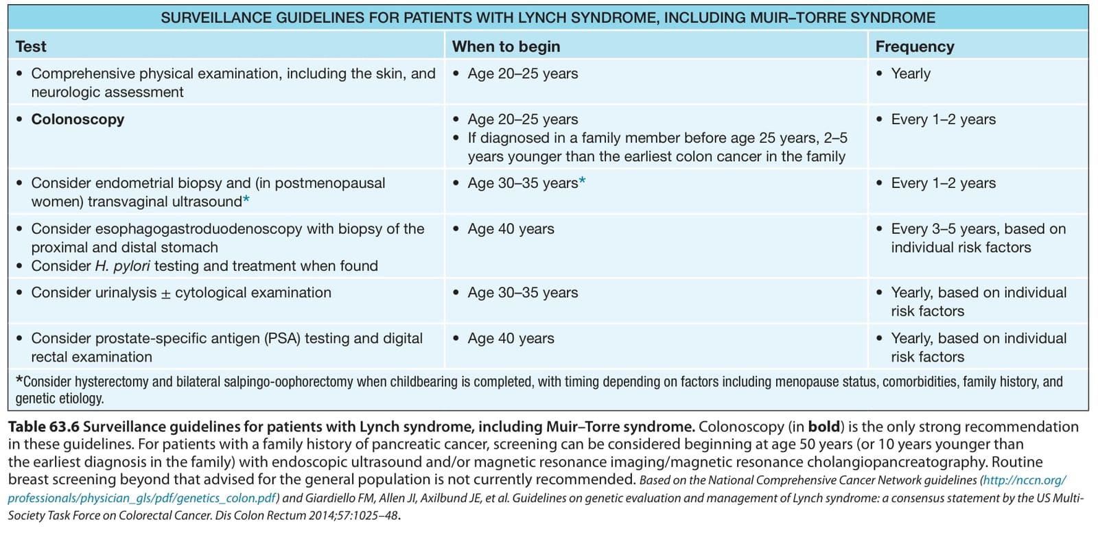
Table 63.6 Surveillance guidelines for patients with Lynch syndrome, including Muir–Torre syndrome. Colonoscopy (in bold) is the only strong recommendation in these guidelines. For patients with a family history of pancreatic cancer, screening can be considered beginning at age 50 years (or 10 years younger than the earliest diagnosis in the family) with endoscopic ultrasound and/or magnetic resonance imaging/magnetic resonance cholangiopancreatography. Routine breast screening beyond that advised for the general population is not currently recommended. Based on the National Comprehensive Cancer Network guidelines (http://nccn.org/ professionals/physician_gls/pdf/genetics_colon.pdf) and Giardiello FM, Allen JI, Axilbund JE, et al. Guidelines on genetic evaluation and management of Lynch syndrome: a consensus statement by the US Multi- Society Task Force on Colorectal Cancer. Dis Colon Rectum 2014;57:1025–48.