ALKAPTONURIA
Synonym: Ochronosis
Key features
An autosomal recessive disorder due to deficiency of homogen- tisate 1,2-dioxygenase
Clinical signs include discoloration of cartilage (e.g. the helices), sclerae, and skin (e.g. the axillae)
Extracutaneous findings include urine that darkens on standing, arthritis, and valvular heart disease
Alkaptonuria is an autosomal recessive inborn error of metabolism in which homogentisic acid (HGA), an intermediate in phenylalanine and tyrosine metabolism, cannot be further metabolized and therefore accumulates in body fluids and tissues. The disorder results from a deficiency of the enzyme homogentisate 1,2-dioxygenase. It is estimated to have a prevalence of ~1 in 250 000 in most ethnic groups, but as high as 1 in 20 000 in Slovakia and the Dominican Republic due to founder effects. The cardinal clinical features of alkaptonuria include urine that turns dark on standing, discoloration of cartilage and other connective tissues, and arthritis (Fig. 63.8). The arthritis often presents in the third or fourth decade of life with chronic back pain and stiffness, and radiographs show flattened and calcified intervertebral discs. Later involvement of large peripheral joints can resemble rheumatoid arthritis clinically but osteoarthritis radiographically. Renal complications include an increased incidence of calculi and occasionally kidney failure. Aortic stenosis is common by the sixth or seventh decade of life and coronary artery calcifications may also develop. Life expectancy is normal.
The dermatologic features of alkaptonuria are rarely noted prior to 10–15 years of age and typically become apparent during adulthood, often in the fourth decade of life. Axillary skin may be one of the initial sites to develop discoloration, which can range in color from blue to yellow to brown. The classic blue–grey color associated with this disorder is often first noted in the helices of the ear and sclerae (see Fig. 63.8). Later, it may affect the palmar and plantar surfaces as well as the entire face. Dermoscopy can show speckled white globular structures overlying a grayish-blue structureless area. Collagenous and elastotic marginal plaques of the hand may also develop, typically during the fourth to sixth decades of life. Features that may present early in life
include brownish discoloration of diapers (due to the dark urine) and cerumen that is brown to black in color. Histopathologic findings of discolored skin include orthohyperkeratosis and irregular, brownishyellow, acellular deposits in the dermis.
Diagnosis of alkaptonuria can be confirmed by urine organic acid analysis. Management includes physical therapy and pain control as needed. Beginning at age 40 years, periodic cardiac assessment with echocardiography is recommended. Administration of oral nitisinone, an inhibitor of HGA production, at a dose of 2–10 mg daily reduces urinary HGA excretion by >95% and has been shown to slow disease progression; a low-protein diet in conjunction with this medication may help to prevent excessively high plasma tyrosine levels.
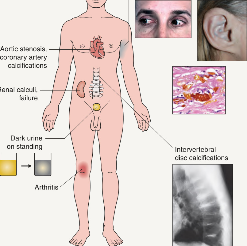
Fig. 63.8 Clinical features of alkaptonuria.Eyes, courtesy William Gahl, MD PhD; ear, courtesy Julie V. Schaffer, MD; radiograph, courtesy Jean L. Bolognia, MD; photomicrograph, courtesy Lorenzo Cerroni, MD.