FABRY DISEASE
Synonym: Anderson–Fabry disease
Key features
X-linked disorder due to a deficiency of α-galactosidase A
Characteristic mucocutaneous finding is multiple angiokeratomas
Additional features include pain and paresthesias of the extremities, hypohidrosis, and renal and coronary insufficiency
Fabry disease is an X-linked lysosomal storage disorder resulting from a defect in glycosphingolipid metabolism due to pathogenic variants in GLA, which encodes the enzyme α-galactosidase A (α-Gal A). It is a pan-ethnic condition that can be identified via newborn screening in ~1 in 7000 male neonates, including both classic severe disease (associated with minimal residual α-Gal A activity) and later-onset phenotypes with milder clinical involvement (cardiac > renal). Female heterozygotes may also be affected, with variable clinical manifestations that often have a later onset. The clinical manifestations result from accumulation of globotriaosylceramide and its deacylated form, globotriaosylsphingosine, in multiple cell types, including vascular endothelial cells, cardiomyocytes, renal podocytes, and neural cells. This leads to sequelae such as apoptosis, inflammation, oxidative stress, ischemia, necrosis, and fibrosis.
Fabry disease has a wide spectrum of clinical findings with variable progression, ranging from severe multi-organ dysfunction in the classic phenotype to minimal symptoms. Classic Fabry disease presents in boys during childhood or adolescence with the cardinal clinical features of acral pain (episodic or chronic), acral paresthesias, hypohidrosis, and angiokeratomas (Fig. 63.9). Characteristic whorled corneal opacities (“cornea verticillata”) are evident in ~75% of both male hemizygotes and female heterozygotes by childhood or adolescence but are rarely of clinical significance. Posterior lenticular opacities and tortuous retinal or conjunctival vessels may also be observed. However, milder forms of Fabry disease with delayed onset often have subtle or absent early findings.
With increasing age, progressive renal insufficiency develops, typically leading to the need for dialysis or transplantation by the fourth or fifth decade of life in untreated men with classic disease. Cardiac sequelae such as hypertrophic cardiomyopathy, arrhythmias, myocardial fibrosis, valvular abnormalities, and coronary insufficiency occur in most men with classic disease by middle age and represent the most common cause of premature death in both men and women with Fabry disease. Manifestations in other organ systems can include transient ischemic attacks and strokes; abdominal pain, diarrhea, and vomiting (especially in children); obstructive lung disease; joint pain and osteopenia; and hearing loss. Characteristic facial features such as periorbital fullness, bushy eyebrows, a broad nasal bridge, thick lips, prognathism, and prominent earlobes have been noted in men with Fabry disease. Although complications usually occur at a later age in female heterozygotes than in men with classic disease, some women do develop early symptoms followed by progressive organ involvement.
Angiokeratomas are one of the earliest signs of Fabry disease, typically beginning to appear during childhood or adolescence. They develop in most male patients and ~30% of female heterozygotes. Angiokeratomas present as punctate, dark red to blue–black macules or papules (Fig. 63.10). The lesions do not blanch with pressure and may become slightly keratotic as they enlarge. The angiokeratomas of Fabry disease, referred to as angiokeratoma corporis diffusum, tend to be concentrated between the umbilicus and the knees but can develop at any cutaneous site (see Fig. 63.10); oral and conjunctival lesions are also seen. Less frequent vascular findings include linear telangiectasias favoring the face, lips, and oral mucosa; episodic acral vasospasm resembling Raynaud phenomenon, typically accompanied by pain and paresthesias; and peripheral edema and lymphedema. Hypohidrosis is an early and almost constant feature of Fabry disease that can result in heat intolerance. Decreased body hair has also been described.
Histologically, angiokeratomas are composed of dilated capillaries in the uppermost dermis, partially enclosed by elongated rete ridges (see Ch. 114); hyperkeratosis may be seen in older lesions. In the skin, there is evidence of lipid storage in endothelial cells, pericytes, arteriolar smooth muscle, and arrector pili muscle. This can be detected by a lipid stain such as Sudan black B, or highlighted by periodic acid Schiff (PAS) staining. Lipid accumulation may also be observed in sweat gland epithelium (see Ch. 39) and perineural cells. Ultrastructural examination demonstrates characteristic cytoplasmic inclusions in all of these cell types.
The diagnosis of Fabry disease can be established by demonstration of deficient α-gal A activity in plasma, leukocytes, dried blood spots on filter paper, or cultures of amniocytes or chorionic villi obtained from prenatal testing. However, enzyme activity is within the normal range in up to a third of female heterozygotes. Molecular analysis of GLA can be helpful in: (1) determining disease status in females; (2) predicting disease severity in male patients via genotype–phenotype correlation; and (3) facilitating prenatal/preimplantation diagnosis and testing of at-risk family members. Elevated plasma or urinary levels of globotriaosylsphingosine (lyso-Gb3), which correlate with disease severity, can confirm the diagnosis in individuals with a GLA variant of uncertain significance identified by genetic testing. Although most commonly seen in association with Fabry disease, angiokeratoma corporis diffusum can also be observed in several other lysosomal storage disorders, as outlined in Table 63.7. Scrotal and vulvar angiokeratomas are also commonly found in older adults (see Ch. 114).
Enzyme-replacement therapy (ERT) with recombinant agalsidase-α or β is available (the latter FDA-approved) and is administered biweekly by intravenous infusion. Accumulated data indicate that it is effective in arresting progression of the disorder, especially when initiated early in the disease process. Current guidelines recommend considering ERT in all symptomatic patients, and at 7–10 years of age in asymptomatic boys with GLA pathogenic variants associated with classic disease. Some authors recommend X-chromosome inactivation (XCI) analysis for asymptomatic girls ≥7 years of age and consideration of ERT in those with a skewed XCI pattern favoring expression of a GLA pathogenic variant that has been associated with classic disease. Baseline assessment for organ involvement is also recommended by age 5 years and 12–15 years for asymptomatic boys and girls, respectively. Controlled studies have found that treatment with migalastat, an oral pharmacologic chaperone that stabilizes specific mutant forms of α-galactosidase, may reduce renal glycosphingolipid accumulation, left ventricular hypertrophy, and gastrointestinal symptoms in Fabry disease patients with suitable mutations. Other interventions include gabapentin or carbamazepine for neuropathic pain and angiotensinconverting enzyme inhibitors to reduce proteinuria.
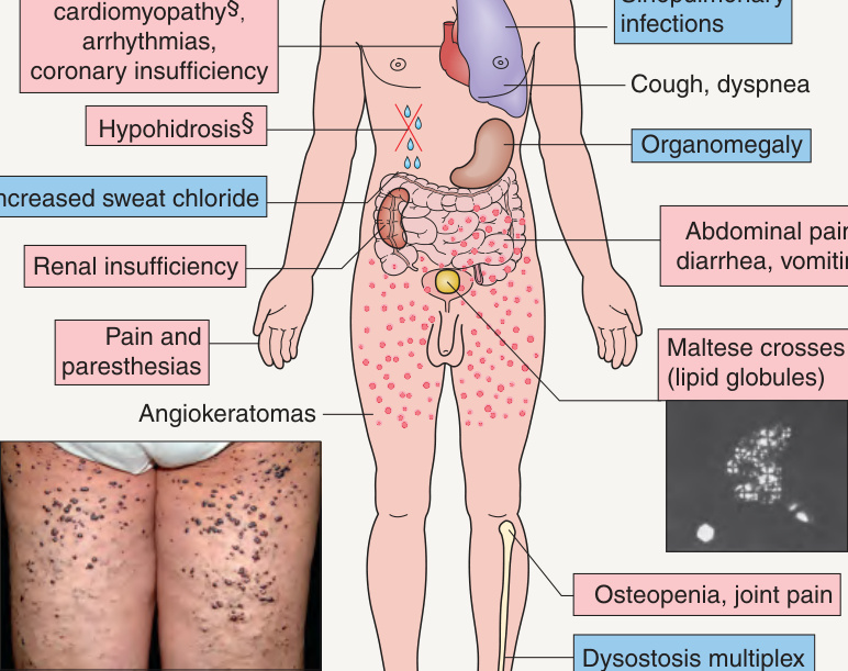
**Fig. 63.9 Clinical features of Fabry disease and fucosidosis. Traditionally, polarizing microscopy of urinary sediment revealing birefringent lipid globules (“Maltese crosses”) was utilized as a marker of Fabry disease. *Also lenticular in Fabry disease. Milder in Fabry disease. §Occasionally observed in fucosidosis. Angiokeratomas of Fabry disease, courtesy Luis Requena, MD; photomicrograph of Maltese crosses, courtesy Robert J. Desnick, MD PhD.
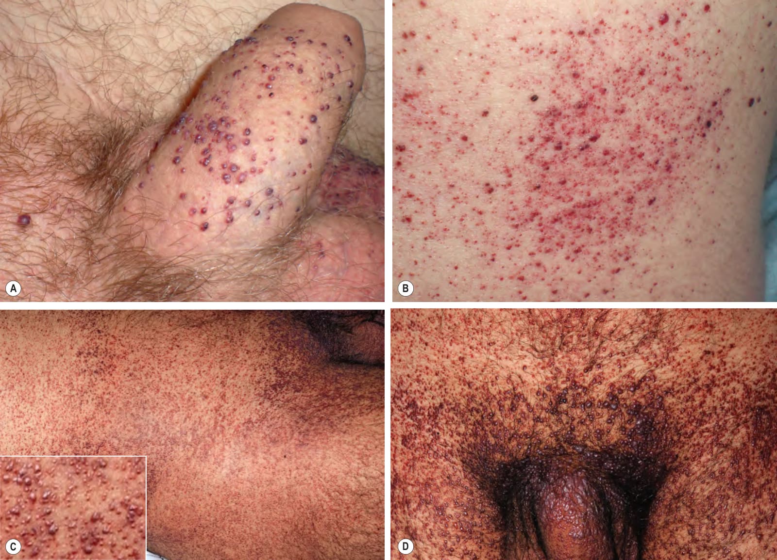
Fig. 63.10 Angiokeratomas of Fabry disease. Multiple dark red to red–brown macules and papules located: (A) on the penis, an uncommon site for angiokeratomas in the general population, as well as on the scrotum; (B) on the lower trunk; and (C, D) in a more extensive “bathing trunk” distribution. A,B, Courtesy Paula Luna, MD; C,D, Courtesy Edward Cowen, MD.
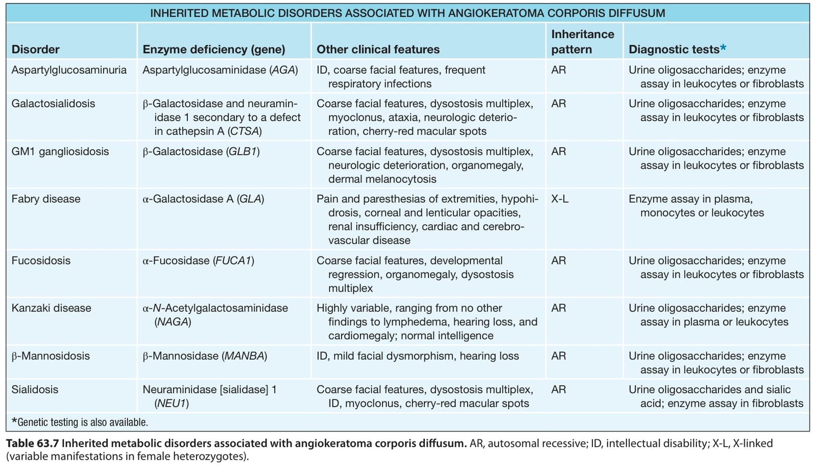
Table 63.7 Inherited metabolic disorders associated with angiokeratoma corporis diffusum. AR, autosomal recessive; ID, intellectual disability; X-L, X-linked (variable manifestations in female heterozygotes).