FUCOSIDOSIS
Key features
Autosomal recessive disorder due to deficiency of α-fucosidase
Multiple angiokeratomas of the skin and oral mucosa
Extracutaneous findings include coarse facial features, dysostosis multiplex, and progressive mental and motor deterioration
Fucosidosis is a rare autosomal recessive lysosomal storage disorder resulting from pathogenic variants in FUCA1, which encodes the enzyme α-fucosidase. In the past, patients were classified into two groups: type I, a more severe form with onset in the first year of life; and type II, a milder form with onset in the second year or later. However, a continuous spectrum of severity is now recognized. The extracutaneous manifestations include coarse facial features, short stature, organomegaly, dysostosis multiplex, and neurologic deterioration with associated hypomyelination (see Fig. 63.9). Patients may also have corneal opacities, retinal vascular abnormalities, and frequent sinopulmonary infections.
Angiokeratomas virtually indistinguishable from those associated with Fabry disease are found in approximately one-third of fucosidosis patients <10 years of age and 85% of those 10–19 years of age. The lesions are distributed primarily on the trunk and lower extremities but can also develop within the mouth. Other reported cutaneous findings include telangiectasias, acrocyanosis, nevus anemicus, xerosis, and elevated sweat chloride levels. The light microscopy findings of the angiokeratomas are similar to those seen in Fabry disease. Electron microscopic examination of clinically normal skin may reveal emptyappearing storage vesicles in endothelial cells, melanocytes, eccrine glands, and fibroblasts.
The diagnosis of fucosidosis is supported by demonstration of characteristic abnormalities on urine oligosaccharide analysis and established by an α-fucosidase assay utilizing leukocytes or cultured skin fibroblasts. Prenatal diagnosis is possible via enzyme analysis or genetic testing. There is no definitive treatment for fucosidosis, and the mortality rate is high, especially after the second decade of life. Hematopoietic stem cell transplantation has been attempted with some evidence of clinical improvement, and enzyme replacement therapy is under investigation in a canine model.
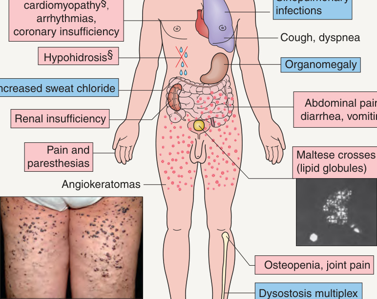
**Fig. 63.9 Clinical features of Fabry disease and fucosidosis. Traditionally, polarizing microscopy of urinary sediment revealing birefringent lipid globules (“Maltese crosses”) was utilized as a marker of Fabry disease. *Also lenticular in Fabry disease. Milder in Fabry disease. §Occasionally observed in fucosidosis. Angiokeratomas of Fabry disease, courtesy Luis Requena, MD; photomicrograph of Maltese crosses, courtesy Robert J. Desnick, MD PhD.
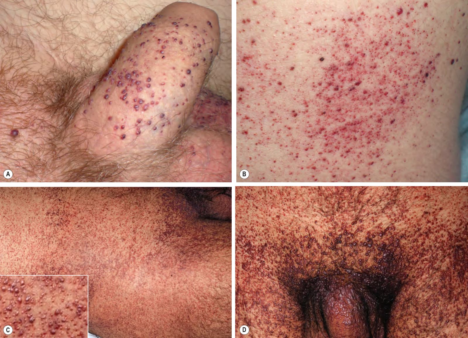
Fig. 63.10 Angiokeratomas of Fabry disease. Multiple dark red to red–brown macules and papules located: (A) on the penis, an uncommon site for angiokeratomas in the general population, as well as on the scrotum; (B) on the lower trunk; and (C, D) in a more extensive “bathing trunk” distribution. A,B, Courtesy Paula Luna, MD; C,D, Courtesy Edward Cowen, MD.