GAUCHER DISEASE
Key features
Autosomal recessive disorder due to deficiency of glucocerebrosidase
Extracutaneous findings include hepatosplenomegaly, bone disease, cytopenias, and highly variable neurologic manifestations
Cutaneous manifestations of type 1 disease include yellow–brown pigmentation, easy tanning, and petechiae
A subset of patients with type 2 (acute neuronopathic) Gaucher disease have ichthyosiform skin at birth
Gaucher disease is a relatively common autosomal recessive lysosomal storage disorder in which the glycolipid glucocerebroside accumulates as a result of a deficiency of the enzyme glucocerebrosidase (β-glucosidase). There are three clinical subtypes of the disorder, although the acute and chronic neuronopathic forms (types 2 and 3, respectively) are thought to exist on a continuum. Type 1 is by far the most common form of Gaucher disease and has a particularly high incidence of ~1 in 850 births in the Ashkenazi Jewish population.
Type 1 Gaucher disease is distinguished by a lack of primary involvement of the CNS, although Parkinson disease-like symptoms develop by the eighth decade of life in ~10% of affected individuals.
Presenting findings in type 1 may include hepatosplenomegaly, thrombocytopenia, anemia, and bone pain. The cutaneous manifestations of type 1 Gaucher disease are infrequent and relatively nonspecific. They include diffuse brown or yellow–brown pigmentation of the skin and easy tanning. In addition, petechiae and ecchymoses may be observed due to the thrombocytopenia that results from infiltration of the bone marrow with glycolipid-filled macrophages (Gaucher cells) or from hypersplenism.
Type 2 Gaucher disease, the acute neuronopathic form of the disorder, is a severe and rapidly progressive condition that usually results in death before 2 years of age. Clinical manifestations develop within the first year of life and include hepatosplenomegaly, severe hypertonia, opisthotonus, oculomotor apraxia, and progressive neurologic deterioration. A subgroup of patients with this type of Gaucher disease presents at birth with a collodion baby phenotype or other ichthyosiform skin findings, suggesting that Gaucher disease should be considered in the differential diagnosis of congenital ichthyosis. Studies in a mouse model indicate that glucocerebrosidase is required to generate functionally competent membranes for normal epidermal barrier function. Electron microscopy of skin samples from patients with type 2 Gaucher disease, including those without clinically evident ichthyosis, shows immature lamellar membranes with characteristic electron-dense clefts. The residual enzymatic activity present in patients with types 1 and 3 Gaucher disease is apparently adequate to protect them, since similar epidermal abnormalities are not observed in these forms. In fact, no specific cutaneous abnormalities have been described in patients with type 3 (chronic neuronopathic) Gaucher disease.
The diagnosis of Gaucher disease is established by demonstration of a glucocerebrosidase deficiency in leukocytes or cultured skin fibroblasts. Carrier detection is possible by genetic analysis, and prenatal diagnosis can be accomplished via chorionic villus sampling (CVS) or amniocentesis. Biomarkers with utility in determining prognosis and monitoring response to therapy include chitotriosidase, CC chemokine ligand 18 (CCL18), glucosylsphingosine, and ferritin. There are two forms of specific treatment for Gaucher disease: (1) intravenous recombinant enzyme-replacement therapy for types 1 (when symptomatic) and 3; and (2) substrate reduction therapy with miglustat or eliglustat, inhibitors of glucosylceramide synthase, for type 1. These treatments can improve organomegaly, hematologic abnormalities, and (to a variable degree) bone disease, but not the CNS manifestations of Gaucher disease.
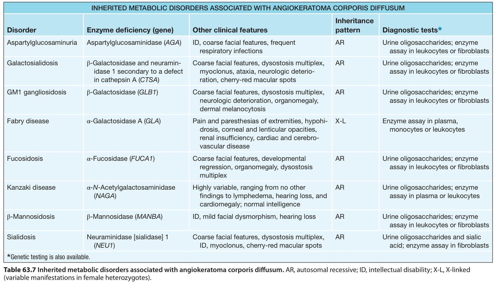
Table 63.7 Inherited metabolic disorders associated with angiokeratoma corporis diffusum. AR, autosomal recessive; ID, intellectual disability; X-L, X-linked (variable manifestations in female heterozygotes).