WERNER SYNDROME
Synonym: Progeria of the adult
Key features
Occurs in 1 in 1 million births, with a higher incidence in Japan
Autosomal recessive disorder due to mutations in WRN (RECQL2), which encodes a DNA helicase
Premature aging with the appearance of canities, cataracts, osteo- porosis, diabetes mellitus, and atherosclerosis during the second decade
Additional features include sclerodermoid changes, keratoses and ulcerations over bony prominences, short stature, and vascular and soft tissue calcification
Similar facies to progeria patients, with a pinched appearance and micrognathia
Increased risk of meningiomas, sarcomas, and (particularly in
Japanese patients) thyroid carcinomas and melanomas
Introduction
Werner syndrome is a genetic disorder of accelerated aging with an onset in the second decade of life. This rare progeroid syndrome has a higher incidence in certain populations in Japan. Although it is often referred to as progeria of the adult, Werner syndrome has some distinctive clinical findings not normally associated with aging, such as hypogonadism, laryngeal atrophy, and osteosclerosis of the distal extremities. The discovery of underlying mutations in WRN (RECQL2) has allowed investigation into the molecular aspects of Werner syndrome.
History
In 1904, Otto Werner described a family with two brothers and two sisters between the ages 36 and 40 years who displayed clinical features of premature aging. In 1934, Oppenheimer and Kugel reported two similar cases and established the eponym Werner syndrome.
Epidemiology
Werner syndrome is a rare autosomal recessive disorder with an overall incidence of 1 in 1 million births. However, its incidence in Japan is higher and may approach 1 in 3500 in some communities owing to high rates of consanguinity. It was the study of several large Japanese families that led to identification of the gene responsible for this disorder. Werner syndrome has been reported in all races and affects both sexes equally.
Pathogenesis
Werner syndrome is caused by mutations in WRN (RECQL2), which encodes a homolog of the Escherichia coli RecQ DNA helicase. Almost all of the mutations identified to date predict a truncated protein, and over half of Japanese patients are homozygous for a specific splice-site mutation.
The Werner protein (WRN) has both exonuclease and helicase activities, and it plays a role both in optimizing DNA repair, particularly via base excision, and in suppressing illegitimate recombination. Therefore, loss of WRN activity results in genomic instability. The accumulation of senescent cells with decreasing replicative capacity and an increasing number of mutations is thought to lead to the clinical findings of premature aging and an increased risk of malignancy. Decreased heterochromatin stability is also thought to play a role in the accelerated cellular senescence of Werner syndrome as well as physiologic aging. Loss of function of WRN may occur in the normal population as a function of age.
A subset of patients have “atypical Werner syndrome” and lack pathogenic changes in WRN. Rather, they have heterozygous missense mutations in LMNA (see HGPS section) that affect the heptad repeat region and are predicted to interfere with protein– protein interactions. Compared to patients with WRN mutations, those with atypical Werner syndrome tend to have an earlier onset of disease and more severe age-related manifestations. Atypical Werner syndrome might therefore be better classified as a late-onset form of HGPS.
Clinical Features
In most affected individuals, growth progresses normally until the beginning of the second decade, when short stature and thin limbs become noticeable. Graying of the hair may first appear during childhood but characteristically develops in late adolescence or the early twenties, often accompanied by hair thinning. While other findings of Werner syndrome typically become evident during the second and third decades of life, diagnosis is often delayed until the fourth or fifth decade.
The typical patient is short, with an average height of 5 ft (1.5 m), and has spindly limbs but central obesity. The hands and feet are small, and the face is thin with a pinched appearance, prominent eyes, a beaked nose, circumoral radial furrows, taut lips, protuberant teeth, and micrognathia (Fig. 63.14). The voice is high-pitched and raspy.
Cutaneous changes include atrophy (epidermal, dermal and subcutaneous), scale, mottled hyperpigmentation, and tightness reminiscent of scleroderma. These findings are most prominent on the face, forearms, hands, legs, and feet. Nails may be dystrophic, hypoplastic, or absent, and plantar hyperkeratosis is common. Thick keratoses
develop over pressure points such as the fingers, toes, ankles, elbows and, occasionally, the ears. Removal of these keratoses by accidental or deliberate trauma leaves painful progressive ulcers. Ulcers may be resistant to therapy and prone to infection because of ischemia related to peripheral vascular disease. Dystrophic soft tissue calcification and osteomyelitis can also complicate chronic ulcers.
Other characteristic findings in Werner syndrome that reflect accelerated aging include bilateral cataracts, type 2 diabetes mellitus, hyperlipidemia, generalized atherosclerosis, osteoporosis, and less often hypertension. Additional features not normally associated with aging include hypogonadism, laryngeal atrophy, and osteosclerosis of the extremities. Affected individuals have an increased risk of meningiomas, soft tissue sarcomas, and osteosarcoma. Japanese patients are also at increased risk for thyroid follicular carcinomas and melanomas, especially in acral and mucosal locations.
The average lifespan is ~54 years, and death is usually related to cardiovascular and cerebrovascular disease. Some authors have postulated that heterozygous carriers may have higher rates of malignancy and myocardial infarction than the general population.
Pathology and Laboratory Findings
The epidermis is hyperkeratotic and atrophic, with focal hypermelanosis of the basal layer. Appendages are decreased in number and atrophic, and there is fibrosis and variable hyalinization of the dermis. The fat is also atrophic and often replaced by hyalinized connective tissue. Vessels may show changes typical of diabetic angiopathy.
Differential Diagnosis
A clinical diagnosis can usually be made when the characteristic skin findings, premature canities, “bird-like” facies, short stature, and bilateral cataracts are recognized. More detailed clinical diagnostic criteria are available at https://dlmp.uw.edu/research-center/werner/ diagnostic-criteria, and genetic testing can be utilized to confirm the diagnosis if clinical features are inconclusive. There is considerable overlap with mandibular hypoplasia, deafness, progeroid features, and lipodystrophy (MDPL) syndrome caused by mutations in POLD1, which encodes DNA polymerase δ (see Table 63.9); this polymerase interacts with the Werner helicase during DNA replication and repair. However, MDPL is differentiated by frequent hearing impairment, absence of cataracts, and no predisposition to malignancy. In addition to other premature aging syndromes outlined in Table 63.9, Werner syndrome must be distinguished from ataxia–telangiectasia (see Ch. 60), prolidase deficiency (which features facial dysmorphism, telangiectasias, recalcitrant leg ulcers, and premature canities), and disorders associated with plantar keratoderma and scleroderma-like skin changes, such as Huriez syndrome (see Ch. 58).
Treatment
Genetic counseling should be provided, and prenatal diagnosis can be offered to affected families. Skin ulcers may prove resistant to therapy and should be treated aggressively and early, including with skin grafting; severe recalcitrant disease may lead to lower limb amputation. Case reports have suggested that etidronate can ameliorate painful soft tissue calcifications. Management of diabetes mellitus and hyperlipidemia with proper diet and appropriate medications (e.g. thiazolidinediones, metformin, dipeptidyl peptidase 4 inhibitors [“gliptins”], lipid-lowering agents) can help to reduce complications, including atherosclerosis. Vitamin C supplementation was found to reverse age-related metabolic abnormalities and increase the lifespan in a mouse model of Werner syndrome, suggesting that vitamin C might be beneficial for Werner syndrome patients. Other potential treatments that are under investigation include mTOR inhibitors and CRISPR/ Cas9-mediated gene correction.
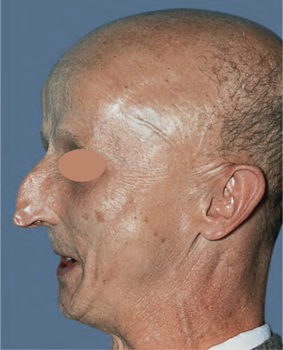
Fig. 63.14 Werner syndrome. Characteristic features include a beaked nose, taut skin, prominent veins, and micrognathia. Courtesy Ronald P. Rapini, MD.
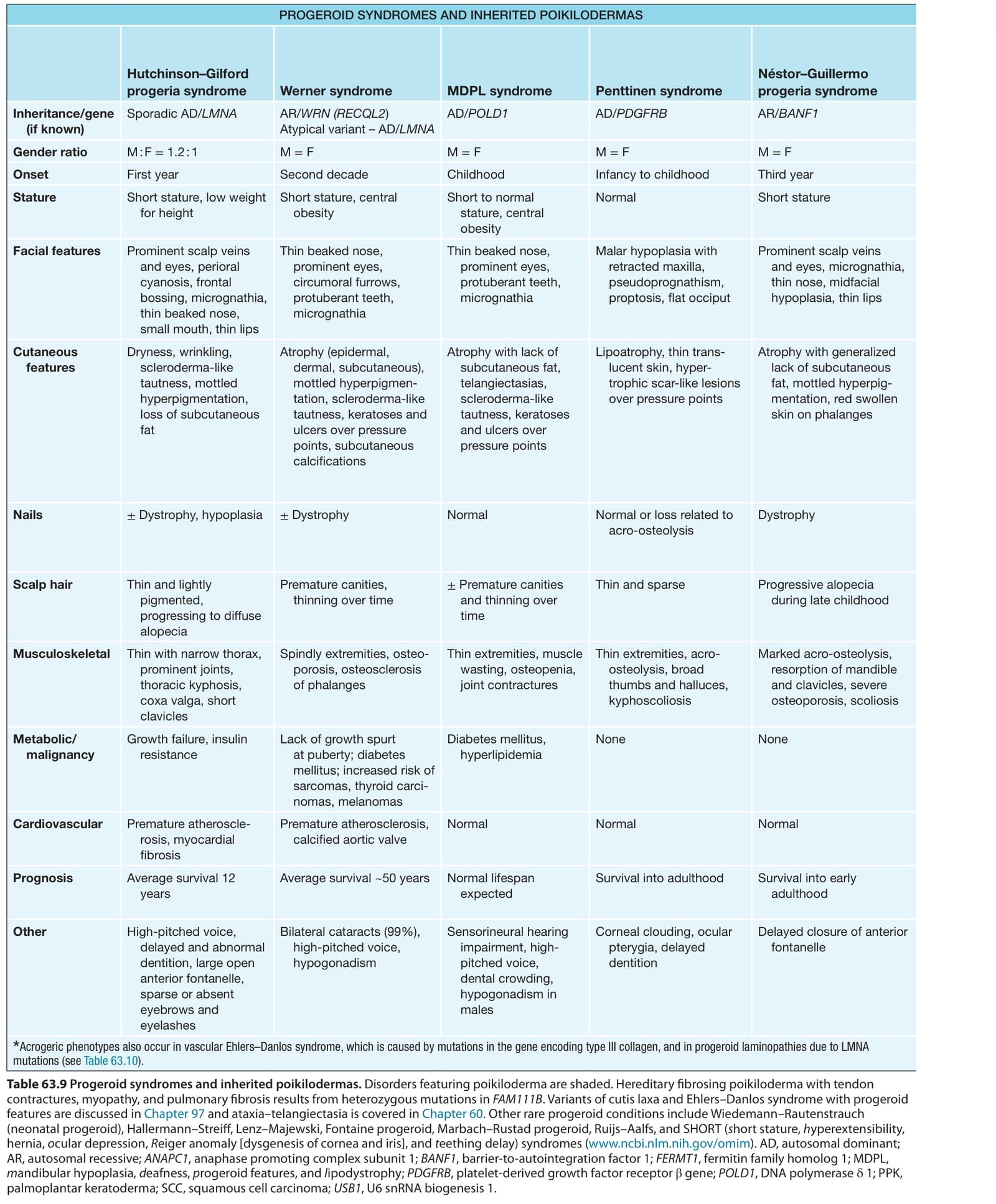
Table 63.9 Progeroid syndromes and inherited poikilodermas. Disorders featuring poikiloderma are shaded. Hereditary fibrosing poikiloderma with tendon contractures, myopathy, and pulmonary fibrosis results from heterozygous mutations in FAM111B. Variants of cutis laxa and Ehlers–Danlos syndrome with progeroid features are discussed in Chapter 97 and ataxia–telangiectasia is covered in Chapter 60. Other rare progeroid conditions include Wiedemann–Rautenstrauch (neonatal progeroid), Hallermann–Streiff, Lenz–Majewski, Fontaine progeroid, Marbach–Rustad progeroid, Ruijs–Aalfs, and SHORT (short stature, hyperextensibility, hernia, ocular depression, Reiger anomaly [dysgenesis of cornea and iris], and teething delay) syndromes (www.ncbi.nlm.nih.gov/omim). AD, autosomal dominant; AR, autosomal recessive; ANAPC1, anaphase promoting complex subunit 1; BANF1, barrier-to-autointegration factor 1; FERMT1, fermitin family homolog 1; MDPL, mandibular hypoplasia, deafness, progeroid features, and lipodystrophy; PDGFRB, platelet-derived growth factor receptor β gene; POLD1, DNA polymerase δ 1; PPK, palmoplantar keratoderma; SCC, squamous cell carcinoma; USB1, U6 snRNA biogenesis 1.