ECTODERMAL DYSPLASIAS
Encarna Guillén-Navarro, Teresa Martínez-Menchón and Virginia P. Sybert
Key features
These genetic disorders share the central feature of abnormalities in at least two of the major ectodermally derived structures – hair, teeth, nails, and sweat glands
Other ectodermal structures, such as mucous and sebaceous glands, as well as non-ectodermally derived structures may also be affected
Different forms of ectodermal dysplasia are distinguished based on the types of ectodermal abnormalities, associated non-ectodermal anomalies, and mode of inheritance, as well as the underlying genetic defect
Ectodysplasin, WNT, and p63 signaling pathways have been impli- cated in the pathogenesis of ectodermal dysplasias
Some inherited abnormalities limited to one ectodermal structure
(e.g. hair, nails) have a genetic basis related to that of ectodermal dysplasias, e.g. hypodontia due to heterozygous WNT10A mutations versus ectodermal dysplasia syndromes due to biallelic mutations in the same gene
Ectodermal dysplasias have long been recognized as a distinct group of inherited disorders that affect ectodermal appendages. Early descriptions of affected individuals were made by Danz in 1792, Wedderburn in 1838, and Darwin in 1875. The term “hereditary ectodermal dysplasia” was subsequently introduced by Weech in 1929. Ectodermal dysplasias are characterized by abnormalities in two or more of the major ectodermal structures – hair, teeth, nails, and sweat glands; other ectodermal structures, such as sebaceous and mucous glands, can also be affected.
Multiple ectodermal dysplasia classification schemes have been proposed over the years, including a descriptive clinical categorization by Pinheiro and Freire-Maia, a clinical–genetic model by Priolo and Lagana, and a functional classification by Lamartine. Over the past two decades, the molecular basis of many ectodermal dysplasias has been elucidated, leading to better understanding of processes of cell signaling involved in the induction and development of ectodermal structures as well as their interactions with mesodermal structures. International conferences were held in 2008, 2012, and 2017 with the aim of developing a classification system for ectodermal dysplasias that integrates clinical, genetic, and functional/pathway-based data and is fluid enough to incorporate new discoveries. The current ectodermal dysplasia classification system recognizes approximately 100 conditions in which defects in ectodermal structures represent the major clinical features133,133a. Disorders recognized as within other genodermatosis groups (e.g. ichthyoses, keratodermas, skin fragility disorders) and complex syndromes with major non-ectodermal manifestations are not included in this classification.
Table 63.12 p63-related ectodermal dysplasia syndromes.TP63 mutations also occur in subsets of autosomal dominant (AD) non-syndromic split-hand/foot malformation (SHFM) and isolated cleft lip/palate. ADULT, acro-dermato-ungual-lacrimal-tooth; AEC, ankyloblepharon–ectodermal defects–cleft lip/palate; EEC, ectrodactyly–ectodermal dysplasia–clefting; GER, gastroesophageal reflux; GU, genitourinary; SAM, sterile alpha motif.
Tables 63.11, 63.12 and 63.13 summarize the key clinical and genetic features of selected ectodermal dysplasias with a known molecular basis. Several classic ectodermal dysplasias are discussed in detail in this chapter. Ectodermal dysplasias that are discussed in other chapters include: Goltz syndrome (Ch. 62);
oculodentodigital dysplasia and Basan, Johanson–Blizzard, scalp– ear–nipple, and ulnar–mammary syndromes (Ch. 64); Naegeli– Franceschetti–Jadassohn syndrome and dermatopathia pigmentosa reticularis (Ch. 67); and Coffin–Siris and Zimmermann–Laband syndromes (Ch. 70).
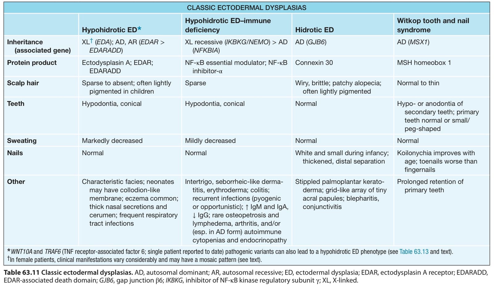
Table 63.11 Classic ectodermal dysplasias. AD, autosomal dominant; AR, autosomal recessive; ED, ectodermal dysplasia; EDAR, ectodysplasin A receptor; EDARADD, EDAR-associated death domain; GJB6, gap junction β6; IKBKG, inhibitor of NF-κB kinase regulatory subunit γ; XL, X-linked.
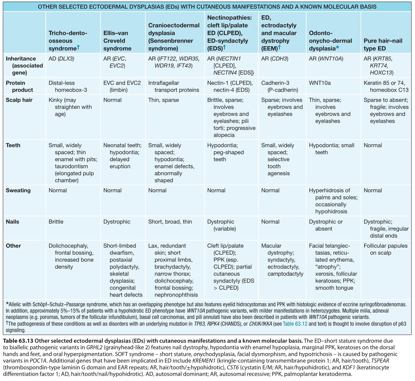
Table 63.13 Other selected ectodermal dysplasias (EDs) with cutaneous manifestations and a known molecular basis. The ED–short stature syndrome due to biallelic pathogenic variants in GRHL2 (grainyhead-like 2) features nail dystrophy, hypodontia with enamel hypoplasia, marginal PPK, keratoses on the dorsal hands and feet, and oral hyperpigmentation. SOFT syndrome – short stature, onychodysplasia, facial dysmorphism, and hypotrichosis – is caused by pathogenic variants in POC1A. Additional genes that have been implicated in ED include KREMEN1 (kringle-containing transmembrane protein 1; AR, hair/tooth), TSPEAR (thrombospondin-type laminin G domain and EAR repeats; AR, hair/tooth/±hypohidrotic), CST6 (cystatin E/M; AR, hair/hypohidrotic), and KDF1 (keratinocyte differentiation factor 1; AD, hair/tooth/nail/hypohidrotic). AD, autosomal dominant; AR, autosomal recessive; PPK, palmoplantar keratoderma.