HYPOHIDROTIC ECTODERMAL DYSPLASIA
Synonyms: Anhidrotic ectodermal dysplasia Christ–Siemens– Touraine syndrome
Hypohidrotic ectodermal dysplasia (HED) refers to a group of disorders that share the following features: sparse or absent hair; missing or peg-shaped teeth; and decreased ability to sweat. The most common form is X-linked, and traditionally the term HED has referred to this condition. However, clinically similar conditions with autosomal dominant and autosomal recessive inheritance can result from molecular defects that affect the same pathway as in X-linked HED.
Epidemiology
The X-linked form of HED is estimated to affect 0.5–2 in 10 000 live-born boys and occurs in all ethnic groups. The autosomal dominant and recessive forms are much less common.
Pathogenesis
HED is caused by pathogenic variants in genes that affect the ectodysplasin signal transduction pathway140,140a (Fig. 63.15). Epithelial cells in the developing tooth, hair follicle, and eccrine gland utilize this pathway during morphogenesis, and errors in signaling result in aplasia, hypoplasia, or dysplasia of these structures. Activation of the ectodysplasin pathway at a critical time during development leads to translocation of the nuclear factor-κB (NF-κB) transcription factor into the nucleus of these epithelial cells, thereby altering the expression of a variety of target genes. The change in gene expression likely has an effect on both cellular proliferation and survival.
The EDA gene that is altered in X-linked HED encodes a soluble ligand, ectodysplasin A. It is secreted by a subset of epithelial cells and binds to its receptor (ectodysplasin-A receptor; EDAR) on another group of epithelial cells. Pathogenic variants in EDAR can cause autosomal recessive or autosomal dominant HED. Less often, autosomal recessive and autosomal dominant HED is caused by pathogenic variants in EDARADD (EDAR-associated death domain), which encodes an intracellular adapter protein that assists in transducing the signal from the activated receptor to the nucleus. A dominant-negative mutation in TRAF6
(TNF receptor-associated factor 6), which encodes a protein that interacts with EDARADD, has also been reported in a patient with HED. In addition, mutations in WNT10A are responsible for a subset (~5%–15%) of patients with an HED phenotype (see below and Table 63.12).
Clinical Features
Affected newborns may present with a collodion-like membrane or with marked skin scaling. Scalp hair is sparse to absent (Fig. 63.16), and, when present, is usually lightly pigmented. Hair may darken at puberty and secondary sexual hairs are typically normal, although body hair is usually sparse or absent. Most male patients are unable to sweat to a detectable degree. This can lead to elevation of core temperature in warm environments or with exertion, and symptomatic hyperthermia is a major problem. Affected infants often present with fever of unknown origin, and hyperthermia during the first few years of life can be fatal when HED is not recognized. The skin is smooth and dermatoglyphics may be effaced because of absent eccrine pores. Atopy is a major cause of morbidity, with eczema affecting nearly two-thirds of HED patients. Periorbital wrinkling and hyperpigmentation are common. Sebaceous hyperplasia of the face, clinically resembling milia, can develop over time (see Fig. 63.16E). The nails are usually normal.
Both the primary and secondary dentition are affected. Teeth may be absent or reduced in number and abnormally shaped (e.g. peg-shaped). Dental radiographs are helpful in determining the extent of hypodontia, and prenatal tooth germ sonography can reliably identify affected fetuses between 18 and 28 weeks of gestation. Individuals with HED typically have an alteration in their facial appearance characterized by a saddle nose, full everted lips and frontal bossing (see Fig. 63.16). Nasal secretions and cerumen are thick and viscous, and recurrent respiratory tract infections are common. The voice is frequently hoarse or raspy. Affected infants often develop gastroesophageal reflux and feeding problems. Unilateral or bilateral amastia is an occasional feature.
Female patients with the X-linked form of HED fall into one of three categories: (1) carriers with no detectable clinical features; (2) limited involvement with findings such as decreased hair density, one or more peg-shaped or missing teeth, patchy distribution of sweat glands along the lines of Blaschko, and relative hyperpigmentation of the skin that lacks adnexa (most evident on the back); or (3) full-blown features of the disorder (Fig. 63.17). Although this variability has been classically linked to the random nature of X-inactivation (see Ch. 62), hypomorphic EDA variants, genetic modifiers, and additional biologic mechanisms may also play a role.
Pathology
A skin biopsy is usually not necessary, although a scalp or palmar biopsy specimen lacking eccrine structures was traditionally considered diagnostic of HED. Microscopic examination of hairs often shows small or variable shaft diameters and parallel dark bands of various lengths resembling a “bar code”.
Differential Diagnosis
In patients with clinical features of HED, options to confirm the diagnosis include single gene testing or a next-generation sequencing (NGS)-based multigene panel including EDA, EDAR, EDARADD, TRAF6, and WNT10A. The majority of affected individuals, either male or female, have the X-linked form. Homozygous or compound heterozygous pathogenic variants in the WNT10A gene can give rise to an HED phenotype without associated facial dysmorphism, in addition to causing odonto-onycho-dermal dysplasia and Schöpf–Schulz– Passarge syndrome (see Table 63.13 and Fig. 55.6). Heterozygous WNT10A pathogenic variants also represent a common etiology of mild HED or isolated hypodontia. At birth, HED can be confused with an ichthyosis if a collodion-like membrane is present (see Ch. 57, Table 57.4). Recurrent fevers in an infant may lead to consideration of infectious diseases before the features of HED are recognized. HED with immune deficiency (HED-ID) due to pathogenic variants in the IKBKG (NEMO) gene can be differentiated by the clinical and laboratory features of the associated immune system abnormalities and confirmed by genetic analysis (see below).
Treatment
Controlling ambient temperatures and external methods of cooling, e.g. wet T-shirts, wet headbands and cooling vests, are critical to prevent hyperthermia in children with HED. Regular use of moisturizers is helpful for dry skin. Dentures can be fitted in children as young as 3 years of age, and dental restoration through implants should be employed for older patients. Multidisciplinary care is required for treatment of other manifestations, which range from nasal concretions, asthma and recurrent respiratory infections to weight deficits and reduced salivary secretion. Patient organizations such as the National Foundation for Ectodermal Dysplasias (www.nfed.org) are also an important aspect of care, providing information and support to affected families.
Prenatal administration of recombinant EDA protein is a promising approach to correct the X-linked HED phenotype. This method has been successfully utilized in murine and canine models. Intra-amniotic administration of recombinant Fc-EDA fusion protein (constant domain of IgG1 plus receptor-binding portion of EDA) to three human fetuses with X-linked HED at 26 ± 31 weeks’ gestation had positive effects on tooth development and restored sweating ability in the infants.
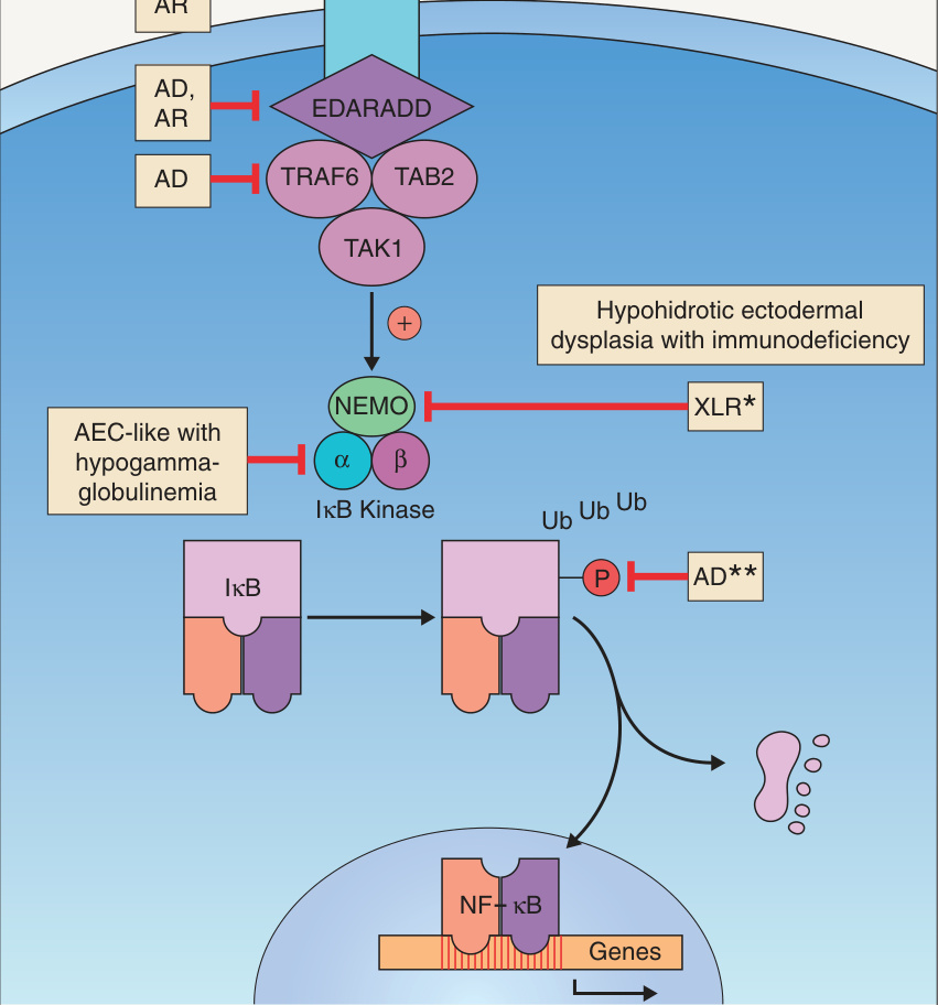
**Fig. 63.15 The ectodysplasin signal transduction pathway. The IκB kinase α subunit (IKKα), which is also known as CHUK (component of IκB kinase complex), is upregulated by p63; this explains the ankyloblepharon–ectodermal defects– cleft lip/palate (AEC) syndrome-like phenotype in individuals with pathogenic variants in this gene. *Allelic with incontinentia pigmenti. A hypermorphic mutation in the gene encoding IκBα that prevents this protein’s phosphorylation (and subsequent ubiquitination/degradation) can result in continued inhibition of NF-κB and a phenotype of hypohidrotic ectodermal dysplasia with immunodeficiency. AD, autosomal dominant; AR, autosomal recessive; EDA, ectodysplasin A; EDAR, EDA receptor; EDARADD, EDAR-associated death domain adapter protein; NEMO, NF-κB essential modulator (also known as IKBKG, inhibitor of NF-κB kinase regulatory subunit γ); XL(R), X-linked (recessive).
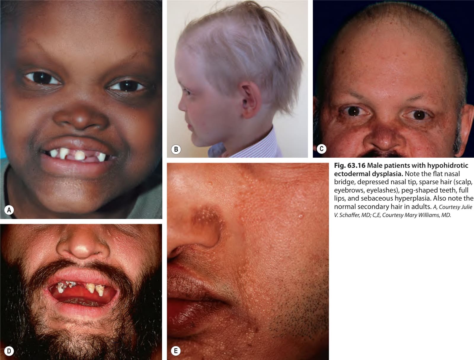
Fig. 63.16 Male patients with hypohidrotic ectodermal dysplasia. Note the flat nasal bridge, depressed nasal tip, sparse hair (scalp, eyebrows, eyelashes), peg-shaped teeth, full lips, and sebaceous hyperplasia. Also note the normal secondary hair in adults. A, Courtesy Julie V. Schaffer, MD; C,E, Courtesy Mary Williams, MD.
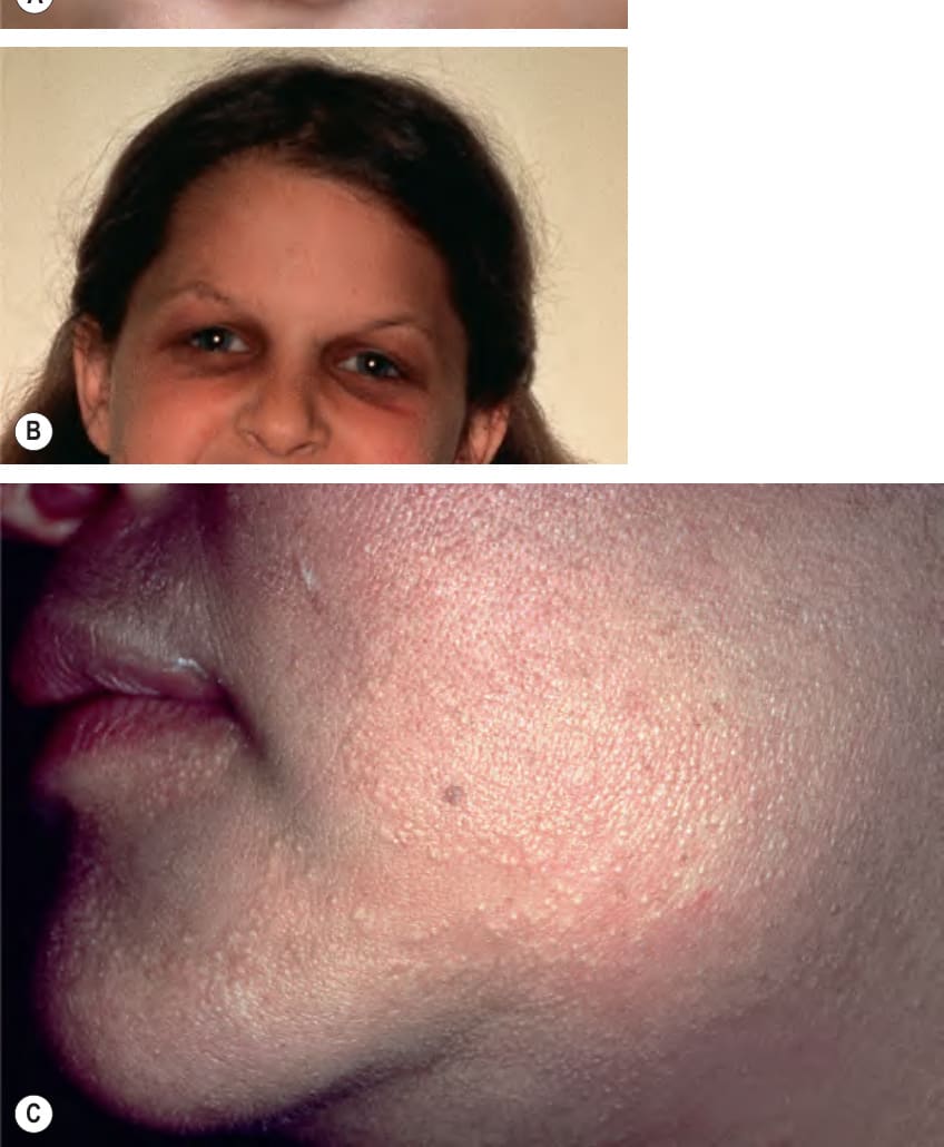
Fig. 63.17 Female patients with X-linked hypohidrotic ectodermal dysplasia.
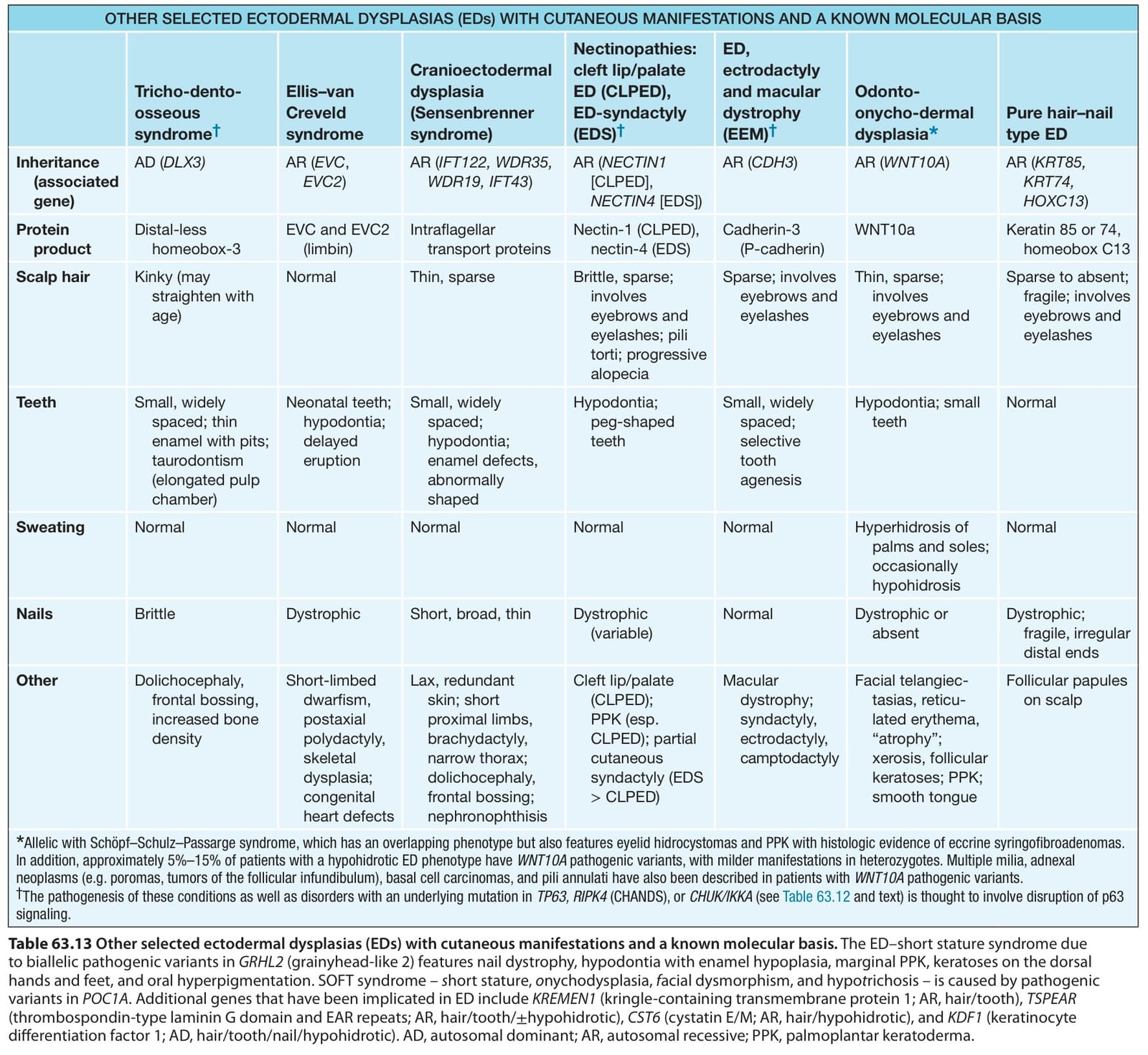
Table 63.13 Other selected ectodermal dysplasias (EDs) with cutaneous manifestations and a known molecular basis. The ED–short stature syndrome due to biallelic pathogenic variants in GRHL2 (grainyhead-like 2) features nail dystrophy, hypodontia with enamel hypoplasia, marginal PPK, keratoses on the dorsal hands and feet, and oral hyperpigmentation. SOFT syndrome – short stature, onychodysplasia, facial dysmorphism, and hypotrichosis – is caused by pathogenic variants in POC1A. Additional genes that have been implicated in ED include KREMEN1 (kringle-containing transmembrane protein 1; AR, hair/tooth), TSPEAR (thrombospondin-type laminin G domain and EAR repeats; AR, hair/tooth/±hypohidrotic), CST6 (cystatin E/M; AR, hair/hypohidrotic), and KDF1 (keratinocyte differentiation factor 1; AD, hair/tooth/nail/hypohidrotic). AD, autosomal dominant; AR, autosomal recessive; PPK, palmoplantar keratoderma.