HIDROTIC ECTODERMAL DYSPLASIA
Synonym: Clouston syndrome
Hidrotic ectodermal dysplasia was first described in a French-Canadian kindred and the ancestry of many affected individuals was traced to a single founder. Subsequently, the disorder has been reported in individuals of varying ethnic backgrounds.
Pathogenesis
Hidrotic ectodermal dysplasia is an autosomal dominant condition that is caused by missense mutations in the GJB6 gene, which encodes the connexin 30 protein and is thought to be regulated by the p63 transcription factor. Connexins oligomerize to form gap junctions that are important for communication between cells (see Ch. 58), and GJB6 is expressed in keratinocytes. Pathogenic variants in genes that encode other connexins cause skin disorders ranging from Vohwinkel and keratitis–ichthyosis–deafness (KID) syndromes (GJB2) to erythrokeratodermia variabilis (GJB3 and GJB4) (see Table 58.5). Pathogenic variants in GJB6 can also cause non-syndromic, autosomal dominant, digenic (together with a GJB2 mutation) deafness, with normal teeth and hair in affected individuals; less often the inheritance pattern is autosomal
recessive. A GJB6 mutation has also been described in a patient with a KID syndrome-like phenotype that included congenital atrichia.
Clinical Features
Hidrotic ectodermal dysplasia is characterized by the triad of hypotrichosis, nail dystrophy, and palmoplantar keratoderma, with normal teeth and sweating. The hair is wiry, brittle, and pale; patchy alopecia is common. Both hair loss and nail changes may progress over time. In affected infants, the nails are typically milky white and smaller than normal with gradual thickening throughout childhood. In adults, the nail plates grow slowly, are thick, and separate from the nail bed distally (Fig. 63.18). Keratoderma with stippling of the palms and soles can also be progressive (see Ch. 58). Tiny papules in a grid-like array, corresponding to eccrine acrosyringia, or larger papules coalescing in a cobblestone-like pattern may extend from the palms and soles onto the dorsal surface of the digits, especially distally (see Fig. 63.18 and Fig. 58.11); the latter corresponds to where the dermatoglyphics are more prominent. Similar papules may also occur on the extensor surfaces of the extremities. Oral leukoplakia has been described, and sparse eyelashes may predispose patients to conjunctivitis and blepharitis.
Pathology
Histologically, the thickened palms and soles have orthohyperkeratosis with a normal granular layer. Eccrine syringofibroadenomatosis, which is characterized by proliferation of ductal structures within a fibrovascular stroma, may be observed when papular lesions are biopsied. The hair does not have specific microscopic changes.
Differential Diagnosis
The hair abnormalities in hidrotic ectodermal dysplasia distinguish it from pachyonychia congenita, which may have similar nail findings. A hidrotic ectodermal dysplasia-like phenotype plus deafness has been reported in a few patients with a GJB2 mutation, and the dominant deafness–onychodystrophy syndrome results from pathogenic variants in ATP6V1B2.
Treatment
The National Foundation for Ectodermal Dysplasias can provide support to affected individuals and families. Ablation of the nail matrix is occasionally necessary for pain relief, and patients with substantial alopecia may choose to use hairpieces. Management of hyperkeratotic palms and soles is difficult; similar strategies to those used for the palmoplantar keratodermas, e.g. α-hydroxy acids, urea, soaking and paring, can be employed.
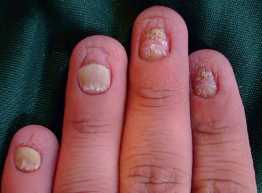
Fig. 63.18 Hidrotic ectodermal dysplasia (Clouston syndrome). Note the thickened, shortened nails with distal separation and tiny papules in a regular distribution on the fingertips.