ANKYLOBLEPHARON–ECTODERMAL DEFECTS– CLEFT LIP/PALATE SYNDROME
Synonyms: Hay–Wells syndrome Rapp–Hodgkin syndrome
This autosomal dominant ectodermal dysplasia, first described in 1976, has been reported in ethnically and geographically diverse individuals and families.
Pathogenesis
The ankyloblepharon–ectodermal defects–cleft lip/palate (AEC) syndrome is caused by TP63 mutations that typically affect the sterile alpha motif (SAM) domain of the p63 protein. p63, a transcription factor, is expressed within basal keratinocytes of the skin, which have a high proliferative capacity that is lost in differentiated keratinocytes. The p63 protein has multiple isotypes, with the SAM domain present in only some forms. This helps to explain the phenotypic differences between AEC syndrome and ectodermal dysplasia–ectrodactyly–clefting (EEC) syndrome (see Table 63.12 and below); the latter is usually due to mutations within the DNA-binding domain of p63.
Clinical Features
This disorder is evident at birth. Up to 90% of affected infants present with erythroderma, peeling skin, or erosions, which can result in lifethreatening infectious complications (Fig. 63.20A). The scalp is almost always involved, and a chronic, erosive scalp dermatitis is a characteristic finding (Fig. 63.20B,C). The scalp hair is wiry and usually lightly pigmented and sparse, often with patchy alopecia. Some degree of nail dystrophy is typically evident, with findings ranging from hyperconvex, thickened nail plates to anonychia. Patients may have decreased sweating and heat intolerance.
Patients often develop abnormal granulation tissue and recurrent bacterial infections of the skin. Cribriform and stellate scarring tends to occur in a shawl-like distribution on the shoulders and upper trunk. Progressive reticulated hyperpigmentation frequently appears in inter-triginous areas, and hypopigmentation of the scalp and face may be seen in children with darkly pigmented skin.
Congenital strands of tissue between the eyelids (ankyloblepharon adnatum filiforme) are observed in approximately three-quarters of affected individuals (Fig. 63.20D). These may lyse spontaneously, even prior to birth, or require surgical correction. The lacrimal ducts may be atretic. Almost all patients with AEC syndrome are born with a cleft palate with or without a cleft lip. Malformed external ears and recurrent
Small primary teeth, some conical, in a 7-year-old boy. Radiographs showed an absence of nearly all of the secondary teeth. Courtesy Julie V. Schaffer, MD.
otitis media with secondary conductive hearing loss are common. Dental abnormalities include hypodontia and misshapen (e.g. conical) teeth, and most patients have maxillary hypoplasia.
Gastroesophageal reflux develops in the majority of AEC patients, while one-quarter of affected children fail to thrive and require gastrostomy placement. Hypospadias affects up to 80% of male patients, and supernumerary nipples and/or ectopic breast tissue have been described. Limb abnormalities are more common in AEC syndrome than once thought; these malformations, which are typically less severe than the defects seen in EEC syndrome (see below), can include syndactyly (partial or complete), camptodactyly, brachydactyly, and even ectrodactyly (Fig. 63.20E).
Pathology
Hair shaft abnormalities such as pili torti and pili trianguli et canaliculi may be observed. Histopathologic changes are nonspecific and often include mild epidermal atrophy and dermal melanophages.
Differential Diagnosis
Rapp–Hodgkin syndrome, once considered to be a distinct entity, is now thought to fall within the AEC spectrum. Affected individuals have heterozygous pathogenic variants in TP63, generally affecting the SAM domain of p63. Other than a lack of ankyloblepharon, clinical findings overlap with those of AEC syndrome, including maxillary hypoplasia, cleft palate ± lip, nail dystrophy, and abnormalities of the teeth and hair. The clinical features of AEC also overlap with those of curly hair–ankyloblepharon–nail dysplasia syndrome (CHANDS), which is caused by biallelic mutations in the receptor-interacting serinethreonine kinase 4 gene (RIPK4) that is a direct transcriptional target of p63. An AEC/EEC-like syndrome presenting with hypogammaglobulinemia as well as ankyloblepharon, cleft lip/palate, conical teeth, sparse hair, nail dystrophy, syndactyly/ectrodactyly, and hypoplastic genitalia can result from pathogenic variants in CHUK/IKKA (component of inhibitor of NF-κB kinase complex/inhibitor of NF-κB kinase subunit α; see Fig. 63.15), which is activated by p63. The peeling erythroderma of neonates with AEC syndrome can lead to a misdiagnosis of epidermolysis bullosa or ichthyosis.
Treatment
Surgical repair of oral clefting is usually necessary. Treatment for the erosive scalp dermatitis should be gentle and non-occlusive, with surveillance for and treatment of secondary infections. Debridement often causes erosions to worsen, and skin grafts typically fail. Ulceration and granulation tissue may be very difficult to treat, and healing tends to be slow. A recent study found that topical application APR-246/eprenetapopt/ PRIMA-1MET, which was developed to activate mutant p53 in cancer cells, led to substantial re-epithelialization of erosions on the hands and scalp that had been present for 9–15 years in two patients with AEC.
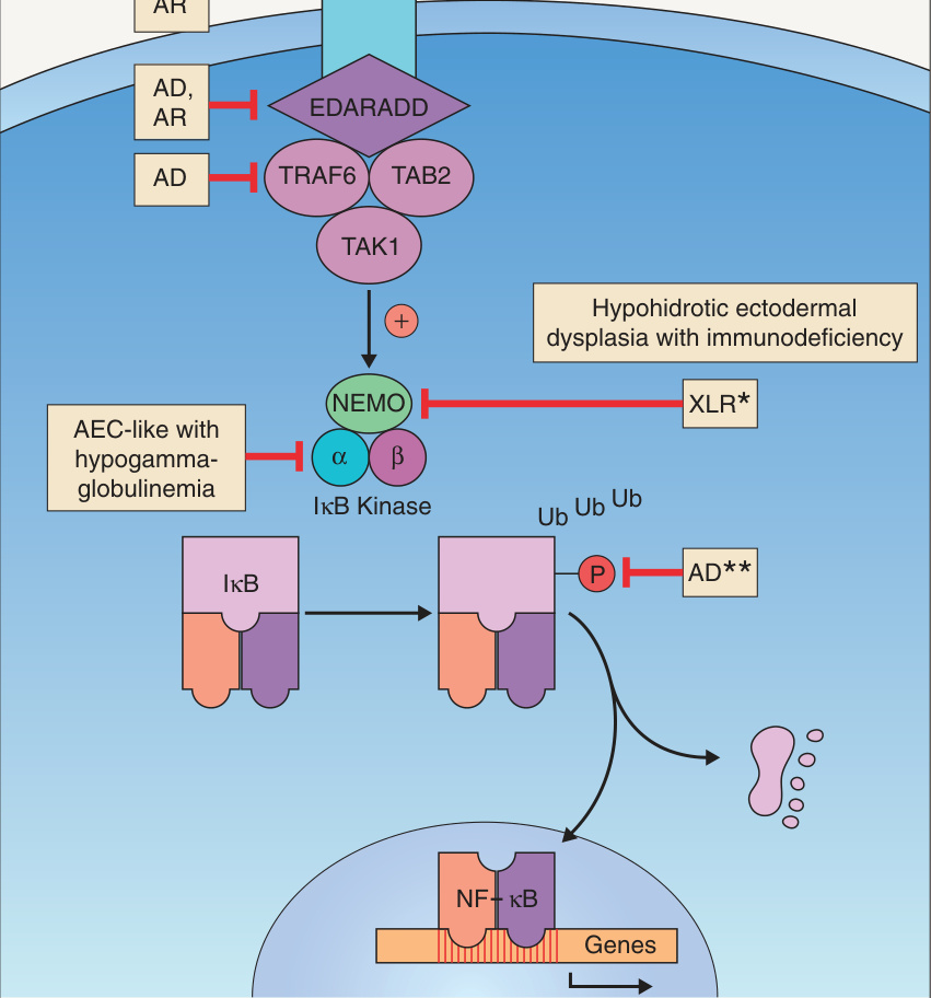
**Fig. 63.15 The ectodysplasin signal transduction pathway. The IκB kinase α subunit (IKKα), which is also known as CHUK (component of IκB kinase complex), is upregulated by p63; this explains the ankyloblepharon–ectodermal defects– cleft lip/palate (AEC) syndrome-like phenotype in individuals with pathogenic variants in this gene. *Allelic with incontinentia pigmenti. A hypermorphic mutation in the gene encoding IκBα that prevents this protein’s phosphorylation (and subsequent ubiquitination/degradation) can result in continued inhibition of NF-κB and a phenotype of hypohidrotic ectodermal dysplasia with immunodeficiency. AD, autosomal dominant; AR, autosomal recessive; EDA, ectodysplasin A; EDAR, EDA receptor; EDARADD, EDAR-associated death domain adapter protein; NEMO, NF-κB essential modulator (also known as IKBKG, inhibitor of NF-κB kinase regulatory subunit γ); XL(R), X-linked (recessive).
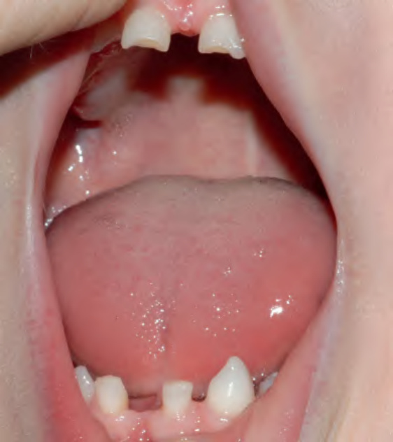
Fig. 63.19 Witkop tooth and nail syndrome.
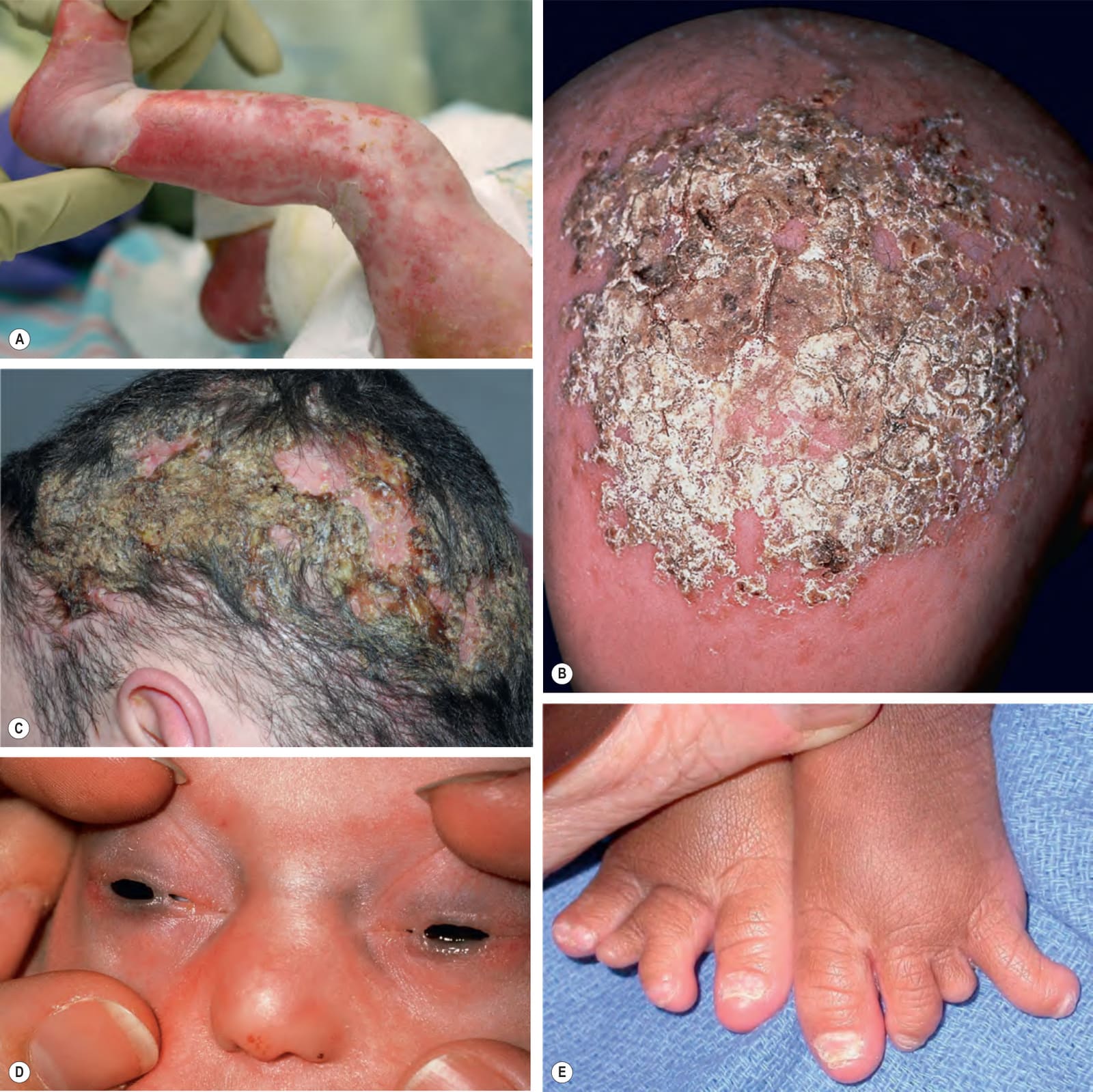
Fig. 63.20 Ankyloblepharon–ectodermal defects–cleft lip/palate syndrome.A Extensive erosions in an infant who succumbed to sepsis. B, C Thick scale-crust and alopecia of the scalp. D Strands of tissue between the eyelids (ankyloblepharon adnatum filiforme). E Digital abnormalities (including syndactyly, brachydactyly) and nail dystrophy (yellowing and thickening of the nail plate). B, Courtesy Jean L. Bolognia, MD; C, Courtesy Julie V. Schaffer, MD.