ECTODERMAL DYSPLASIA–ECTRODACTYLY– CLEFTING SYNDROME
Synonym: Split hand–split foot–ectodermal dysplasia–clefting syndrome
First described by Eckholdt and Martens in 1804, this disorder occurs in all ethnic groups and has been reported worldwide.
Pathogenesis
Ectodermal dysplasia–ectrodactyly–clefting (EEC) syndrome is an autosomal dominant disorder caused by pathogenic variants in the region of TP63 that encodes the protein’s DNA-binding domain. These mutations disrupt the ability of the p63 transcription factor, which is expressed in proliferating epidermal basal cells, to regulate gene expression. TP63 pathogenic variants also occur in several related autosomal dominant disorders, including non-syndromic split hand/foot malformation (minority of affected families), limb–mammary syndrome
and ADULT (acro-dermato-ungual-lacrimal-tooth) syndrome, as well as in AEC syndrome (see Table 63.12 and above). Patients with EEC syndrome usually have missense mutations within the p63 DNA-binding domain that differ from those seen in these other disorders.
Clinical Features
The ectodermal changes in this highly variable condition may be quite mild. The scalp hair is usually light in color and coarse. It may be sparse and grow slowly. The secondary sexual hair may also be affected. Scalp folliculitis and dermatitis are rare manifestations. Approximately 80% of affected individuals have transverse ridging, pitting, and slow growth of the nail plates. Xerotic skin and palmoplantar hyperkeratosis can occur, and sweating is usually normal.
Clefting of the palate/lip occurs in ~50% of affected individuals, and additional oral abnormalities include enamel hypoplasia, hypodontia, and premature loss of secondary teeth. Lacrimal duct defects leading to blepharitis and dacryocystitis are frequently present, and limbal stem cell deficiency can result in photophobia and keratopathy as well as corneal ulceration and scarring. Secondary conductive hearing loss is also common, and choanal atresia is an occasional manifestation.
Ectrodactyly, a specific split-hand/foot malformation due to failure of normal development of the central digits, represents a defining feature of EEC syndrome (Fig. 63.21). The ectrodactyly is frequently
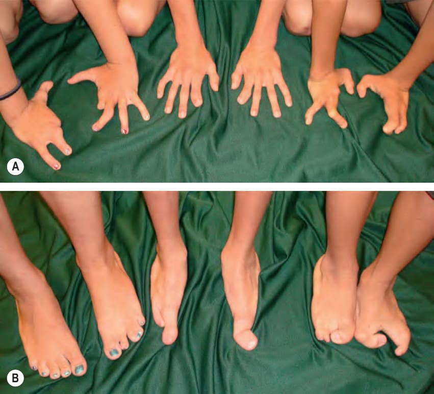
Fig. 63.21 Ectodermal dysplasia–ectrodactyly–clefting syndrome. Note the characteristic split-hand (A) and foot (B) deformities in these three siblings.
asymmetric, usually with more severe involvement of the feet than the hands. Intrafamilial variability is marked. Genitourinary abnormalities are an under-appreciated manifestation of EEC syndrome and may be associated with dysplasia of the urogenital epithelium; renal and urogenital malformations affect more than one-third of patients and can result in hydronephrosis.
The presence of split-hand/foot malformations and the absence of ankyloblepharon or cutaneous erosions help to distinguish EEC syndrome from AEC syndrome. The findings that differentiate limb– mammary syndrome and ADULT syndrome from EEC are presented in Table 63.12. Other ectodermal dysplasias that feature digital abnormalities include ectodermal dysplasia–ectrodactyly–macular dystrophy (EEM) syndrome, cleft lip/palate ectodermal dysplasia, oculodentodigital dysplasia, and ulnar–mammary syndrome (see Table 63.13 and Table 64.6). Although Goltz syndrome (focal dermal hypoplasia) can present with split-hand/foot and other digital malformations, nail dystrophy, sparse hair, dental anomalies and (occasionally) cleft lip/ palate, it is easily recognized based on the highly characteristic skin findings as well as ocular and additional skeletal (e.g. osteopathia striata) defects (see Ch. 62). Lastly, the popliteal pterygium syndrome, which is caused by pathogenic variants in the interferon regulatory factor 6 gene (IRF6) that is transcriptionally activated by p63, has features overlapping with EEC (see Ch. 64).
As for the other ectodermal dysplasias with oral clefting and lacrimal abnormalities, management involves a multidisciplinary approach. Limb defects may be ameliorated by surgical interventions. All affected individuals should be screened with a renal ultrasound. Potential approaches to therapy include small interfering RNAs (siRNAs) that selectively silence mutant TP63 alleles and the p53-activating compound APR-246/eprenetapopt/PRIMA-1MET, which was found in vitro to rescue impaired epidermal and corneal differentiation in cells from EEC patients.
The authors wish to thank Peter H. Itin, MD and Alanna F. Bree, MD for their valuable contributions to this chapter in the previous editions. The Ectodermal Dysplasia section of this chapter has been supported by the Instituto de Salud Carlos III (ISCIII), Spanish Ministry of Economy and Competitiveness (grant numbers PI/14/01259 and PI/17/00796 and PI/21/01082), FEDER funds and AADE (Spanish Association of patients with ectodermal dysplasia).
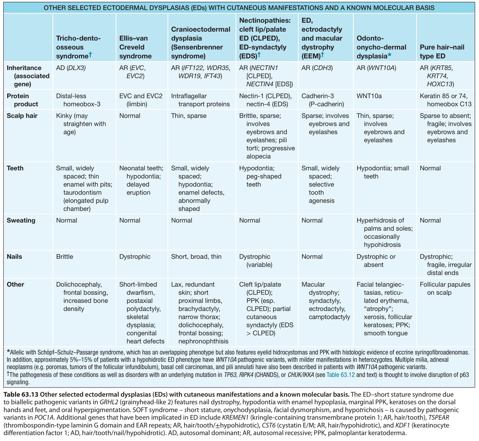
Table 63.13 Other selected ectodermal dysplasias (EDs) with cutaneous manifestations and a known molecular basis. The ED–short stature syndrome due to biallelic pathogenic variants in GRHL2 (grainyhead-like 2) features nail dystrophy, hypodontia with enamel hypoplasia, marginal PPK, keratoses on the dorsal hands and feet, and oral hyperpigmentation. SOFT syndrome – short stature, onychodysplasia, facial dysmorphism, and hypotrichosis – is caused by pathogenic variants in POC1A. Additional genes that have been implicated in ED include KREMEN1 (kringle-containing transmembrane protein 1; AR, hair/tooth), TSPEAR (thrombospondin-type laminin G domain and EAR repeats; AR, hair/tooth/±hypohidrotic), CST6 (cystatin E/M; AR, hair/hypohidrotic), and KDF1 (keratinocyte differentiation factor 1; AD, hair/tooth/nail/hypohidrotic). AD, autosomal dominant; AR, autosomal recessive; PPK, palmoplantar keratoderma.