APLASIA CUTIS CONGENITA
Synonyms: Cutis aplasia Congenital absence of skin Congenital scars
Aplasia cutis congenita (ACC) is a condition in which localized or widespread areas of skin are absent or scarred at birth. It may occur as an isolated defect, in association with other developmental anomalies, or as a feature of a variety of disorders (Tables 64.3 & 64.4). There is no single underlying cause of ACC, which simply represents a physical finding that reflects an abnormality in intrauterine skin development. The many possible etiologies include genetic factors, vascular compromise, trauma, teratogens, and intrauterine infections.
As a result of its causal heterogeneity, the clinical appearance of ACC at birth is extremely variable, ranging from an erosion or deep ulceration to a scar due to healing in utero to a discrete ovoid defect covered by a membrane. The latter, referred to as membranous aplasia cutis, is the most common form. Occurring primarily on the scalp and often surrounded by a hair collar (see Figs. 64.3 & 64.4), such lesions may represent a forme fruste of a neural tube defect. However, membranous aplasia cutis can also be seen along the embryonic fusion lines of the face (Table 64.4; Figs. 64.2 & 64.17). Membranous aplasia cutis typically presents at birth as a sharply marginated (“punched-out”), oval or round defect covered by a thin, translucent, glistening epithelial membrane. Lesions may contain serous fluid in the neonatal period, resulting in a bullous appearance (see Fig. 64.4). Eventually, they tend to flatten and transform into an atrophic scar (Fig. 64.18A). Dermoscopy typically shows a lack of follicular openings, telangiectatic vessels, and radially oriented hair follicles extending horizontally at the periphery (“starburst-like”). Hypertrophic scarring can be a manifestation of an autosomal dominant form of scalp ACC in which mutations in the ribosomal GTPase BMS1 result in a cell cycle defect.
A second major type of ACC consists of stellate or angulated lesions, which are thought to result from vascular abnormalities and/or intra-uterine ischemic events. Examples include irregular midline scalp defects in Adams–Oliver syndrome and extensive, symmetric lesions on the trunk and/or extremities associated with fetus papyraceus, placental infarction, or other forms of vascular insufficiency (Fig. 64.19). These defects usually appear as ulcers of variable depth with a raw, hemorrhagic, or granulating base (Fig. 64.18B,C).
Other patterns of ACC include a lack of skin overlying embryologic malformations, either obvious or occult, and large erosions with jagged borders on the extremities of neonates with epidermolysis bullosa. The latter likely result from the mechanical trauma of fetal movements in a setting of increased skin fragility. The morphology and distribution of the skin defects (see Fig. 64.19) as well as the presence or absence of associated abnormalities represent important clues to the etiology of ACC. Table 64.3 presents a classification scheme for ACC, and Table 64.4 describes several syndromes associated with ACC. However, because ACC is a physical finding that may result from any intrauterine event that disrupts skin development, not all cases can be neatly categorized. Unusual presentations of ACC range from an isolated defect on the ventral penis to an extreme form with absence of skin and subcutaneous tissue over >90% of the body surface area associated with extracutaneous anomalies (“systemic” ACC; see Table 64.3).
The scalp is the most common site for ACC, accounting for >85% of solitary lesions. The majority of cases of scalp ACC, whether membranous or irregular and scar-like, are located at or near the vertex, in proximity to the parietal hair whorl. Approximately 25% of patients have more than one lesion, and multiple membranous lesions may occur in a linear arrangement. Most defects are 1–2 cm in diameter, although the size can range from 0.5 to >10 cm. A capillary malformation (see above, Midline lesions of the scalp) is sometimes present in the skin surrounding membranous ACC, and dilated scalp veins may also be evident, especially in patients with Adams–Oliver syndrome (see Table 64.3). Scalp ACC involves the underlying skull in 10%–30% of cases, with increased risk when the lesion has a midline vertex location, hair collar, size >5 cm, and associated capillary stain. Large, irregular lesions are more likely to extend to deeper structures and may affect the dura and/or leptomeninges.
Histologic features of membranous aplasia cutis include an atrophic flattened epidermis, replacement of the dermis by loose connective tissue, and an absence of adnexal structures. Findings in other types of ACC vary but lesions that have healed generally show scarring, a lack of adnexa, and in some cases fragmented elastic tissue. Although histologic examination may occasionally be helpful, the diagnosis of ACC is primarily clinical.
Cutaneous defects noted at birth may initially be erroneously attributed to obstetric trauma, such as injuries from forceps or fetal scalp electrodes. Evaluation of a patient with ACC is directed by the personal, obstetric, and family history as well as the physical examination; in a neonate, it may include specific investigations such as examination of the placenta and viral studies for herpes simplex and varicella zoster. In patients with large, deep, irregular or membranous lesions of the scalp (Fig. 64.18B,E), especially when there is a hair collar and/or midline vertex location, imaging studies are indicated to assess for underlying bone defects, vascular anomalies, or brain malformations. Lastly, elevated α-fetoprotein in midtrimester maternal serum and amniotic fluid as well as elevated acetylcholinesterase in the amniotic fluid may represent early signs of ACC, although they are neither sensitive nor specific for this condition.
Most small lesions of ACC heal within the first few months of life, leaving an atrophic (see Fig. 64.18A) or, less often, hypertrophic (“lumpy”) scar. In such instances, daily cleansing and application of petrolatum or a topical antibiotic ointment until healing is complete are often the only interventions required. Underlying skull defects also tend to resolve spontaneously during infancy. However, potential complications of deep ACC of the scalp include life-threatening sagittal sinus hemorrhage/thrombosis and meningitis. Because the risk of complications increases if the period of healing is prolonged, early surgical repair of large stellate scalp lesions or those associated with a dural defect or exposure of the sagittal sinus is recommended. Preoperative imaging studies are required to identify underlying vascular structures. Even without surgical intervention, residual scarred, hairless areas generally become less conspicuous as the child grows. If desired for cosmetic reasons, excision or hair transplants can be performed later in life.
Atrophic cutaneous defect extending from the lateral eyebrow to the anterior hairline in a patient with Setleis syndrome. Note the upward-slanting eyebrows and “leonine” facial appearance. Courtesy Seth J. Orlow, MD, PhD.
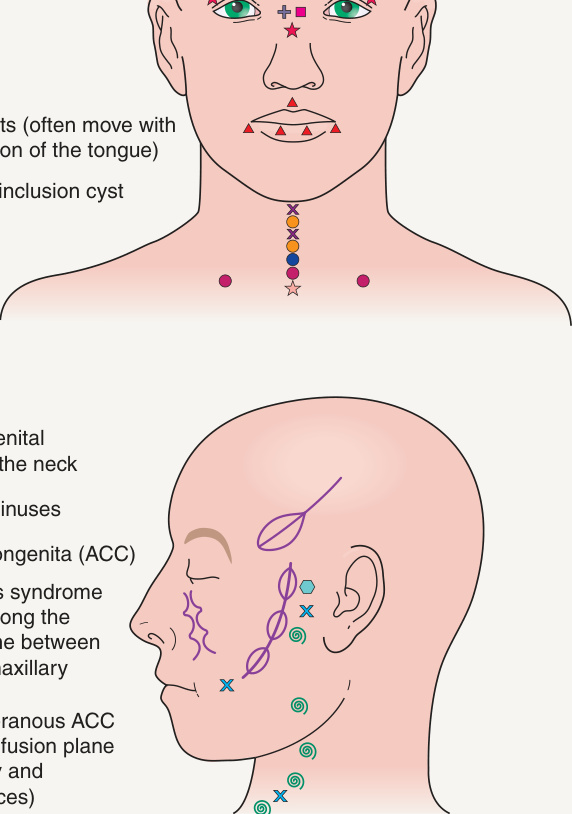
Fig. 64.2 Common sites of developmental anomalies of the face and neck.
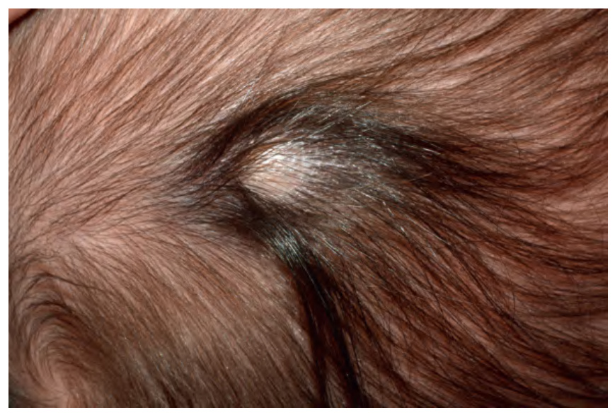
Fig. 64.3 Hair collar sign. Membranous aplasia cutis congenita with a hair collar.
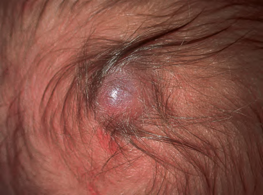
Fig. 64.4 Hair collar sign. Membranous aplasia cutis congenita with a bullous appearance and a surrounding hair collar.
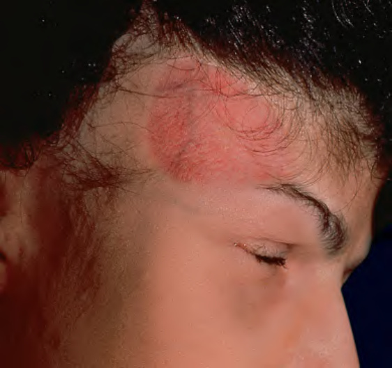
Fig. 64.17 Facial aplasia cutis congenita.
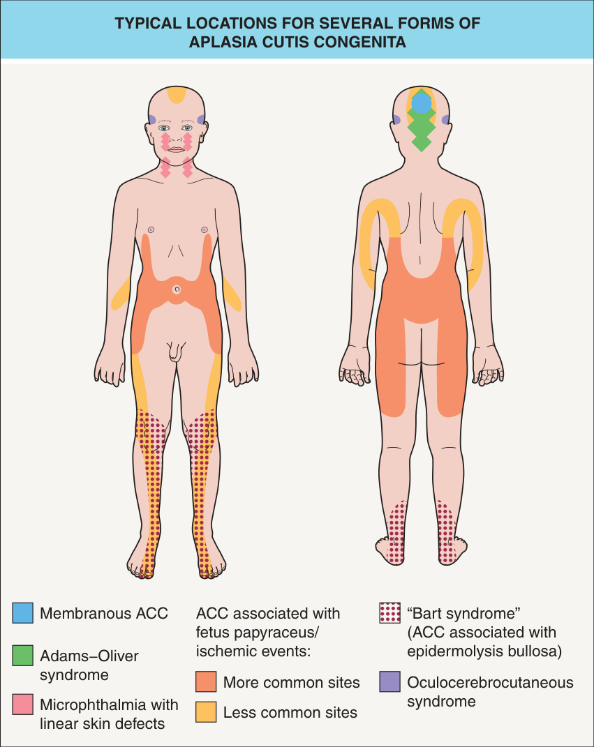
Fig. 64.19 Typical locations for several forms of aplasia cutis congenita (ACC).
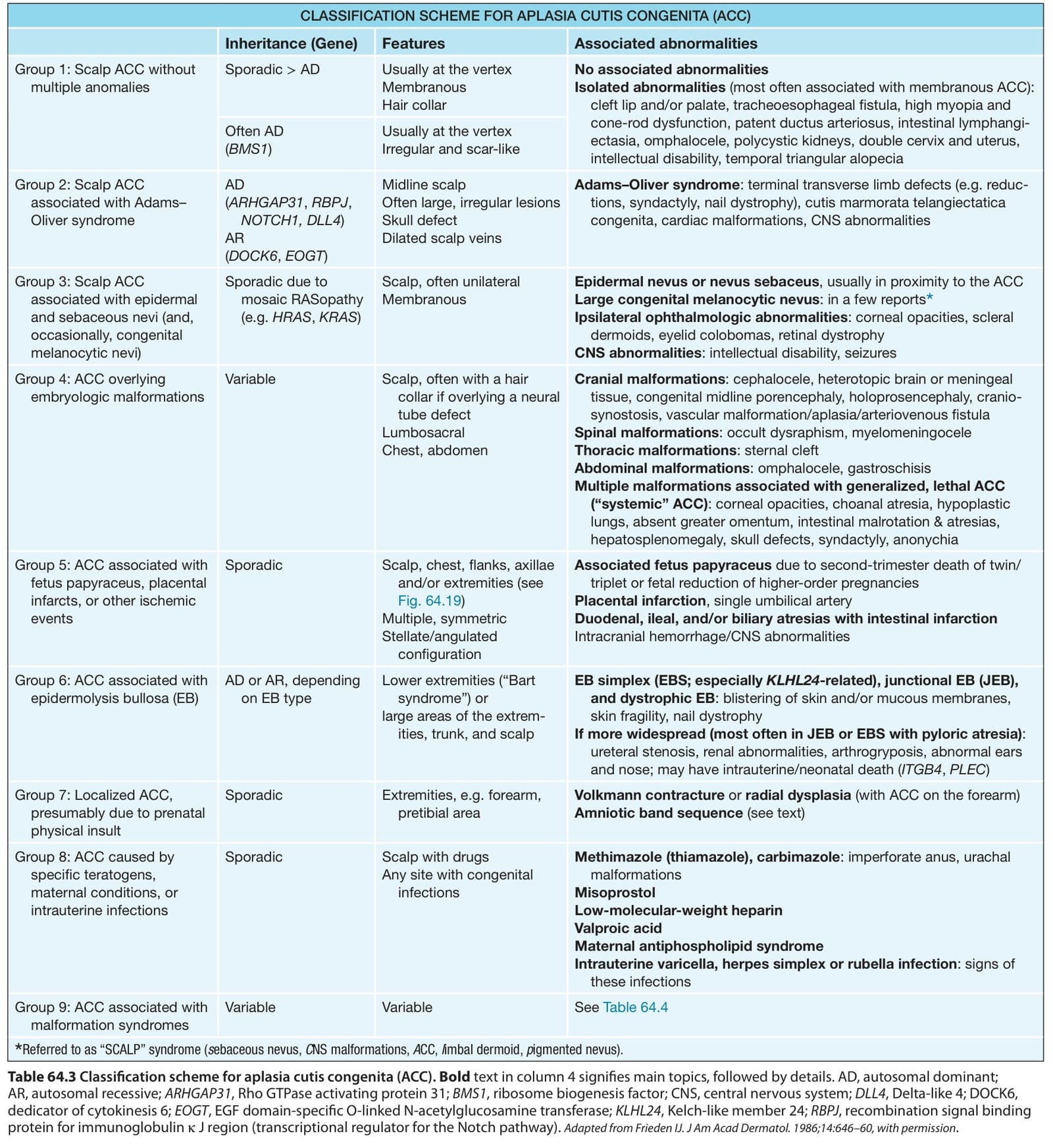
Table 64.3 Classification scheme for aplasia cutis congenita (ACC).Bold text in column 4 signifies main topics, followed by details. AD, autosomal dominant; AR, autosomal recessive; ARHGAP31, Rho GTPase activating protein 31; BMS1, ribosome biogenesis factor; CNS, central nervous system; DLL4, Delta-like 4; DOCK6, dedicator of cytokinesis 6; EOGT, EGF domain-specific O-linked N-acetylglucosamine transferase; KLHL24, Kelch-like member 24; RBPJ, recombination signal binding protein for immunoglobulin κ J region (transcriptional regulator for the Notch pathway). Adapted from Frieden IJ. J Am Acad Dermatol. 1986;14:646–60, with permission.
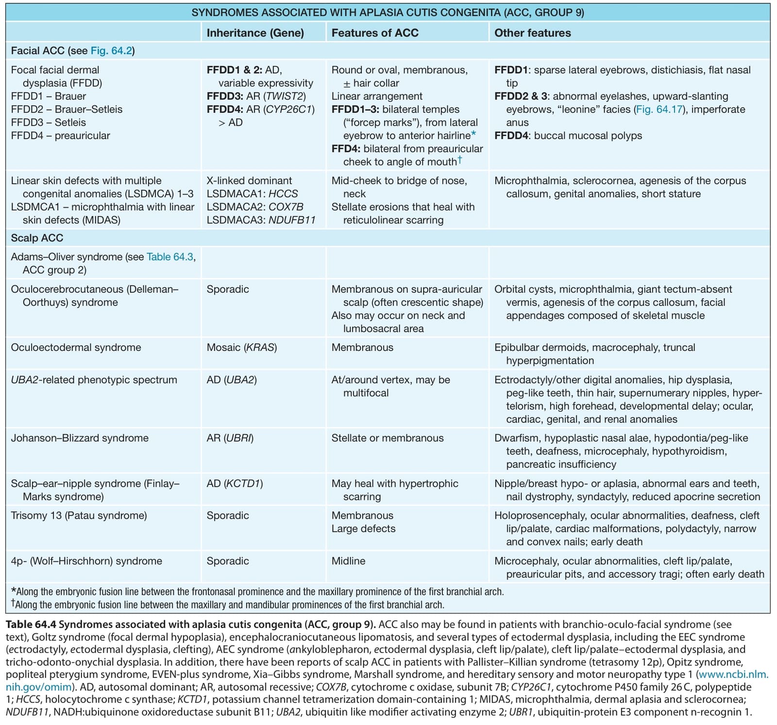
Table 64.4 Syndromes associated with aplasia cutis congenita (ACC, group 9). ACC also may be found in patients with branchio-oculo-facial syndrome (see text), Goltz syndrome (focal dermal hypoplasia), encephalocraniocutaneous lipomatosis, and several types of ectodermal dysplasia, including the EEC syndrome (ectrodactyly, ectodermal dysplasia, clefting), AEC syndrome (ankyloblepharon, ectodermal dysplasia, cleft lip/palate), cleft lip/palate–ectodermal dysplasia, and tricho-odonto-onychial dysplasia. In addition, there have been reports of scalp ACC in patients with Pallister–Killian syndrome (tetrasomy 12p), Opitz syndrome, popliteal pterygium syndrome, EVEN-plus syndrome, Xia–Gibbs syndrome, Marshall syndrome, and hereditary sensory and motor neuropathy type 1 (www.ncbi.nlm. nih.gov/omim). AD, autosomal dominant; AR, autosomal recessive; COX7B, cytochrome c oxidase, subunit 7B; CYP26C1, cytochrome P450 family 26 C, polypeptide 1; HCCS, holocytochrome c synthase; KCTD1, potassium channel tetramerization domain-containing 1; MIDAS, microphthalmia, dermal aplasia and sclerocornea; NDUFB11, NADH:ubiquinone oxidoreductase subunit B11; UBA2, ubiquitin like modifier activating enzyme 2; UBR1, ubiquitin-protein E3 component n-recognin 1.