Congenital Lip Pits
Synonyms: Lip sinuses Congenital sinuses of the lower lip Midline sinuses of the upper lip
Congenital lip pits are divided into three types based on their location: (1) commissural; (2) upper lip; and (3) lower lip. Commissural lip pits are by far the most common, occurring in 1%–2% of newborns. Lip pits likely result from defective embryologic fusion of facial processes with epithelial entrapment. They usually represent the opening of a blind sinus tract, which can extend inward through the orbicularis oris muscle to a depth of >10 mm. This tract occasionally communicates with the ducts of underlying minor salivary glands, and saliva or mucus may drain from the ostium. Lesions are sometimes associated with swelling of the lip or, particularly if located on the philtrum, recurrent infection.
See Fig. 64.2 for Brauer lines/Setleis syndrome, preauricular membranous ACC, and microphthalmia with linear skin defects.
Commissural lip pits are typically found bilaterally in the mucosal surface at the angles of the mouth (Fig. 64.20A). Although most often an isolated anomaly, they may be inherited in an autosomal dominant fashion along with preauricular pits and hearing impairment in patients with a form of branchio-otic syndrome linked to chromosome 1q31. Commissural lip pits also have been described in association with alveolar synechiae, ankyloblepharon filiforme adnatum (congenital adhesions between the upper and lower eyelids), and ectodermal defects.
Upper lip pits, also known as midline sinuses of the upper lip, are rare lesions that are typically located along the philtrum. Although usually an isolated defect, they may be associated with hypertelorism and other dysmorphic facial features. In addition, paramedian upper lip pits may occur (Fig. 64.20B) and are bilateral in patients with the branchio-oculo-facial syndrome (see below, Branchial Cleft Sinuses and Fistulae).
Lower lip pits may be an isolated finding or seen as a part of van der Woude syndrome, which is characterized by paramedian pits in the vermilion portion of the lower lip, cleft lip and/or palate, and hypodontia. The lip pits are usually bilateral, and they may be located on the apex of a conical elevation (Fig. 64.20C). Van der Woude syndrome, which occurs in ~1–3 per 100 000 live births, is an autosomal dominant disorder caused by mutations in the interferon regulatory factor 6 (IRF6) or grainyhead-like 3 (GRHL3) gene. The popliteal pterygium syndrome is also due to IRF6 mutations; in addition to lower lip pits, findings include cleft lip and/or palate, syngnathia, popliteal webbing, nail dysplasia, syndactyly, and genital anomalies. Lastly, lower lip pits are occasionally seen in oral–facial–digital syndrome type I (Table 64.5) and Kabuki syndrome (www.ncbi.nlm.nih.gov/omim).
Histologically, the sinus tracts associated with lip pits are lined by stratified squamous epithelium; associated salivary or mucous glands are occasionally observed. The evaluation of patients with lip pits should include a family history and a physical examination to exclude associated anomalies. If lip pits of any type are symptomatic or cosmetically undesirable, surgical excision is the treatment of choice. The extent of the sinus can be estimated preoperatively by injection of a radio-opaque contrast medium.
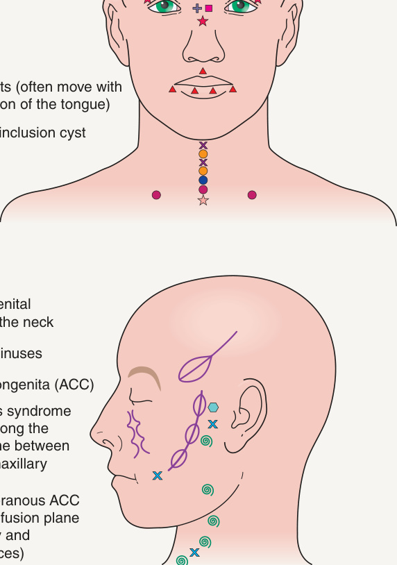
Fig. 64.2 Common sites of developmental anomalies of the face and neck.
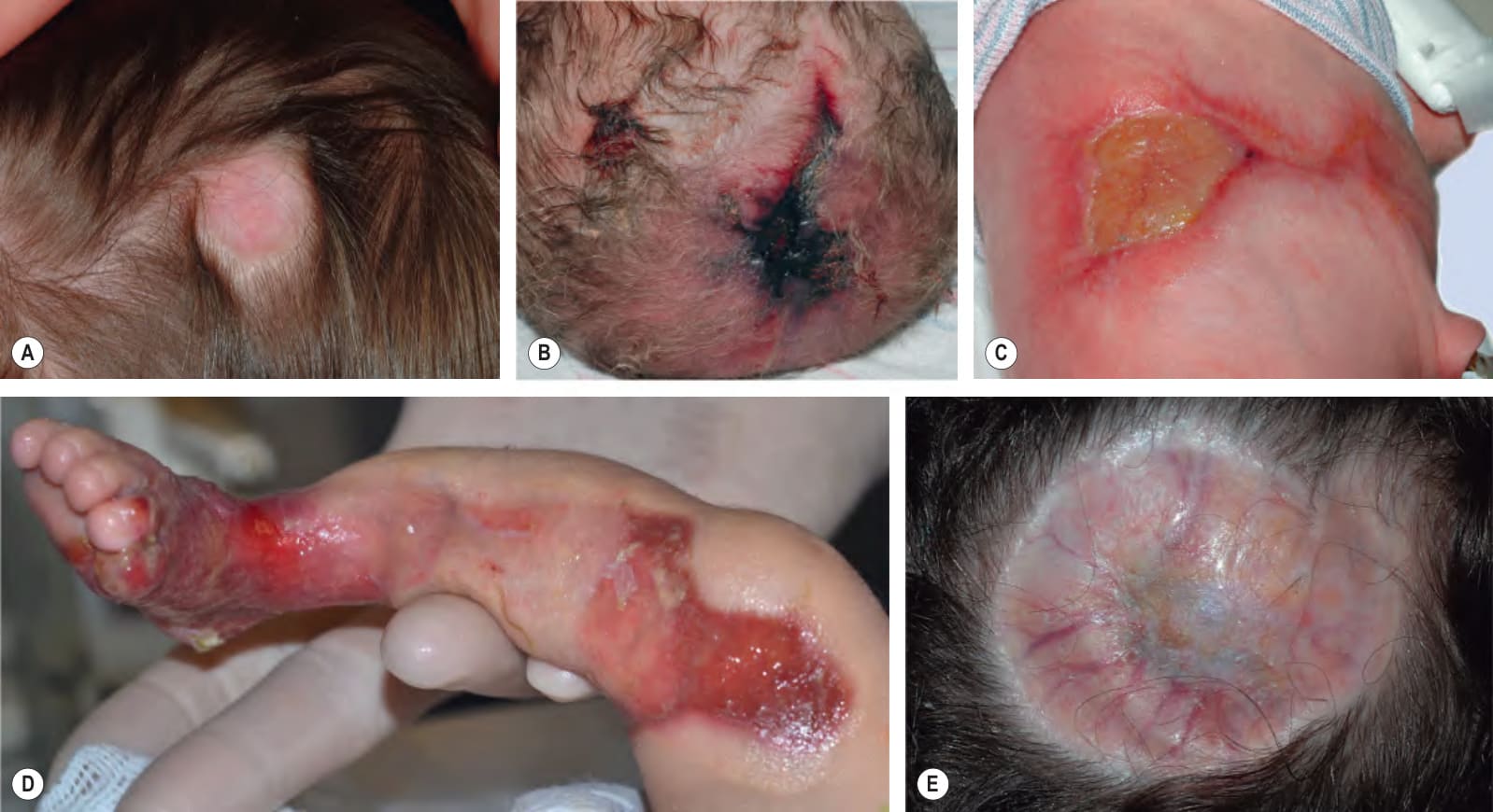
Fig. 64.18 Aplasia cutis congenita (ACC).A This hairless, round, scarred plaque on the scalp of a 6-month-old was present at birth. B Stellate ACC on the midline scalp of a neonate. This hemorrhagic lesion was associated with an underlying skull defect and cerebrovascular anomalies. C Stellate ACC on the lateral trunk of a neonate born of an initial sextuplet gestation for which fetal reduction was performed. The lesions had a bilateral, symmetric distribution. D ACC on the leg in a neonate with epidermolysis bullosa (“Bart syndrome”). E Membranous ACC associated with a large defect of the underlying skull in a patient with Goltz syndrome. A, Courtesy Anthony J. Mancini, MD; D, Courtesy Emily Berger, MD.
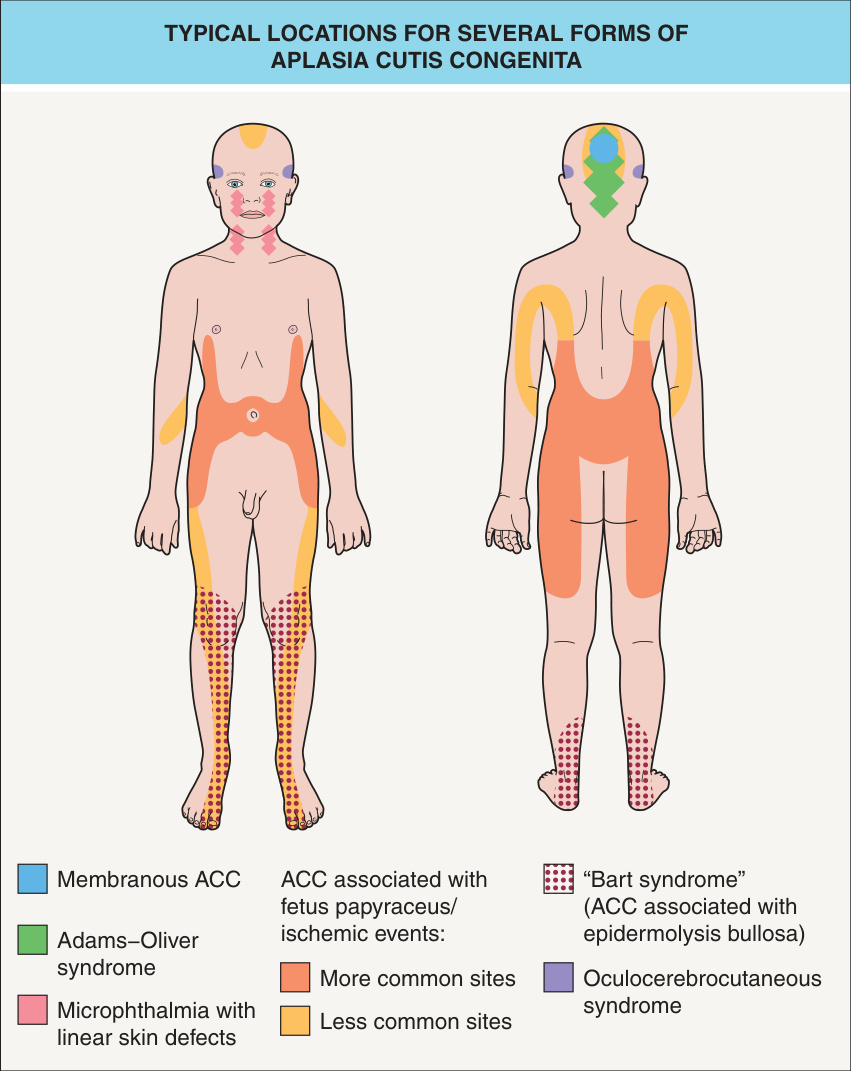
Fig. 64.19 Typical locations for several forms of aplasia cutis congenita (ACC).
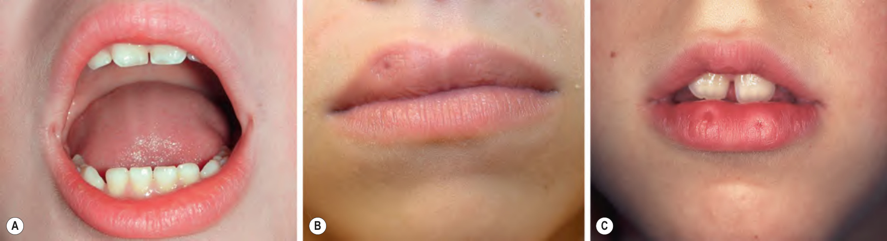
Fig. 64.20 Lip pits.A Bilateral commissural lip pits. B Unilateral right-sided upper lip pit. C Bilateral paramedian lower lip pits, each located on the apex of a conical elevation. All were isolated anomalies.
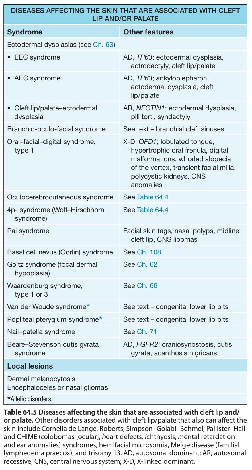
Table 64.5 Diseases affecting the skin that are associated with cleft lip and/ or palate. Other disorders associated with cleft lip/palate that also can affect the skin include Cornelia de Lange, Roberts, Simpson–Golabi–Behmel, Pallister–Hall and CHIME (colobomas [ocular], heart defects, ichthyosis, mental retardation and ear anomalies) syndromes, hemifacial microsomia, Meige disease (familial lymphedema praecox), and trisomy 13. AD, autosomal dominant; AR, autosomal recessive; CNS, central nervous system; X-D, X-linked dominant.