INTRODUCTION
In order to understand the underlying pathophysiology of cutaneous disorders of hypopigmentation and hyperpigmentation, as well as the process of normal physiologic pigment production, an appreciation of the structure and function of the melanocyte is required. A classic example of basic pathogenesis is type 1 oculocutaneous albinism (OCA1), in which pigmentary dilution of the skin, hair, and eyes is due to a reduction or absence of tyrosinase activity secondary to mutations in both copies of the tyrosinase gene (TYR). Physiologic pigmentation is influenced by variants in multiple genes. For example, melanocytes in individuals with red hair often express variants of the melanocortin 1 receptor (MC1R). As a consequence of the altered amino acid sequences of the variant MC1Rs, their cell surface expression and inter-actions with melanocyte stimulating hormone (MSH) can be affected, leading to an increase in the production of pheomelanin as opposed to eumelanin. Based upon population genetics, genes that are mutated in OCA (e.g. TYR, OCA2, TYRP1, SLC45A2, SLC24A5) also influence normal pigment variation (Table 65.1).
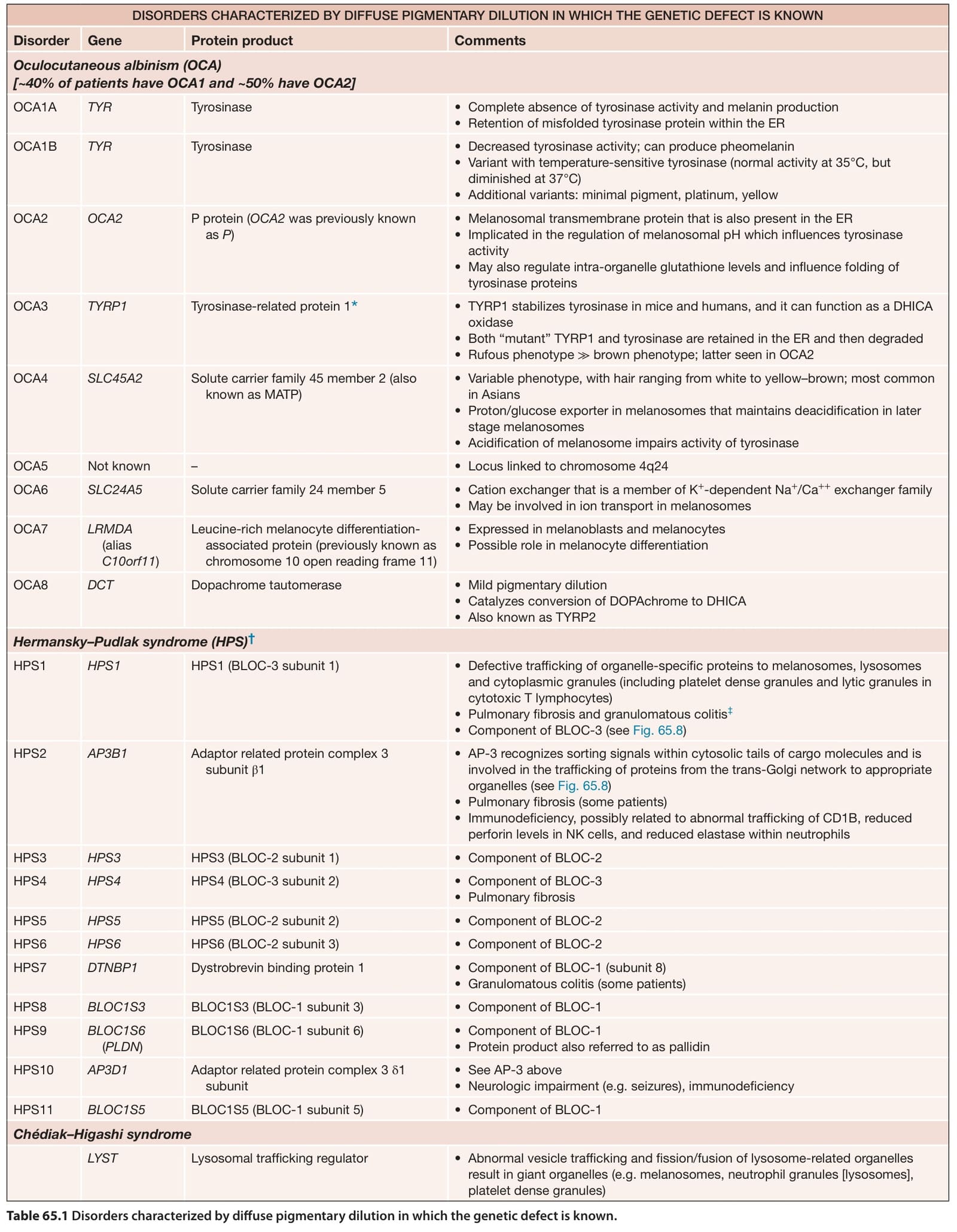
Table 65.1 Disorders characterized by diffuse pigmentary dilution in which the genetic defect is known.