POSTINFLAMMATORY HYPERPIGMENTATION
Synonym: Postinflammatory hypermelanosis
Key features
Extremely common, especially in individuals with more darkly pigmented skin
Develops after inflammation or injury to the skin, but preceding inflammation may be transient or subclinical
The increased melanin can be primarily within the dermis (e.g.
following lichen planus) or in the epidermis (e.g. following acne or atopic dermatitis)
Epidermal hyperpigmentation fades more readily than dermal hyperpigmentation
Introduction
PIH represents an acquired excess of melanin pigment following cutaneous inflammation or injury. It can occur anywhere on the skin surface, including the mucous membranes and the nail unit. PIH is extremely common and can have significant cosmetic and psychosocial consequences.
Epidemiology
PIH can occur at any age and there is no sex preference. Individuals with darkly pigmented skin tend to have a greater frequency, severity, and duration of PIH than those with lighter complexions.
Pathogenesis
In the epidermal form of PIH, there is increased melanin production and/or transfer to keratinocytes. Inflammatory mediators (e.g. prostaglandins E and D, IL-33) that enhance pigment production may play a role in this process. In dermal hyperpigmentation, melanin enters (“falls into”) the dermis via a damaged basement membrane, where it is phagocytosed by and subsequently resides within dermal macrophages (referred to as melanophages). Macrophages may also migrate into the epidermis, phagocytose melanosomes, and then return to the dermis. Melanin within dermal melanophages tends to persist for long periods of time (e.g. years).
Clinical Features
Asymptomatic hyperpigmented macules and patches range in color either from tan to dark brown (epidermal melanin) or from gray–blue to gray–brown (dermal melanin). Primary lesions of the underlying inflammatory disorder may be admixed with the hyperpigmentation or present elsewhere. When primary lesions are absent, the size, shape, and distribution pattern of the hyperpigmented lesions may provide clues to the underlying etiology (Table 67.2). PIH can be exacerbated by continued inflammation, trauma, exposure to ultraviolet (UV) irradiation, or treatment-related irritation.
Therapeutic options are discussed in cited chapters.
Disorders that commonly lead to epidermal PIH include acne, insect bites, pyodermas, atopic dermatitis, psoriasis, and pityriasis rosea (Fig. 67.2A–C,E). In contrast, dermal PIH is associated with dermatoses characterized by degeneration of the basal layer of the epidermis and inflammation at the dermal–epidermal junction, such as lichen planus, lichenoid drug reactions, lupus erythematosus, and fixed drug eruptions (Fig. 67.2D). Upon treatment of the underlying disorder, epidermal PIH generally resolves over time, although fading may require months or years in darkly pigmented individuals. Dermal melanosis tends to persist longer and is sometimes permanent.
Pathology
Epidermal PIH is characterized by increased pigment in keratinocytes and dermal PIH by melanophages within the superficial dermis.
hyperpigmentation. Hyperpigmentation can also result from keratinocytic (epidermal) disorders (see Table 67.1).
Differential Diagnosis
The size, shape, and distribution of the hyperpigmented lesions can provide clues to the inflammatory disease or injury that preceded PIH (see Table 67.2), and a thorough skin examination may detect active primary lesions. A history of prior inflammatory lesions and medication use (including prescription, over-the-counter, and alternative products) should be obtained. Disorders such as erythema dyschromicum perstans, melasma, tinea (pityriasis) versicolor, and atrophoderma of Pasini and Pierini should be considered in patients without evidence of preceding inflammation by history or on examination. Occasionally, a biopsy may assist in establishing the diagnosis.
Treatment
Provided that the underlying dermatosis is successfully treated, PIH eventually improves in most patients, especially those with epidermal hypermelanosis. Sun protection, including daily application of a broad-spectrum sunscreen, can help to prevent accentuation of the pigmentation by UV exposure. Topical hydroquinone (2%–4%) may lead to lightening over 3–6 months if the increase in pigmentation is limited to the epidermis. As with melasma (see below), use of a topical retinoid and corticosteroid together with hydroquinone may be more effective than monotherapy. Topical azelaic, α-hydroxy, L-ascorbic, tranexamic, and kojic acids as well as cysteamine, arbutin, licorice extracts, mequinol, niacinamide, N-acetyl glucosamine, and soy are additional potential therapeutic options. However, it is important to avoid irritant contact dermatitis from topical agents, as this may exacerbate the hyperpigmentation. Chemical peels (e.g. with salicylic or glycolic acid) and laser therapy may be of benefit but can also result in hyper- or hypopigmentation, especially in patients with darkly pigmented skin. Laser therapy (e.g. 1927-nm non-ablative fractional thulium fiber laser, Q-switched Nd:YAG, ruby, and alexandrite lasers) are variably successful in removing dermal pigment (see Ch. 137).
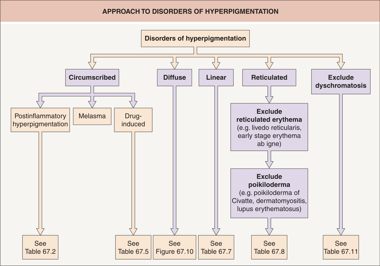
Fig. 67.1 Approach to disorders of
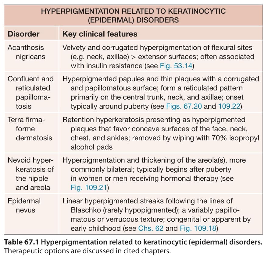
Table 67.1 Hyperpigmentation related to keratinocytic (epidermal) disorders.
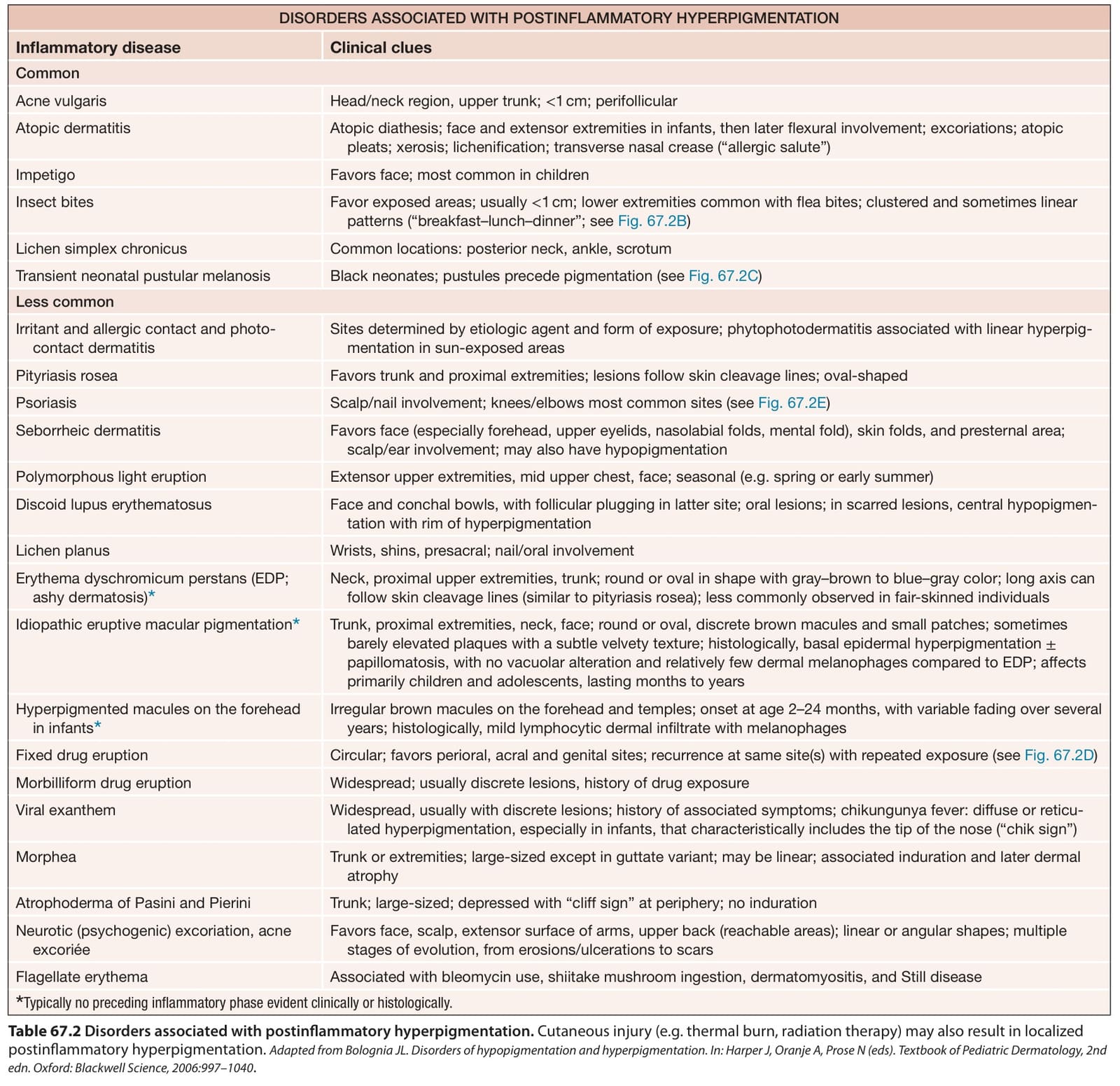
Table 67.2 Disorders associated with postinflammatory hyperpigmentation. Cutaneous injury (e.g. thermal burn, radiation therapy) may also result in localized postinflammatory hyperpigmentation. Adapted from Bolognia JL. Disorders of hypopigmentation and hyperpigmentation. In: Harper J, Oranje A, Prose N (eds). Textbook of Pediatric Dermatology, 2nd edn. Oxford: Blackwell Science, 2006:997–1040.