ERYTHEMA DYSCHROMICUM PERSTANS
Synonyms: Ashy dermatosis Dermatosis cenicienta
Key features
Most common in individuals with skin phototypes III–IV
Gray–brown to blue–gray macules and patches in a symmetric distribution
Favors the neck, trunk, and proximal extremities
A consistently effective treatment is not currently available
Introduction
Ramirez first described erythema dyschromicum perstans (EDP) in 1957, referring to affected individuals as los cenicientos (ashen ones). This is an asymptomatic, slowly progressive eruption that is characterized by circumscribed areas of dermal pigmentation.
Epidemiology
Although EDP occurs worldwide, it is most common in Latin America. There is no sex preference. The disorder usually presents during the second to third decade, but it occasionally develops in younger children or older adults.
Pathogenesis
The etiology of EDP is not known. Although it has been postulated that a cell-mediated immune reaction to an ingestant, contactant, or microorganism underlies the discrete areas of pigmentary incontinence, no causal agent has consistently been identified. In most patients, a trigger is never found. However, there have been reports of EDP developing in association with the ingestion of ammonium nitrate, oral X-ray contrast media, and medications (e.g. benzodiazepines, penicillin, proton pump inhibitors, epidermal growth factor receptor inhibitors, cyclin-dependent kinase 4/6 inhibitors); exposure to various pesticides, fungicides or toxins; endocrinopathies such as thyroid disease; and whipworm and HIV infections. The presence of the HLA-DR4 allele may represent a risk factor for EDP in Mexican patients.
Clinical Features
Oval, circular, or irregularly shaped macules and patches that are 0.5 to 3 cm in diameter and slate-gray to blue–brown in color develop gradually in a symmetric pattern and may coalesce (Fig. 67.3A). The initial site of involvement is often the trunk, with subsequent spread to the neck, proximal upper extremities, and sometimes the face. The mucous membranes are spared, and the palms, soles, and scalp are rarely affected. Occasionally, early lesions have a thin, raised, erythematous border, which tends to resolve over a few months, and the long axis of lesions may follow skin cleavage lines. Peripheral hypopigmentation may be seen in older lesions. Characteristic dermoscopic features of EDP include pigmentation in the crista cutis and small brown to gray dots, described as having a “Wagyu beef-like” appearance.
EDP is usually asymptomatic but can be mildly pruritic. The disease progresses slowly and typically does not regress in adults. However, in one study of 33 prepubertal children, the majority of whom were White children, the disease spontaneously resolved in two-thirds, with no recurrences over the ensuing 2–3 years.
Pathology
In early (“active”) lesions, vacuolar degeneration of the basal cell layer, a perivascular mononuclear cell infiltrate and melanophages in the upper dermis, and increased epidermal melanin are seen (Fig. 67.3B). Colloid bodies (apoptotic basal keratinocytes) and dermal hemosiderin may be present. In later (“inactive”) lesions, the number of melanophages in the superficial dermis is increased but hydropic changes within the basal cell layer are absent and the dermal mononuclear cell infiltrate is minimal or absent.
Differential Diagnosis
Idiopathic eruptive macular hyperpigmentation (IEMH) features discrete brown macules and small patches with a distribution similar to that of EDP but with more prominent epidermal pigment. Some reports have noted a velvety surface and papillomatosis histologically (see Table 67.2). Considering the substantial clinical and histologic overlap of EDP, IEMH, lichen planus pigmentosus, and pigmented contact dermatitis (see Table 67.3), the umbrella term acquired dermal macular hyperpigmentation has been proposed for this group of disorders. The differential diagnosis of EDP also includes PIH secondary to a lichenoid drug eruption (see Ch. 11), lichen planus, or a generalized fixed drug eruption as well as mastocytosis and (less often) infectious diseases such as leprosy or pinta.
Treatment
There is no consistently effective therapy for EDP. Topical corticosteroids and hydroquinone are generally of no benefit. Successful treatment with topical tacrolimus, narrowband UVB phototherapy, and oral dapsone, clofazimine, or isotretinoin has been reported in small series.
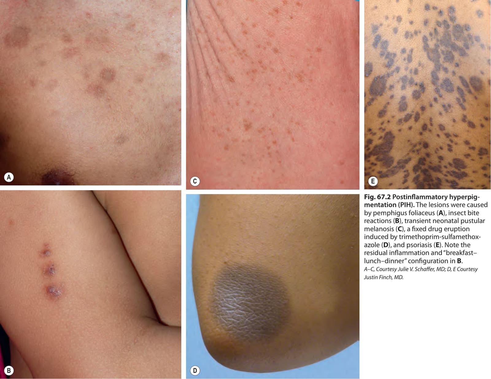
Fig. 67.2 Postinflammatory hyperpig- mentation (PIH). The lesions were caused by pemphigus foliaceus (A), insect bite reactions (B), transient neonatal pustular melanosis (C), a fixed drug eruption induced by trimethoprim-sulfamethoxazole (D), and psoriasis (E). Note the residual inflammation and “breakfast– lunch–dinner” configuration in B. A–C, Courtesy Julie V. Schaffer, MD; D, E Courtesy Justin Finch, MD.
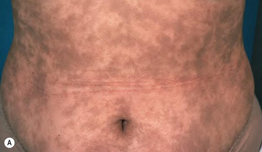
Fig. 67.3 Erythema dyschromicum perstans (ashy dermatosis).A Multiple gray–brown macules and patches on the abdomen; note the coalescence of lesions. The “ashy” color is characteristic. B Histopathologic features include vacuolar degeneration of the basal layer, a sparse perivascular lymphocytic infiltrate, and prominent dermal melanophages (see inset). B, Courtesy Lorenzo Cerroni, MD.
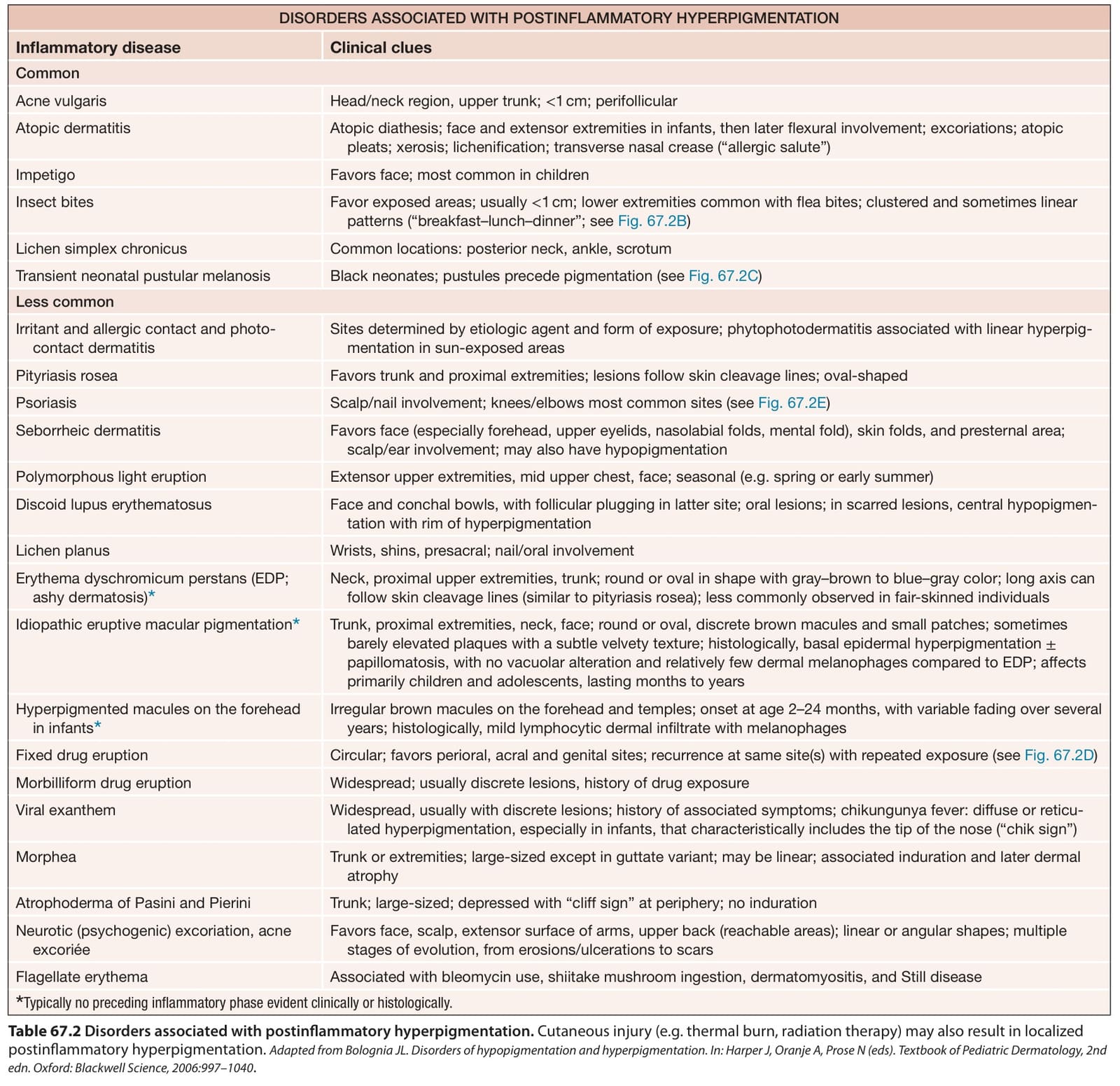
Table 67.2 Disorders associated with postinflammatory hyperpigmentation. Cutaneous injury (e.g. thermal burn, radiation therapy) may also result in localized postinflammatory hyperpigmentation. Adapted from Bolognia JL. Disorders of hypopigmentation and hyperpigmentation. In: Harper J, Oranje A, Prose N (eds). Textbook of Pediatric Dermatology, 2nd edn. Oxford: Blackwell Science, 2006:997–1040.
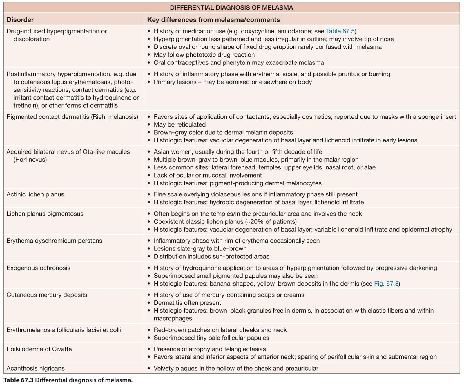
Table 67.3 Differential diagnosis of melasma.