MELASMA
Synonyms: Chloasma Mask of pregnancy
Key features
At least 90% of patients are women
Increased prevalence in individuals who are Hispanic, or of Asian or
African descent
Most common location is the face, followed by the forearms
Symmetric patches of hyperpigmentation with irregular borders due to increased melanin within the epidermis and/or dermis
Introduction and Epidemiology
Melasma is a common acquired disorder characterized by symmetric, hyperpigmented patches with an irregular outline, occurring most commonly on the face. It is most prevalent among young to middleaged women who are Hispanic, Black, or of Asian or Middle Eastern descent. Exacerbating factors include sun exposure, pregnancy, and use of oral contraceptives.
Pathogenesis
Although the exact pathogenesis of melasma is unknown, it is hypothesized that following exposure to UV irradiation or another inducer, hyperfunctional melanocytes within involved skin produce increased amounts of melanin. The key role of UV irradiation is supported by fading of lesions during winter months and a distribution pattern characterized by involvement of sun-exposed regions with sparing of relatively sun-protected sites such as the philtrum. In addition to oral contraceptive use and hyperestrogenic states, other medications (e.g. phenytoin, phototoxic drugs) and disorders (e.g. autoimmune thyroid disease) have the potential to aggravate melasma. Increased expression of KIT and stem cell factor within lesional epidermis and dermis, respectively, may play a role in the hyperpigmentation of melasma. Furthermore, reduced expression of Wnt-inhibitory factor 1 (WIF-1) and microRNAs may lead to tyrosinase upregulation via increased Wnt signaling13a.
The presence of enhanced blood flow within areas of melasma has also been investigated. An increase in both blood vessel size and density has been observed that correlates with the increase in pigmentation. When compared to perilesional normal skin, increased expression of vascular endothelial growth factor (VEGF) was detected within lesional skin. These findings have led to alternative therapeutic strategies.
Clinical Features
Light to dark brown or brown–gray patches with irregular borders appear primarily on the face (Fig. 67.5). The areas of hypermelanosis are distributed symmetrically in three classic patterns: (1) centrofacial (most common), involving the forehead, cheeks, nose, upper lip (sparing the philtrum and nasolabial folds), and chin; (2) malar, affecting the cheeks and nose; and (3) mandibular, along the jawline. Less common sites include the extensor aspect of the forearms and mid upper chest. Lesions often first appear or are accentuated following exposure to UV irradiation or during pregnancy. In lightly pigmented individuals, this “mask of pregnancy” frequently diminishes or disappears after parturition, but it tends to persist in women with more darkly pigmented skin.
Melasma has classically been subdivided into four types based upon the primary location of the pigment: epidermal, dermal, mixed, or indeterminate (e.g. in patients with very dark skin pigmentation). In theory, lesions with increased epidermal melanin are accentuated and those with increased dermal melanin become less obvious (i.e. blend with uninvolved skin) with Wood’s lamp examination. However, clinicopathologic studies utilizing adjacent uninvolved skin as controls have shown that Wood’s lamp examination does not correlate with histologic findings. Considering that epidermal pigmentation is more likely to respond to topical therapies, further studies are needed to determine the
clinical utility and prognostic significance of Wood’s lamp examination and perhaps optical coherence tomography.
Pathology
Compared to uninvolved adjacent skin, increased melanin deposition is observed in all layers of the epidermis, particularly the basal layer. An increased number of melanophages may also be seen in the upper dermis. Epidermal melanocytes are normal to slightly increased in number, and they are enlarged with prominent dendrites.
Ultrastructurally, lesional melanocytes contain an increased number of melanosomes. In addition, the mitochondria, Golgi apparatus, and rough endoplasmic reticulum are increased in number. These findings support the theory of hyperfunctional melanocytes, presumably stimulated by UV irradiation or hormones (see Pathogenesis above).
Differential Diagnosis
The differential diagnosis of melasma is reviewed in Table 67.3. These disorders are distinguished from melasma based upon historical aspects (e.g. drug ingestion, previous inflammation), color, distribution pattern, histologic features, and, if present, primary inflammatory lesions.
Treatment
The treatment of melasma is summarized in Table 67.4. Diligent sun protection and patient motivation are necessary for any melasma treatment regimen to be successful. For epidermal melasma, 2 months of therapy are typically required to initiate lightening and 6 months of treatment are often needed to achieve satisfactory results.
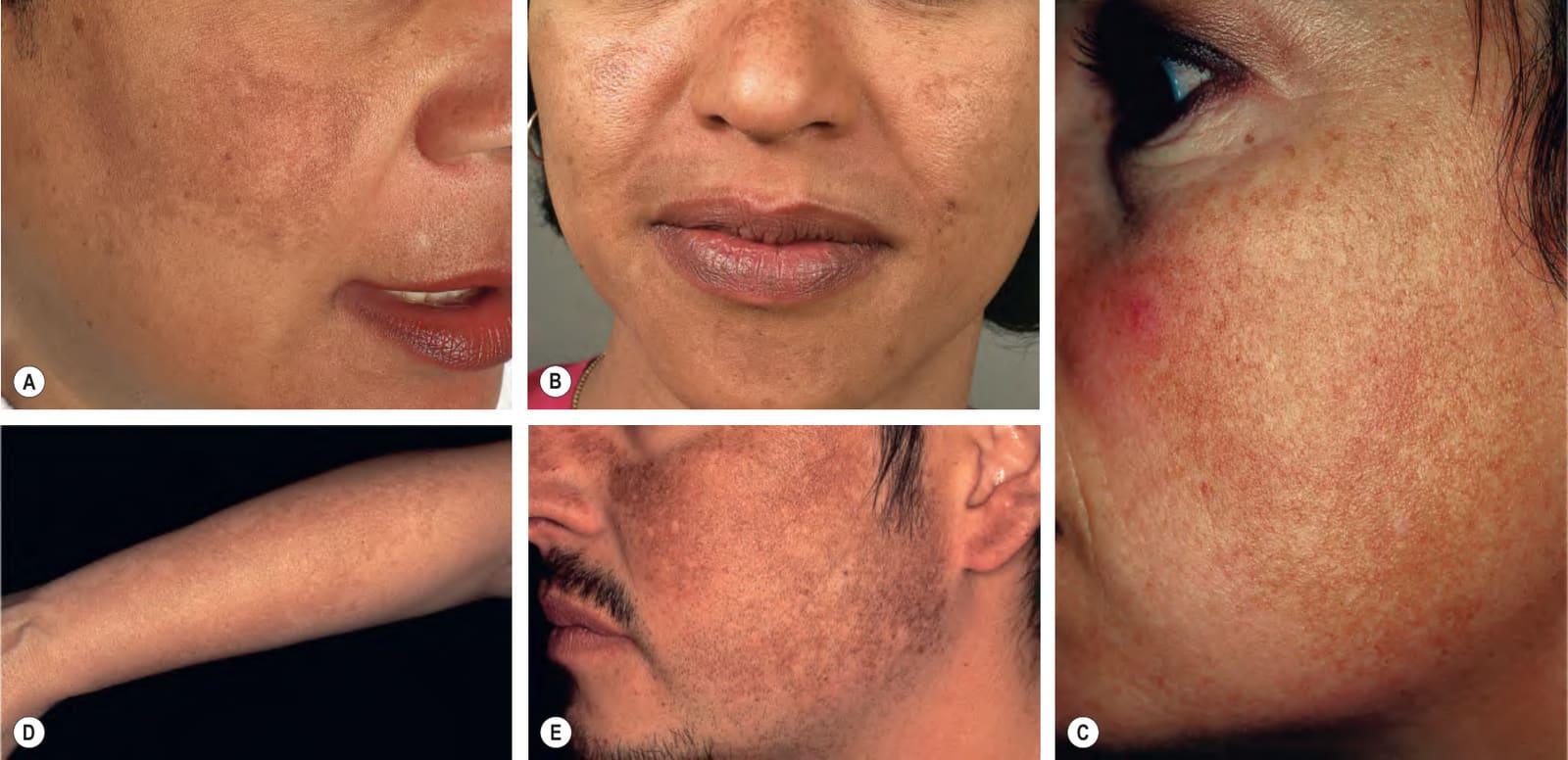
Fig. 67.5 Various forms of melasma and melasma-like hyperpigmentation.A Malar variant. B Mild centrofacial type with sparing of the philtrum. C Extension of the hyperpigmentation onto the mandible. D Involvement of the extensor forearm; note the same irregular outline as is seen on the face. E Melasma-like appearance in a patient with previous acute cutaneous lupus erythematosus. C–E, Courtesy Jean L. Bolognia, MD.
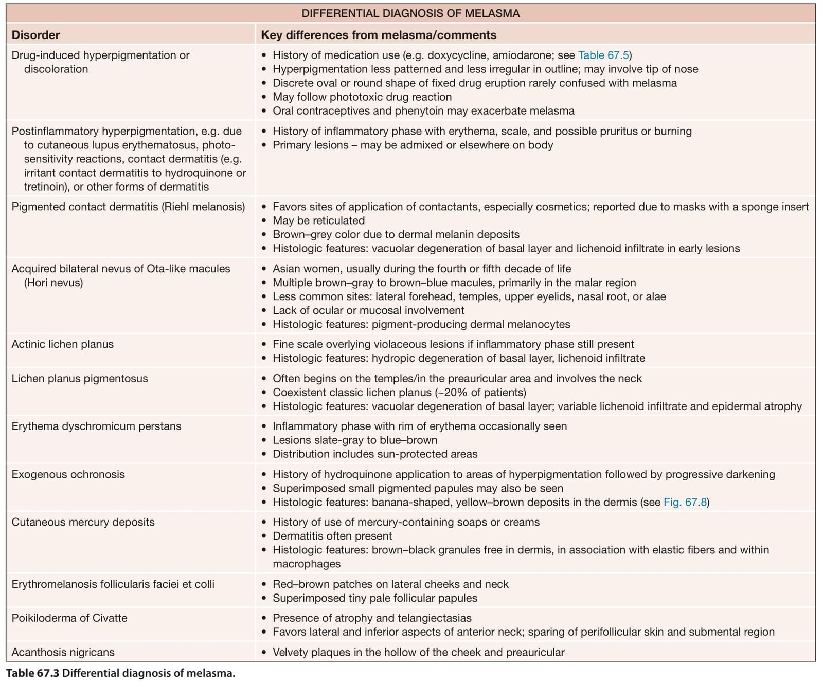
Table 67.3 Differential diagnosis of melasma.
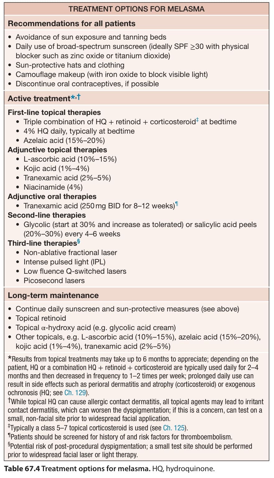
Table 67.4 Treatment options for melasma. HQ, hydroquinone.