PRURIGO PIGMENTOSA
First described by Nagashima in 1971, this disorder is characterized by a markedly pruritic eruption of erythematous papules and papulovesicles on the back, neck, and chest (Fig. 67.21A). Crops of inflammatory lesions develop rapidly and then involute within a week, leaving macular reticulated hyperpigmentation (Fig. 67.21B). Recurrences tend to occur at the same site, and lesions in multiple stages are often
A On the neck and back of a teenage girl. B On the chest of an older woman. A, Courtesy Seth Orlow, MD PhD.
evident. The condition favors young adults, with a female : male ratio of ≥2 : 1; the majority of reported patients have been from East Asia. Prurigo pigmentosa triggered by ketosis due to bariatric surgery, a ketogenic diet, or type 1 diabetes mellitus has been reported in patients with a variety of ethnic backgrounds.
Three histopathologic stages have been identified: (1) neutrophilic exocytosis, spongiosis, papillary dermal edema, and a superficial perivascular infiltrate of neutrophils; (2) intra-/subepidermal vesiculation, necrotic keratinocytes, and a patchy lichenoid infiltrate of predominantly lymphocytes admixed with eosinophils (Fig. 67.21C); and (3) variable parakeratosis, acanthosis, and hyperpigmentation of the epidermis as well as dermal melanophages. Treatment with oral minocycline, doxycycline, or dapsone is often effective for the inflammatory component of prurigo pigmentosa. The condition does not respond to corticosteroids (topical or systemic) or antihistamines. Prurigo pigmentosa associated with ketosis may improve with reintroduction of carbohydrates.
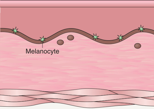
Fig. 67.18 Histologic differences between stage 3 incontinentia pigmenti (IP) and linear and whorled nevoid hypermelanosis (LWNH).
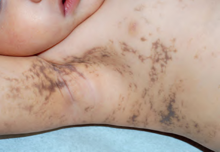
Fig. 67.19 Stage 3 incontinentia pigmenti in a 2-year-old child. Note the characteristic gray–brown color and the distribution along the lines of Blaschko.
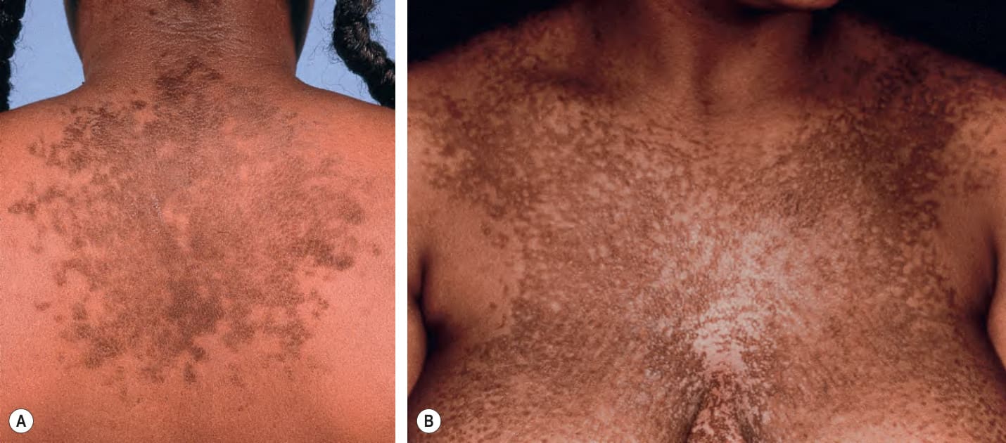
Fig. 67.20 Confluent and reticulated papillomatosis.
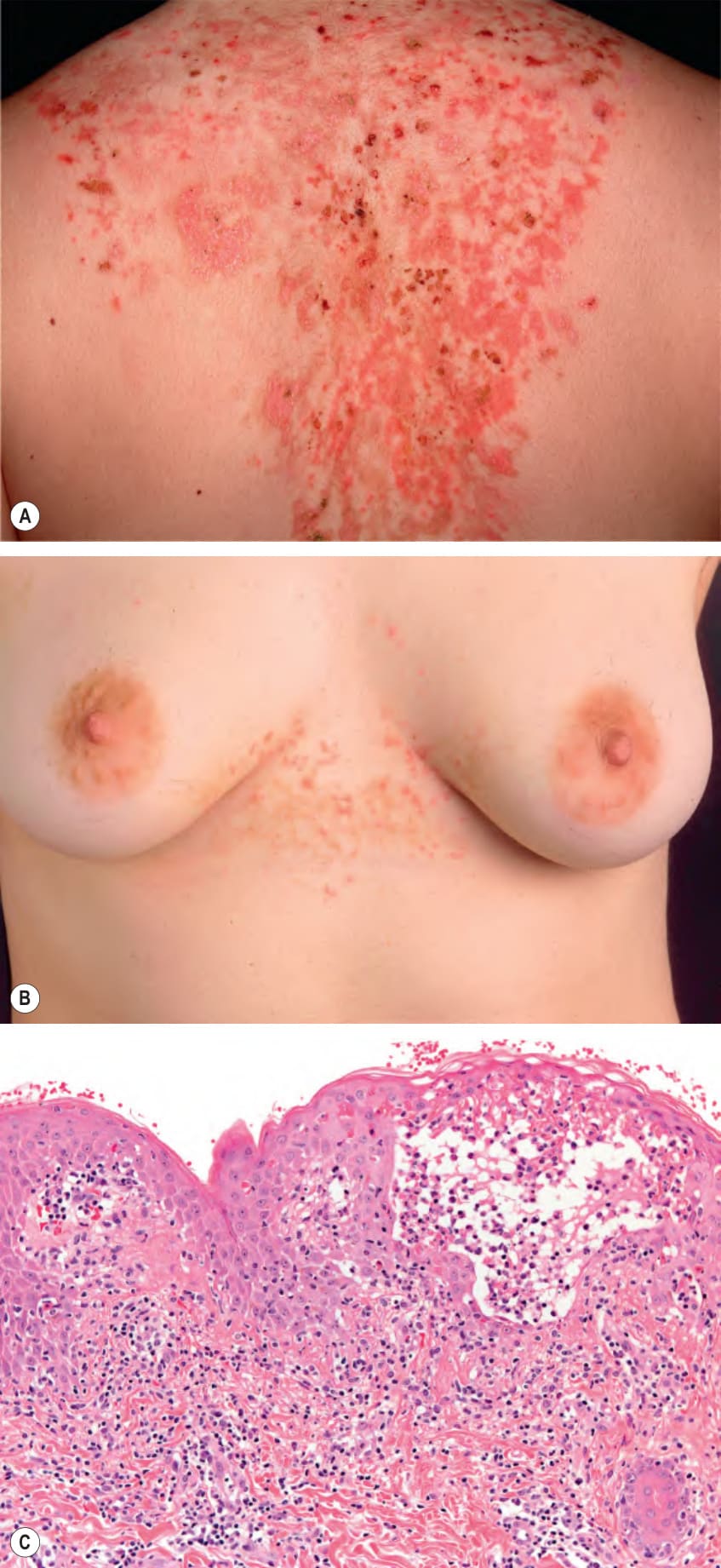
Fig. 67.21 Prurigo pigmentosa. A Eroded and crusted erythematous papules and plaques. A few patches of hyperpigmentation are seen inferiorly. B Erythematous crusted papules admixed with reticulated macular hyperpigmentation. C Histopathologic features include intraepidermal vesiculation, necrotic keratinocytes, and a patchy lichenoid infiltrate. A, Courtesy Luis Requena, MD; B, C, Courtesy Lorenzo Cerroni, MD.
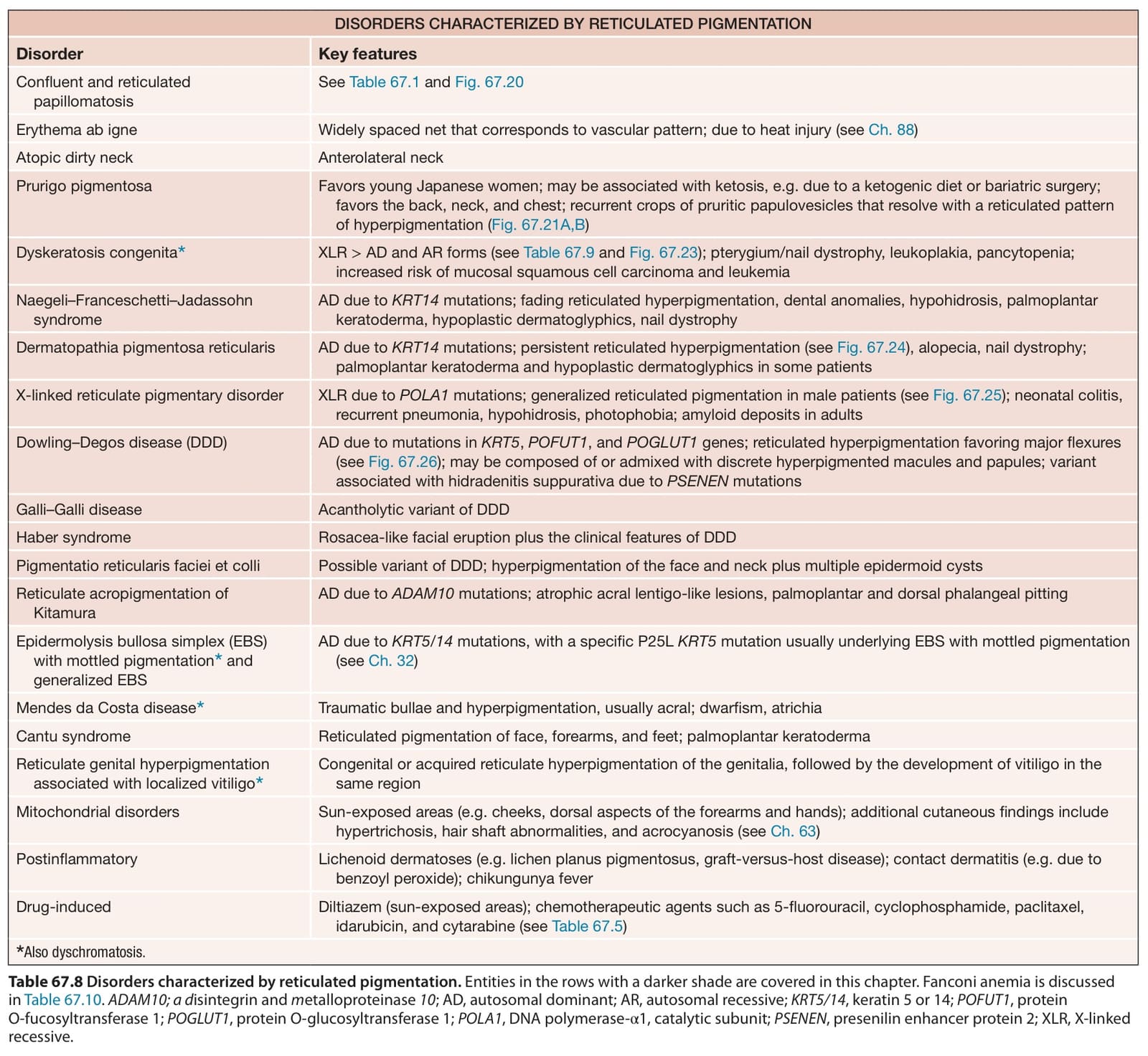
Table 67.8 Disorders characterized by reticulated pigmentation. Entities in the rows with a darker shade are covered in this chapter. Fanconi anemia is discussed in Table 67.10. ADAM10; a disintegrin and metalloproteinase 10; AD, autosomal dominant; AR, autosomal recessive; KRT5/14, keratin 5 or 14; POFUT1, protein O-fucosyltransferase 1; POGLUT1, protein O-glucosyltransferase 1; POLA1, DNA polymerase-α1, catalytic subunit; PSENEN, presenilin enhancer protein 2; XLR, X-linked recessive.